

Color Atlas of Neurology
.pdfDisturbances of Orientation
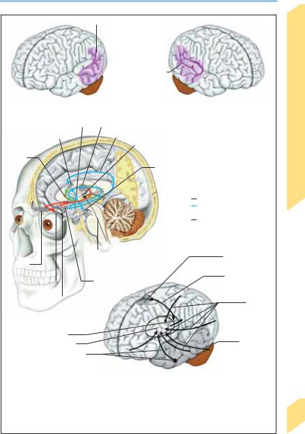
Hemispatial neglect
(left side) (task was to draw a clockface and set it to “quarter past 12”)
Hemispatial neglect (left side)
Behavioral Manifestations of Neurological Disease
133
Rohkamm, Color Atlas of Neurology © 2004 Thieme
All rights reserved. Usage subject to terms and conditions of license.

Behavioral Manifestations of Neurological Disease
134
Disturbances of Memory
Memory
Memory involves the acquisition, storage, recall, and reproduction of information. Memory depends on intact functioning of the limbic system (p. 144) and areas of the brain that are connected to it.
Declarative or explicit memory (i.e., memory for facts and events) can be consciously accessed and depends on intact functioning of the mediobasal portion of the temporal lobe. The duration of information storage may be relatively short (short-term, immediate, and working memory) or long (long-term memory). Verbal (telephone number) or visuospatial information (how to find a street) can be directly recalled from shortterm memory. The entorhinal cortex plays a key role in these memory functions: all information from cortical regions (frontal, temporal, parietal) travels first to the entorhinal cortex and then, by way of the parahippocampal and perirhinal cortex, to the hippocampus. There is also a reciprocal projection from the hippocampus back to the entorhinal cortex. Long-term memory stores events of personal history that occurred at particular times (episodic memory for a conversation, one’s wedding day, last year’s holiday; orbitofrontal cortex) as well as conceptual, non–time-related knowledge (semantic memory for the capital of Spain, the number of centimeters in a meter, the meaning of the word “stethoscope”; subserved by different cortical regions).
Nondeclarative (procedural, implicit) memory, on the other hand, cannot be consciously accessed. Learned motor programs (riding a bicycle, swimming, playing the piano), problem-solving (rules), recognition of information acquired earlier (priming), and conditioned learning (avoiding a hot burner on the stove, sitting still in school) belong to this category. Nondeclarative memory is mediated by the basal ganglia (motor function), neocortex (priming), cerebellum (conditioning), striatum (agility), amygdala (emotional responses), and reflex pathways.
Examination. Only disturbances of declarative memory (amnesia) can be studied by clinical examination. Short-term memory: the acquisition of new information is tested by having the patient repeat a series of numbers or groups of words and asking for this information again
5–10 minutes later. The patient’s orientation (name, place of residence/address, time/date) and long-term memory (place of birth, education, place of employment, family, general knowledge) are also tested by directed questioning.
Memory Disorders (Amnesia)
Forgetfulness. Verbal memory does not decline until approximately age 60, and even then only gradually, if at all. Aging is, however, often accompanied by an evident decline in information processing ability and attention span (benign senescent forgetfulness). These changes occur normally, yet to a degree that varies highly among individuals, and they are often barely measurable. They are far less severe than fullblown dementia, but they may be difficult to distinguish from incipient dementia.
Amnesia. Anterograde amnesia is the inability to acquire (declarative) information, for later recall, from a particular moment onward; retrograde amnesia is the inability to remember (declarative) information acquired before a particular moment (p. 269). Amnestic patients commonly confabulate (i.e., fill in gaps in memory with fabricated, often implausible information); they may be disoriented and lack awareness of their own memory disorder. For individual symptoms and their causes, see Table 13 (p. 365).
Rohkamm, Color Atlas of Neurology © 2004 Thieme
All rights reserved. Usage subject to terms and conditions of license.
Disturbances of Memory
|
|
|
|
|
|
|
|
Word recollection/recognition |
|||||
|
|
|
|
|
|
|
|||||||
|
|
|
|
|
|
|
|
(“verbal memory”) |
|||||
|
|
|
|
|
|
Spatial perception and |
|||||||
|
|
|
|
|
|
orientation; recognition |
|||||||
|
|
|
|
|
|
of familiar faces |
|||||||
|
|
|
|
|
|
(“visuospatial memory”) |
|||||||
Mamillothalamic tract |
|
Temporal cortical representation |
|||||||||||
|
|
|
|
|
Medial thalamic nuclei |
||||||||
|
|
|
|
|
|||||||||
Anterior thalamic |
|
|
|
|
|
|
|
|
|
|
Fornix |
||
|
|
|
|
|
|
|
|
|
|
||||
|
|
|
|
|
|
|
|
||||||
nuclei |
|
|
|
|
|
|
|
|
|
|
Intralaminar |
||
|
|
|
|
|
|
|
|
|
|
||||
Septal |
|
|
|
|
|
|
|
|
|
|
nuclei |
||
|
|
|
|
|
|
|
|
|
|
|
|||
nuclei |
|
|
|
|
|
|
|
|
|
|
Mamillary |
||
|
|
|
|
|
|
|
|
|
|
|
|
|
|
|
|
|
|
|
|
|
|
|
|
|
|
|
body |
Limbic system
Inner ring
Outer ring (Papez circuit)
Medial forebrain bundle
|
|
|
|
Reticular activating |
Premotor cortex |
|||
|
|
|
|
system (RAS) |
||||
Orbito- |
|
|
||||||
|
|
|
|
|
||||
frontal cortex |
|
|
|
Thalamocortical |
||||
Entorhinal |
|
|
|
Hippocampus, |
||||
|
|
|
|
projection |
||||
|
|
|
||||||
region |
|
parahippocampal |
|
|||||
|
|
|
||||||
|
|
Amygdala |
gyrus |
|
|
|||
|
|
|
|
|
|
Cortical |
||
|
|
|
|
|
|
|
|
projections |
Explicit memory* |
|
|
|
|
to the basal |
|||
|
|
|
|
ganglia |
||||
|
|
|
|
|
|
|
|
|
|
|
Thalamus |
|
|
|
|
|
|
|
|
Basal ganglia |
|
|
|
|
Cerebellar |
|
|
|
Substantia nigra, |
|
|
|
|
projections |
|
|
|
|
|
|
|
|
||
|
|
nigrostriatal |
|
|
|
|
|
|
|
|
projection |
|
|
|
Implicit memory* |
||
|
|
|
|
|
|
|
|
|
|
Structure |
|
Function* |
|
|
|||
|
Orbitofrontal cortex |
|
Activation, drive, long-term memory |
|
|
|||
|
Entorhinal area |
|
Visual memory, recognition |
|
|
|
||
|
Amygdala |
|
Emotional memory |
|
|
|||
|
Hippocampus |
|
Spatial memory, spatial orientation |
|
|
|
||
|
Mamillary body/diencephalon |
|
Long-term memory, insight, flexibility |
|
|
|||
|
RAS |
|
Activation |
|
*Model |
|||
|
|
|
|
|
|
|
|
|
Behavioral Manifestations of Neurological Disease
135
Rohkamm, Color Atlas of Neurology © 2004 Thieme
All rights reserved. Usage subject to terms and conditions of license.

Behavioral Manifestations of Neurological Disease
Dementia
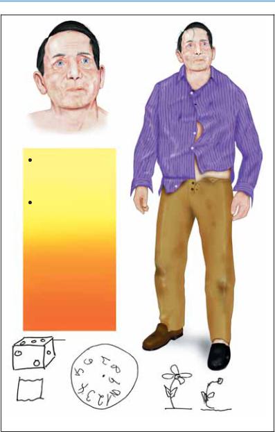
Dementia is a newly occurring, persistent, and progressive loss of cognitive function. Both short-term and long-term memory are impaired, in conjunction with at least one of the following disorders: aphasia, apraxia, agnosia, or impairment of abstract thinking, decisionmaking ability, visuospatial performance, planned action, or personality. Professional, social and interpersonal relationships deteriorate, and the sufferer finds it increasingly difficult to cope with everyday life without help. The diagnosis of dementia requires the exclusion of disturbances of consciousness (e. g., delirium) and of psychiatric disease (e. g., depression, schizophrenia). The differential diagnosis also includes benign senescent forgetfulness (“normal aging,” in which daily functioning is unimpaired) and amnestic disorders. Approximately 90% of all cases of dementia are caused by Alzheimer disease (p. 297) or cerebrovascular disorders; diverse etiologies account for the rest. The physician confronted with a case of incipient dementia must distinguish primary dementia from that secondary to another disease (Table 14, p. 366). The objective is early determination of the etiology of dementia, especially when these are treatable or reversible.
Examination. The patient or another informant should be asked for an account of the duration, type, and extent of problems that arise in every-
day life. The clinical examination is used to ascertain the type and severity of cognitive deficits and any potential underlying disease. Standardized examining instruments are useful for precise documentation and differentiation of the cognitive deficits. Rapid tests for dementia, such as the Mini-Mental Status Examination, mini-syndrome test, and clock/numbers test, are useful for screening. Function-specific neurophysiological tests permit diagnostic assessment of individual aspects of cognition including orientation, attention, concentration, memory, speech, and visual constructive performance. Laboratory tests (ESR1, differential blood count, electrolytes, liver function tests, BUN2, creatinine, glucose, vitamin B12, folic acid, TPHA3, TSH4, and HIV5), EEG, and diagnostic imaging techniques (CT6, MRI7, SPECT8, and PET9) provide further useful information for classification and determination of the cause of dementia. None of these diagnostic techniques alone can pinpoint the etiology of dementia; definitive diagnosis practically always requires multiple tests and examinations. Diagnostic imaging is of particular importance in patients with the subacute onset of cognitive impairment or amnesia (!1 month), fluctuation or acute worsening of symptoms, papilledema, visual field defects, headaches, a recent head injury, known malignancies, epilepsy, a history of stroke, urinary incontinence, or an abnormal gait.
1ESR |
Erythrocyte sedimentation rate |
2BUN |
Blood urea nitrogen |
3TPHA |
Treponema pallidum hemagglutination test |
4TSH |
Thyroid-stimulating hormone |
5HIV |
Human immunodeficiency virus |
6CT |
Computerized tomography |
7MRI |
Magnetic resonance imaging |
8SPECT |
Single photon emission computerized tomography |
9PET |
Positron emission tomography |
136Additional diagnostic tests to be performed as needed: Coagulation profile, serum protein electrophoresis, serum ammonia, parathyroid hormone, cortisol, rheumatoid factor, antinuclear antibodies, blood alcohol, serum/urine drug levels, copper/ceruloplasmin, lactate/pyruvate, hexosaminidase, CSF tests, molecular genetic analysis.
Rohkamm, Color Atlas of Neurology © 2004 Thieme
All rights reserved. Usage subject to terms and conditions of license.
Dementia
|
Disease |
|
Neurological |
|
of |
Loss of cognitive function |
Manifestations |
|
|
Memory impairment |
Behavioral |
(shortand long-term |
|
memory) |
|
Impairment of other |
|
higher cortical functions |
|
|
|
(abstraction, judgment, |
|
arithmetic, aphasia, apraxia, |
|
agnosia, attention) |
|
 Personality change
 Loss of social and occupational skills
Model drawing
|
|
|
|
Model |
|
|
|
Patient’s |
|
|
|
|
|
||||||
|
|
|
|
drawing |
|
|
copy |
|
|
|
|
|
|
|
|
|
|
|
|
Patient’s copy |
Clock face |
|
|
|
|
|
137 |
||
|
|
|
|
||||||
|
|
(patient’s drawing) |
|
|
|
||||
|
|
|
|
Personality change, cognitive impairment |
|
||||
|
|
|
|
|
|
|
|
|
|
Rohkamm, Color Atlas of Neurology © 2004 Thieme
All rights reserved. Usage subject to terms and conditions of license.

|
Pseudo-neurological Disorders |
|
|
Patients with symptoms and signs that are un- |
|
Disease |
||
usual, difficult to classify, or resistant to treat- |
||
ment are often referred to a neurologist for a de- |
||
termination whether the patient’s problem is |
||
Neurological |
“organic” or “psychogenic.” Many such patients |
|
may arise acutely or subacutely, perhaps in re- |
||
|
make a diagnostic and therapeutic odyssey from |
|
|
one medical or paramedical office to another, |
|
|
and have a long list of positive findings to show |
|
|
for it. In other patients, symptoms and signs |
|
of |
peated episodes, creating the impression of a se- |
|
Manifestations |
rious illness. The physician’s primary objectives |
|
must be (1) to identify the possible physical or |
||
|
||
|
psychosocial causes of the problem, and at the |
|
|
same time (2) to avoid unnecessary or danger- |
|
|
ous diagnostic tests. A correct diagnosis requires |
|
|
time, solid knowledge of the relevant anatomy |
|
Behavioral |
and physiology, and the ability to recognize the |
|
psychosocial dynamics that may have given rise |
||
|
||
|
to the patient’s complaints. |
|
|
If detailed neurological examination reveals no |
|
|
abnormality and the symptoms cannot be at- |
|
|
tributed to any neurological disease, the physi- |
|
|
cian should consider potential psychosocial |
|
|
causes. These may be unconscious (e. g., an inner |
|
|
conflict of which the patient is unaware) or con- |
|
|
scious (e. g., a deliberate attempt to acquire the |
|
|
financial benefits and increased attention as- |
|
|
sociated with illness). The underlying cause may |
|
|
be an unresolved social conflict (familial, pro- |
|
|
fessional, financial) or some other mental dis- |
|
|
order (depression, anxiety, obsessive-compul- |
|
|
sive disorder, personality disorder). Organic |
|
|
dysfunction and objective signs of illness are |
|
|
disproportionately mild in relation to the |
|
|
patient’s complaints, unrelated to them, or en- |
|
|
tirely absent. |
|
|
Conversion disorders (previously termed “con- |
|
|
version hysteria”) often present with a single |
|
|
(pseudoneurological) symptom, such as psycho- |
|
|
genic amnesia, stupor, mutism, seizures, paraly- |
|
|
sis, blindness, or sensory loss. It has been |
|
|
theorized that such symptoms serve to resolve |
|
|
unconscious inner conflicts. The diagnosis may |
|
|
be particularly difficult to make in patients who |
|
|
simultaneously suffer from organic neurological |
|
|
or psychiatric disease (e. g., hyperventilation in |
|
|
epilepsy, headaches in depression, paralysis in |
138multiple sclerosis).
Somatoform disorders, according to current
psychiatric terminology, are mental disorders
characterized by “repeated presentation of physical symptoms, together with persistent requests for medical investigations, in spite of repeated negative findings and reassurances by doctors that the symptoms have no physical basis” (ICD-10, WHO, 1992). In somatization disorder, the patient asks for treatment of multiple, recurrent, and frequently changing symptoms, which often affect multiple organ systems (e. g. headaches + bladder dysfunction + leg pains + breathing disorder). In hypochondriacal disorder
(previously termed “hypochondriasis”), the patient is less concerned about the symptoms themselves, and more preoccupied with the supposed presence of a serious disease. The fears persist despite repeated, thorough examination, normal test results, and medical reassurance. Any mild abnormalities that may happen to be found, e. g. of heartbeat, respiration, or intestinal function, or skin changes, only amplify the patient’s anxiety. Persistent somatoform pain disorder involves complaints of “persistent, severe, and distressing pain, which cannot be explained fully by a physiological process or a physical disorder” (ICD-10), though an organic cause of pain is often present as well. The physical impairment that the patient attributes to pain may actually be due to a lack of fulfillment in familial, professional, or social relationships. For these patients, dealing with the pain may become the major “purpose in life.” Malingering is not a mental disorder, but rather the deliberate, premeditated feigning of illness to achieve a goal (e. g., feigning of headaches to obtain opiates).
Simulated or intentionally induced (factitious) symptoms may serve no clear purpose (neither the resolution of an unconscious conflict, nor any obvious kind of gain); they may be present in the simulator (Münchhausen syndrome) or in a child or other person under their care (Münch- hausen-by-proxy). The peregrinating patient (hospital hopper) demands diagnostic tests from one physician after another, but negative test results can never put the patient’s fears to rest. Patients with Ganser syndrome give approximate or fatuous answers to simple questions, possibly creating the impression of dementia.
Rohkamm, Color Atlas of Neurology © 2004 Thieme
All rights reserved. Usage subject to terms and conditions of license.
Pseudo-neurological Disorders
Behavioral Manifestations of Neurological Disease
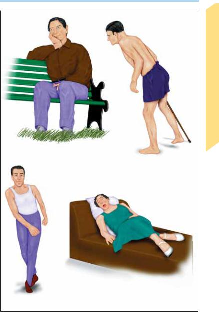
Depression and anxiety
(may give rise to pseudoneurological complaints)
Persistent somatoform pain disorder
Factitious gait disturbance |
Hypochondriacal disorder |
139 |
|
||
|
|
|
Rohkamm, Color Atlas of Neurology © 2004 Thieme
All rights reserved. Usage subject to terms and conditions of license.

Autonomic Nervous System
140
Organization
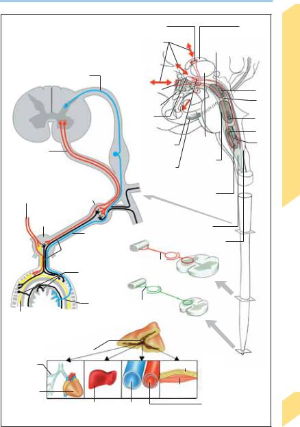
The autonomic nervous system (ANS) is so called because its functions are normally not subject to direct voluntary control. It regulates hormonal and immunological processes as well as the functioning of major organ systems (cardiovascular, respiratory, gastrointestinal, urinary, and reproductive systems).
ANS, Central Portion
Central components of the ANS are found in the cerebral cortex (insular, entorhinal, orbitofrontal, and frontotemporal areas), the hypothalamus, the limbic system, the mid brain (periaqueductal gray substance), the medulla (nucleus of the solitary tract, ventrolateral and ventromedial areas of the medulla), and the spinal cord (various tracts and nuclei, discussed in further detail below).
Afferent connections. Afferent impulses enter the ANS from spinal tracts (anterolateral fasciculus = spinothalamic + spinocerebellar + spinoreticular tracts), brain stem tracts (arising in the reticular formation), and corticothalamic tracts, and from the circumventricular organs. The latter are small clusters of specialized neurons, lying on the surface of the ventricular system, that sense changes in the chemical composition of the blood and the cerebrospinal fluid (i.e., on both sides of the blood–CSF barrier). These organs include the organum vasculosum of the lamina terminalis (in the roof of the third ventricle behind the optic chiasm cytokines/ fever), the subfornical organ (under the fornices between the foramina of Monro angiotensin II/blood pressure and fluid balance), and the area postrema (rostral to the obex on each side of the fourth ventricle cholecystokinin/ gastrointestinal function, food intake).
Efferent connections. Projections from the hypothalamus and brain stem, particularly from the brain stem reticular formation, travel to the lateral horn of the thoracolumbar spinal cord, where they form synapses onto the sympathetic neurons of the spinal cord. The latter, in turn, project preganglionic fibers to the sympathetic ganglia (p. 147). The parasympathetic neurons receive input from higher centers in similar fashion and project in turn to parasympathetic ganglia that are generally located near the end organs they serve. The hypothalamus regulates hormonal function through its regulator hormones as well as efferent neural impulses.
Neurotransmitters. The main excitatory neurotransmitter is glutamate, and the main inhibitory neurotransmitter is γ-aminobutyric acid (GABA). Modulating neurotransmitters include acetylcholine, amines, neuropeptides, purines, and nitric oxide (NO).
ANS, Peripheral Portion (p. 146)
The sympathetic and parasympathetic components are both structurally and functionally segregated.
Spinal nuclei. Sympathetic spinal neurons lie in the lateral horn (intermediolateral and intermediomedial cell columns) of the thoracolumbar spinal cord (T1–L2) and are collectively termed the thoracolumbar system. Parasympathetic spinal neurons lie in the brain stem (with projections along CN III, VII, IX, X) and the sacral spinal cord (S2–S4), and are collectively termed the craniosacral system. The intestine has its own autonomic ganglia, which are located in the myenteric and submucous plexuses (p. 154).
Afferent connections. Afferent impulses to the ANS enter the spinal cord via the dorsal roots, and the brain stem via CN III, VII, IX and X.
Efferent connections. The projecting fibers of the spinal autonomic neurons (preganglionic fibers) exit the spinal cord in the ventral roots and travel to the paravertebral and prevertebral ganglia, where they synapse onto the next neuron of the pathway. The sympathetic preganglionic fibers (unmyelinated; white ramus communicans) travel a short distance to the paravertebral sympathetic chain, and the postganglionic fibers (unmyelinated; gray ramus communicans) travel a relatively long distance to the effector organs. An exception to this rule is the adrenal medulla: playing, as it were, the role of a sympathetic chain ganglion, it receives long preganglionic fibers and then, instead of giving off postganglionic fibers, secretes epinephrine into the bloodstream. The parasympathetic preganglionic fibers are long; they project to ganglia near the effector organs, which, in turn, give off short postganglionic processes.
Neurotransmitters. Acetylcholine is the neurotransmitter in the sympathetic and parasympathetic ganglia. The neurotransmitters of the postganglionic fibers are norepineprhrine (sympathetic) and acetylcholine (parasympathetic). Neuromodulators include neuropeptides (substance P, somatostatin, vasoactive intestinal
Rohkamm, Color Atlas of Neurology © 2004 Thieme
All rights reserved. Usage subject to terms and conditions of license.
|
|
|
|
|
Organization |
|
|
|
Mamillothalamic tract |
|
Anterior |
|
|
|
|
|
|
|
thalamic nucleus |
|
|
|
Cortical afferent and |
Control of visuo-spatial |
|
||
|
|
efferent fibers |
|
orientation by ANS |
|
|
Spinal afferent fibers |
|
|
|
|
|
|
|
|
|
Medial |
|
|
|
|
|
|
fore brain |
|
III |
|
|
|
|
bundle |
|
|
|
|
|
|
|
IV |
System |
|
|
|
|
Hypothalamus |
|
||
|
|
|
|
|
||
|
|
|
Organon |
|
VII |
|
|
|
|
vasculosum |
|
|
|
|
|
|
|
|
Nervous |
|
|
|
|
laminae |
|
IX |
|
|
|
|
terminales |
|
||
|
|
|
Medial |
|
||
|
|
|
Dorsal |
X |
||
Spinal efferent |
|
|
thalamic |
|||
|
|
longitudinal |
nucleus |
|
||
fibers |
|
|
fasciculus |
Control of |
|
Autonomic |
|
|
|
Cingulum |
breathing, |
|
|
|
|
|
|
circulation, |
||
|
|
|
|
sucking, licking, |
||
|
|
|
|
chewing |
|
|
|
Sympathetic |
|
|
Control of |
|
|
Parasympathetic |
trunk |
|
|
vasomotor func- |
|
|
fibers |
|
|
|
tion, breathing, |
|
|
Autonomic |
|
|
cardiac function, |
|
||
|
|
vomiting |
|
|
||
plexus |
|
|
|
Reticular |
|
|
|
|
|
|
|
|
|
|
Viscerosensory pathway |
formation |
|
|
||
|
|
|
|
Area postrema |
|
|
|
Postganglionic |
|
|
|
|
|
|
sympathetic fibers |
Postganglionic |
|
|
|
|
|
|
|
|
|
|
|
|
Myenteric and |
sympathetic fibers |
|
|
|
|
|
|
|
|
|
||
|
submucous |
|
|
|
|
|
|
plexuses |
|
|
|
|
|
|
Viscero- |
Postganglionic |
|
|
|
|
Postganglionic |
sensory |
parasympathetic |
|
|
|
|
pathway |
fibers |
|
|
|
||
parasympathetic fibers |
|
|
Peripheral |
|
|
|
Peripheral pathways, |
|
|
|
|
||
|
|
portion of |
|
|
||
enteric nervous system |
|
|
ANS |
|
|
|
Adrenal medulla |
|
|
|
|
|
|
Dilatation |
|
|
Lipolysis |
Central portion |
|
|
|
|
of ANS |
|
|||
|
|
|
|
|
|
|
Increase in |
|
|
Glycogeno- |
|
|
|
|
|
lysis |
Vasoconstriction (skin, |
|
||
heart rate |
|
|
|
|||
|
Glycogenolysis |
Vasoconstriction |
viscera) and vasodilata- |
|
||
|
|
tion (muscles, |
141 |
|||
|
|
|
|
|
||
|
Effects of catecholamines (epinephrine, norepinephrine) |
coronary arteries) |
|
|||
polypeptides, thyrotropin-releasing hormone, |
lated peptide, neuropeptide Y, galanin, oxytocin, |
cholecystokinin, bombesin, calcitonin-gene-re |
enkephalins) and nitric oxide. |
Rohkamm, Color Atlas of Neurology © 2004 Thieme
All rights reserved. Usage subject to terms and conditions of license.

Hypothalamus
|
The hypothalamus lies in the anterior portion of |
|
|
the diencephalon, below the thalamus and |
|
|
above the pituitary gland. It forms part of the |
|
|
wall and floor of the third ventricle. Among its |
|
|
anatomical components are the preoptic area, |
|
|
infundibulum, tuber cinereum, and mamillary |
|
|
bodies. It is responsible for the control and inte- |
|
|
gration of endocrine function, thermoregulation |
|
|
(p. 152), food intake (p. 154), thirst, cardiovascu- |
|
|
lar function (p. 148), respiration (p. 150), sexual |
|
System |
function (p. 156), behavior and memory (p. 122 |
|
ff), and the sleep–wake rhythm (p. 112). Under |
||
|
||
|
the influence of changes in the external and in- |
|
Nervous |
ternal environment, and the emotional state of |
|
the individual, the hypothalamus controls the |
||
|
||
|
activity of the ANS through its neural and |
|
Autonomic |
humoral outflow. |
|
Neuroendocrine Control |
||
|
(Table 15, p. 367) |
|
|
The neuroendocrine control circuits of the hy- |
|
|
pothalamic-pituitary axis regulate the plasma |
|
|
concentration of numerous hormones. |
|
|
Adenohypophysis (anterior lobe of pituitary |
|
|
gland). Various regulatory hormones (releasing |
|
|
and inhibiting hormones) are secreted by hy- |
|
|
pothalamic neurons into a local vascular net- |
|
|
work, through which they reach the adenohy- |
|
|
pophysis to regulate the secretion of pituitary |
|
|
hormones into the systemic circulation. Among |
|
|
the pituitary hormones, the glandotropic hor- |
|
|
mones (TSH, ACTH, FSH, LH) induce the release |
|
|
of further hormones (effector hormones) from |
|
|
the endocrine glands, which, in turn, affect the |
|
|
function of the end organs, while the aglan- |
|
|
dotropic pituitary hormones (growth hormone, |
|
|
prolactin) themselves exert a direct effect on the |
|
|
end organs. Finally, the plasma concentration of |
|
|
the corresponding effector hormones and |
|
|
aglandotropic pituitary hormones affects the |
|
|
hypothalamic secretion of regulatory hormones |
|
|
in a negative feedback circuit (closed regulatory |
|
|
loop). |
|
|
Neurohypophysis (posterior lobe of pituitary |
|
|
gland). A subset of hypothalamic neurons pro- |
|
|
jects axons to the neurohypophysis. The bulb- |
|
|
like endings of these axons store oxytocin and |
142antidiuretic hormone and secrete them directly into the bloodstream (neurosecretion).
These hormones act directly on their effector
organs. The ensuing effects are sensed by the hypothalamus, thus closing the regulatory loop.
Rohkamm, Color Atlas of Neurology © 2004 Thieme
All rights reserved. Usage subject to terms and conditions of license.