

Color Atlas of Neurology
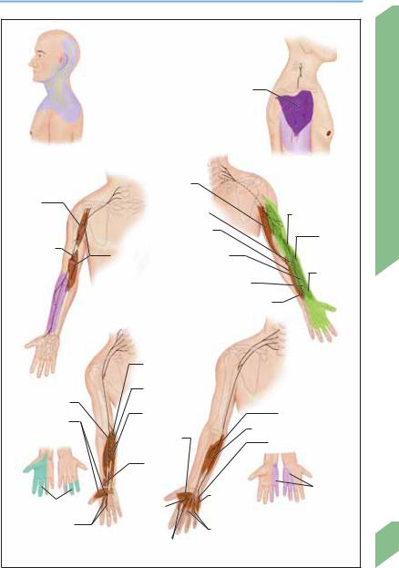
.pdfDermatomes and Myotomes
C 5
T 1
C 6
L 3
C 7
C 8
Rhomboid mm.
Supraspinatus m.
Infraspinatus m.
|
|
|
|
|
|
|
Diaphragm |
|
|
|
|
|
|
|
|
|
|
|
|
||
|
|
C 2 |
|
|
|
Iliopsoas m. |
|
|
||
|
|
|
|
|
|
|
||||
|
|
|
|
|
Gluteus |
|
|
|||
|
|
|
|
|
|
|
|
|
||
|
|
|
|
|
|
|
medius m. |
|
|
|
|
|
C 3 |
|
|
C 2 |
|
|
|
||
|
|
|
|
|
|
|
|
|
||
C 4 |
|
|
|
C 3 |
C 4 |
|
|
|
|
|
|
2 |
|
|
|
|
|
|
|||
|
|
|
|
|
|
|
|
|
||
|
T |
|
|
2 |
|
|
|
|
||
|
|
3 |
T |
|
|
|
|
|||
|
T |
|
|
|
|
|
Gluteus |
|||
|
T4 |
T 3 |
|
|
||||||
|
|
maximus m. |
||||||||
|
T5 |
T 4 |
|
C 5 |
||||||
|
|
|
|
|
||||||
|
T6 |
T 5 |
|
|
|
Adductors |
||||
|
|
7 |
T 6 |
|
|
|
||||
|
T |
|
|
|
|
|
|
|||
|
|
8 |
T 7 |
|
|
|
|
|
||
|
T |
|
|
|
|
Tibialis |
||||
|
T |
|
T 8 |
|
|
|
||||
|
|
9 |
|
|
|
|
|
|
|
|
|
10 |
T |
9 |
|
|
posterior |
||||
T |
11 |
|
|
|
|
|||||
T |
|
T10 |
|
C 6 |
|
m. |
||||
T12 |
|
|
||||||||
|
|
1 |
11 |
|
|
|
|
|||
|
|
L |
2 |
|
|
|
|
|||
|
|
|
T |
12 |
|
|
|
|
||
|
|
L |
|
|
|
|
|
|||
|
|
|
|
T |
L 1 |
T 1 |
|
|
|
|
|
|
|
|
|
|
|
|
|
||
|
|
|
|
|
|
L 2 |
|
|
|
|
|
|
|
|
|
|
|
|
|
|
|
|
|
|
S 2 |
|
L 3 |
|
C 8 |
|
|
|
|
|
|
|
|
Triceps brachii m. |
|
|
|||
S 2 |
C 7 |
|
L 4 |
||
|
Pectoralis m.
Deltoid m.
Biceps brachii m.
Brachioradialis m.
Thenar
muscles
Interossei mm.
Hypothenar muscles
Quadriceps
femoris m.
Tibialis anterior m.
Peroneus longus m.
Extensor hallucis longus m.
Gastrocnemius m.
L 4 |
L 5 |
Myotomes |
|
(left, posterior view; right, anterior view) |
|||
|
|
S 1
L 5
S 1  S 1
S 1
L 4 |
|
L 5 |
L 5 |
|
Dermatomes (left, posterior view; right, anterior view)
Dermatomes and Myotomes
33
Rohkamm, Color Atlas of Neurology © 2004 Thieme
All rights reserved. Usage subject to terms and conditions of license.

Peripheral Nervous System
34
Brachial Plexus
|
|
|
|
|
Hypoglossal n. (XII) |
|
|
|
|
|
|
|
|
|
|
|
Lower trunk (C8/T1) |
|||||||
|
|
|
|
|
|
|
|
|
|
|
|
|
|
|
|
|||||||||
|
|
|
|
|
Great auricular n. |
|
|
|
|
|
|
|
|
|
|
|
|
|
|
|
|
|
|
|
|
|
|
|
|
|
|
|
|
|
|
|
|
|
|
|
|
|
|
|
|
|
|
||
|
|
|
|
|
Lesser occipital n. |
|
|
|
|
|
|
|
|
|
|
|
|
|
|
|
|
C 1 |
||
|
|
|
|
|
|
|
|
|
|
|
|
|
|
|
|
|
|
|
|
|
||||
|
|
|
|
|
Transversus colli n. |
|
|
|
|
|
|
|
|
|
|
|
|
|
|
|
||||
|
|
|
|
|
|
|
|
|
|
|
|
|
|
|
|
|
|
|
|
C 2 |
||||
|
|
|
|
|
|
|
|
|
|
|
|
|
|
|
|
|
|
|
|
|||||
|
|
|
|
|
Ansa cervicalis (from C1 to C3) |
|
|
|||||||||||||||||
|
|
|
|
|
|
|
C 3 |
|||||||||||||||||
|
|
|
|
|
Supraclavicular nn. |
|
|
|
|
|
|
|
|
|
|
|||||||||
|
C 3 |
|
|
|
|
|
|
|
|
|
|
|
|
|
C 4 |
|||||||||
|
|
|
|
|
|
|
|
|
|
|
|
|
|
|
|
|
|
|
|
|
|
|||
|
|
|
|
|
Middle trunk (C7) |
|
|
|
|
|
|
|
|
|
|
|
|
|
|
|
|
|||
|
|
|
|
|
|
|
|
|
|
|
|
|
|
|
|
|
|
|
|
|
C 5 |
|||
|
|
|
|
|
|
|
|
|
|
|
|
|
|
|
|
|
|
|
|
|
||||
|
|
|
|
|
Upper trunk (C5/C6) |
|
|
|
|
|
|
|
|
|
|
|
|
|
||||||
Dia- |
C 4 |
|
|
|
|
|
|
|
|
|
|
|
|
|
|
|
|
C 6 |
||||||
|
|
|
Dorsal scapular n. (C3-C5) |
|
|
|
|
|
|
|
|
|
|
|||||||||||
phragm |
|
|
|
|
|
|
|
|
|
|
|
|
|
|||||||||||
|
|
|
|
|
|
|
|
|
|
|
|
|
|
C 7 |
||||||||||
|
|
|
|
|
Suprascapular n. (C4-C6) |
|
|
|
|
|
|
|
|
|
|
|
||||||||
|
|
|
|
|
|
|
|
|
|
|
|
|
|
|
|
|||||||||
|
|
|
|
|
Subclavian n. (C5/C6) |
|
|
|
|
|
|
|
|
|
|
C 8 |
||||||||
|
|
|
Musculocutaneous |
|
|
|
|
|
|
|
|
|
|
|
|
|
|
|
|
T 1 |
||||
|
|
|
n. (C5-C7) |
|
|
|
|
|
|
|
|
|
|
|
|
|
|
|
|
|
|
|||
|
|
|
Axillary n. (C5/C6) |
|
|
|
|
|
|
|
|
|
|
|
|
|
|
|
|
|
|
|||
|
|
|
Median n. |
|
|
|
|
|
|
|
|
|
|
|
|
|
|
|
|
|
|
|||
|
|
|
(C5-T1) |
|
|
|
|
|
|
|
|
|
|
|
|
|
|
|
|
|
|
|
Medial |
|
|
|
|
Axillary a. |
|
|
|
|
|
|
|
|
|
|
|
|
|
|
|
|
|
pectoral n. |
|||
|
|
|
|
|
|
|
|
|
|
|
|
|
|
|
|
|
|
|
|
(C8/T1) |
||||
|
|
|
|
|
|
|
|
|
|
|
|
|
|
|
|
|
|
|
|
|
|
|
|
|
C 3/C 4 |
|
Radial n. (C5-T1) |
|
|
|
|
|
|
|
|
|
Medial cord |
|
|
Ribs 1 and 2 |
|||||||||
|
|
|
|
|
|
|
|
|
|
|
|
|
|
|||||||||||
|
|
Ulnar n. (C7/8-T1) |
|
|
|
|
|
|
|
|
|
(C8/T1) |
|
|
Long thoracic |
|||||||||
|
|
|
|
|
|
|
|
|
|
|
|
|
|
|
|
|
|
|||||||
|
|
|
|
|
|
|
|
|
|
|
|
|
|
|
|
|
|
|
n. (C5-C7) |
|||||
|
|
Medial cutaneous n. of forearm |
|
|
|
|
|
|
|
|
|
|
|
|
|
|
|
|
|
|||||
|
|
|
|
|
|
|
|
|
|
Lateral cord |
|
|
|
Phrenic n. |
||||||||||
|
|
Medial cutaneous n. of arm |
|
|
|
|
|
|
|
|
|
|
|
|||||||||||
|
|
|
|
|
|
|
|
|
|
|
|
|
||||||||||||
|
|
|
|
|
|
|
|
|
(C5-C7) |
|
|
|
(C3/C4) |
|||||||||||
|
|
Posterior cord (C5-C8) |
|
|
|
|
|
|
|
|
|
|
|
|
Cervicobrachial plexus |
|||||||||
|
|
|
|
|
|
|
|
|
|
|
|
|
|
|
|
|
|
|
||||||
Deltoid m. |
|
|
|
|
|
|
|
|
|
|
|
|
|
|
|
|
|
|
(C = cervical vertebra; |
|||||
|
|
|
|
|
|
|
|
|
|
|
|
|
|
|
|
|
|
|
T = thoracic vertebra) |
|||||
|
|
Biceps |
|
|
Pectoralis |
|
|
|
|
|
|
|
|
|
|
|
|
|
|
|
|
|
|
|
|
|
brachii m. |
major m. |
|
|
|
|
|
|
|
|
|
|
|
|
|
|
|
|
|
|
|||
|
|
|
|
|
|
|
|
|
|
|
|
|
|
|
|
|
|
|
|
|
|
Flexor carpi ulnaris |
||
|
|
|
|
|
Triceps |
|
|
|
|
|
|
|
|
|
|
|
Pronator |
|
|
m. |
||||
|
|
|
|
|
|
|
|
|
|
|
|
|
|
|
|
|
|
|||||||
|
|
|
|
|
|
|
|
|
|
|
|
|
|
|
|
teres m. |
|
|
|
|
||||
|
|
|
|
|
brachii m. |
|
|
|
|
|
|
|
|
|
|
|
|
|
|
|
||||
|
|
|
|
|
|
|
|
|
|
|
|
|
|
|
|
|
|
|
|
|
|
|
||
|
Supraand |
|
|
|
|
|
|
|
|
|
|
Abductor |
|
|
|
|
||||||||
|
infraspinatus |
Extensor |
|
|
|
|
|
|
|
|
|
|
|
|
||||||||||
|
|
|
|
|
|
|
|
|
digiti quinti m. |
|
|
|
|
|||||||||||
|
mm. |
|
|
|
carpi radialis |
|
|
|
|
|
|
|
|
|
|
|
|
|||||||
|
|
|
|
|
|
|
|
|
|
|
|
|
|
|
|
|
|
|
|
|
|
|||
|
|
|
|
|
m. |
|
|
|
|
|
|
|
|
Interos- |
|
|
|
|
|
|
||||
|
|
|
|
|
|
|
|
|
|
|
|
|
|
|
|
|
|
|
|
|
||||
|
|
|
|
|
|
|
|
|
|
|
|
|
|
|
|
|
|
|
|
|||||
|
|
|
|
|
Abductor |
|
|
|
|
|
|
|
|
sei mm. |
|
|
|
|
||||||
|
|
|
|
|
pollicis |
|
|
|
|
|
|
|
|
|
|
|
|
|
|
|
|
|
|
|
|
Brachioradialis m. |
brevis m. |
|
|
|
|
|
|
|
|
Flexor carpi |
|
|
|
|
|||||||||
|
|
|
|
|
Opponens |
|
|
|
|
|
|
|
|
radialis m. |
|
|
|
|
||||||
|
|
|
|
|
|
|
|
|
|
|
|
|
|
|
|
|
|
|
|
|
|
|
||
|
|
|
|
|
pollicis m. |
|
|
|
|
|
|
|
|
Flexor policis |
|
|
|
|
||||||
|
|
|
|
|
|
|
|
|
|
|
|
|
|
|
|
|
||||||||
|
|
|
|
|
|
|
|
|
|
|
|
|
|
|
brevis m. |
|
|
|
|
|||||
C 5 |
|
C 6 |
|
|
C 7 |
|
|
|
|
|
|
|
|
C 8 |
|
|
|
|||||||
(Dermatome: blue) |
(Dermatome: dark red) |
(Dermatome: violet) |
(Dermatome: light red) |
|||||||||||||||||||||
|
|
|
|
|
|
|
|
|
|
|
|
|
|
|
|
|
|
|
|
|
|
|
|
|
Rohkamm, Color Atlas of Neurology © 2004 Thieme
All rights reserved. Usage subject to terms and conditions of license.
Nerves of the Upper Limb
Cervical plexus
(C1-C4, cutaneous distribution)
|
C 5 |
Coraco- |
C 6 |
C 7 |
|
brachialis m. |
|
Brachialis m.
Biceps brachii m.
Musculocutaneous n.
C 5
C 6
C 7
C 8
T 1
C 5
C 6
Deltoid m.
|
C 5 |
Axillary nerve |
|
C 6 |
|
|
C 7 |
|
Triceps |
C 8 |
|
brachii m. |
T 1 |
|
|
|
|
Supinator m. |
|
Brachioradialis m. |
|
|
|
Extensor carpi |
|
Extensor |
ulnaris m. |
|
|
|
|
carpi radialis |
Extensor digitorum |
longus m. |
|
|
||
communis m. |
|
|
|
|
Abductor |
Extensor pollicis longus m. |
pollicis |
|
|
|
longus m. |
Branches to extensor digiti quinti, extensor pollicis brevis, and extensor indicis mm.
C 8 |
Radial n. |
|
T 1 |
||
|
|
Flexor carpi |
|
|
|
radialis m. |
|
|
|
Palmaris |
|
|
Pronator teres m. |
longus m. |
|
|
|
|
|
|
|
Flexor digitorum |
|
Flexor carpi ulnaris m. |
Abductor pollicis |
superficialis m. |
Flexor digitorum profundus m. |
|
brevis, flexor pollicis |
|
||
brevis, and opponens |
Flexor pol- |
|
Abductor digiti quinti m. |
pollicis mm. |
licis brevis m. |
|
|
|
Pronator |
|
|
|
quadratus |
|
|
|
m. |
|
Cutaneous |
|
|
|
|
Cutaneous distribution |
|
Flexor brevis |
distribution |
Adductor |
and opponens |
|
|
|
digiti quinti m. |
|
|
|
pollicis m. |
|
|
|
|
|
|
Lumbrical mm. 1-3 |
|
Lumbrical mm. 3 + 4 |
|
|
|
||
|
Dorsal and palmar interosseous mm. |
|
|
Median nerve |
|
|
Ulnar nerve |
Peripheral Nervous System
35
Rohkamm, Color Atlas of Neurology © 2004 Thieme
All rights reserved. Usage subject to terms and conditions of license.
Lumbar Plexus |
|
|
|
|
|
|
Subcostal n. |
Psoas major m. |
T 12 |
|
|
|
||
|
|
Iliohypogastric n. |
|
|
|
|
Ilioinguinal n. |
|
L 1 |
|
Rectus |
|
|
|
|
|
|
|
|
|
femoris m. |
Femoral n. |
|
L 2 |
|
Vastus lateralis m. |
Lateral cutane- |
|
L 3 |
|
|
|
||
|
|
ous n. of thigh |
|
|
|
Vastus medialis m. |
|
|
|
|
|
|
|
|
|
|
Genitofemoral n. |
|
L 4 |
|
|
|
|
|
System |
|
Obturator n. |
|
S 1 |
Lumbosacral trunk (peroneal n.) |
|
|||
|
|
Gluteal n. |
|
L 5 |
|
|
|
|
|
Nervous |
Lumbosacral trunk (tibial n.) |
|
S 2 |
|
|
S 3 |
|||
|
|
|
||
|
Pudendal n. (from |
|
S 4 |
|
|
|
S 5 |
||
|
coccygeal plexus) |
|
|
|
Peripheral |
|
Obturator n. |
|
|
|
|
|
|
|
|
|
Sciatic n. (peroneal |
|
|
|
|
and tibial n.) |
|
Adductor |
|
|
|
|
|
|
|
|
|
magnus m. |
|
|
Vastus |
|
|
|
|
lateralis n. |
|
|
|
|
Vastus |
|
|
L 3 |
|
intermedius |
|
|
(Dermatome: |
|
m. |
|
|
red; iliopsoas, |
Vastus |
Rectus femoris m. |
|
|
adductor longus, |
medialis |
|
|
|
adductor mag- |
m. |
Vastus medialis m. |
|
|
nus mm. not |
|
|
|
|
shown) |
|
|
Sartorius m. |
|
|
|
|
Gracilis m. |
|
|
|
Lumbosacral plexus |
|
|
Tibialis |
|
Extensor hallucis longus m. |
|
|
|
|
|
|
|
anterior m. |
|
Extensor digitorum brevis m. |
|
|
|
|
|
||
|
|
|
Gastrocnemius m. |
|
L 4 |
|
(medial and lateral heads) |
||
|
|
|
|
|
(Dermatome: green) |
|
|
|
|
|
|
|
Soleus m. |
|
|
L 5 |
|
S 1 |
|
36 |
(Dermatome: green; gluteus |
|
||
medius m. not shown) |
(Dermatome: yellow; gluteus |
|||
|
|
|
maximus not shown) |
|
Rohkamm, Color Atlas of Neurology © 2004 Thieme
All rights reserved. Usage subject to terms and conditions of license.

|
|
|
|
Nerves of the Lower Limb |
|
||
|
|
Psoas major m. |
|
|
|
|
|
|
L 1 |
Iliacus m. |
|
|
|
L 2 |
|
|
|
|
|
L 3 |
|
||
|
L 2 |
Inguinal lig. |
|
|
|
|
|
|
|
|
|
|
|
||
|
L 3 |
|
|
|
|
|
|
|
|
Iliohypogastric n. |
|
|
|
|
|
|
L 4 |
|
|
|
|
|
|
|
L 5 |
Cutaneous distribution |
|
|
|
System |
|
|
|
|
|
|
|
||
|
|
Genitofemoral n. |
|
|
|
||
|
|
(genital branch) |
|
|
|
Nervous |
|
|
|
Genitofemoral n. |
|
|
|
||
|
|
(femoral branch) |
|
|
|
Peripheral |
|
|
|
Ilioinguinal n. |
|
|
|
||
|
|
|
|
|
|
||
|
|
|
Lateral cutaneous n. of thigh |
|
|||
Cutaneous innervation of the groin |
|
|
|
|
|
|
|
(left, in men; right, in women) |
|
|
Sciatic n. |
L 4 |
|
||
|
|
|
L 4 |
L 5 |
|
||
|
|
|
|
|
S 1 |
|
|
|
Iliacus m. |
|
L 5 |
|
|
|
|
|
|
Adductor |
S 2 |
|
|||
|
|
|
S 1 |
S 3 |
|
||
L 1 |
|
|
magnus m. |
|
|||
|
|
S 2 |
|
|
|||
L 2 |
Psoas |
|
|
|
|
|
|
L 3 |
major m. |
Sciatic n. |
|
Semiten- |
|
|
|
L 4 |
Pectineus |
|
dinosus m. |
|
|
||
|
|
|
|
|
|
|
|
|
m. |
Biceps |
|
Semimem- |
|
|
|
|
|
femoris m. |
|
|
|
||
|
|
|
branosus m. |
|
|
||
|
Anterior |
(short head) |
|
|
|
||
|
|
|
|
|
|
||
|
cutaneous |
Anterior |
|
Biceps |
|
|
|
|
branches |
|
|
|
|
||
|
|
femoris m. |
|
|
|
||
|
|
tibial m. |
|
|
|
|
|
|
|
|
(long head) |
|
|
||
|
Sartorius m. |
|
|
|
|
||
|
Common |
|
|
|
|
|
|
|
|
|
|
|
|
|
|
|
Rectus |
peroneal n. |
|
Tibial n. |
|
|
|
|
femoris m. |
Long |
|
Flexor digitorum |
|
||
|
|
|
|
||||
|
Vastus |
peroneal m. |
|
longus m. |
|
|
|
|
intermedius m. |
Extensor |
|
|
|
|
|
|
|
|
Peroneus |
|
|
|
|
|
Vastus |
digitorum |
|
|
|
|
|
|
|
brevis m. |
|
|
|
||
|
lateralis m. |
longus m. |
|
|
|
|
|
|
|
|
|
|
|
||
|
Vastus |
Saphenous n. |
|
Gastro- |
|
|
|
|
|
cnemius m. |
|
|
|||
|
medialis m. |
|
|
|
|
||
|
Intermediate |
|
|
|
|
|
|
|
|
|
|
|
|
|
|
|
|
dorsal |
|
|
|
|
|
|
Saphenous n. |
cutaneous n. |
|
|
|
|
|
Femoral nerve |
Femoral nerve |
Sciatic nerve, |
|
|
Sciatic nerve, |
|
|
|
(cutaneous distribution) |
peroneal nerve |
|
tibial nerve |
37 |
||
|
|
|
(purple: cutaneous |
(purple: cutaneous |
|||
|
|
|
distribution) |
|
|
distribution) |
|
Rohkamm, Color Atlas of Neurology © 2004 Thieme
All rights reserved. Usage subject to terms and conditions of license.

38
Rohkamm, Color Atlas of Neurology © 2004 Thieme
All rights reserved. Usage subject to terms and conditions of license.

Normal and Abnormal
Function of the Nervous
2 System
!Neural Pathways
!Pathophysiology
!Major Syndromes
Rohkamm, Color Atlas of Neurology © 2004 Thieme
All rights reserved. Usage subject to terms and conditions of license.

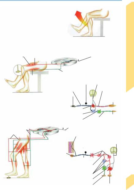
Reflexes
|
Reflexes are involuntary and relatively stereo- |
|
|
typed responses to specific stimuli. Afferent |
|
|
nerve fibers conduct the impulses generated by |
|
|
activated receptors to neurons in the central |
|
|
nervous system, which fire impulses that are |
|
|
then transmitted through efferent nerve fibers |
|
|
to the cells, muscles, or organs that carry out the |
|
|
reflex response. The pathway as a whole is |
|
|
known as the reflex arc. Receptors are found at |
|
|
the origin of all sensory pathways—in the skin, |
|
|
mucous membranes, muscles, tendons, and pe- |
|
|
riosteum, as well as in the retina, inner ear, ol- |
|
|
factory mucosa, and taste buds. A reflex re- |
|
|
sponse may involve the somatic musculature or |
|
|
the internal organs. Most reflexes are relatively |
|
Function |
independent of the state of consciousness. An |
|
interruption of the reflex arc at any point |
||
|
||
|
weakens or abolishes the reflex. Intrinsic reflexes |
|
|
are those whose receptors and effectors are lo- |
|
Motor |
cated in the same organ (e. g., the quadriceps re- |
|
flex), while the receptors and effectors of extrin- |
||
|
||
|
sic reflexes are in different organs (e. g., the |
|
|
oculovestibular reflex). Reflexes are important |
|
|
for normal function (e. g., for postural control |
|
|
and goal-directed movement), and an impaired |
|
|
reflex is an important objective finding in clini- |
|
|
cal neurological examination. |
|
|
Intrinsic Muscle Reflexes (Phasic Stretch |
|
|
Reflexes, Tendon Reflexes) |
|
|
Intrinsic muscle reflexes are triggered by stretch |
|
|
receptors within the muscle (annulospiral nerve |
|
|
endings of muscle spindles). The impulses |
|
|
generated at the receptors are conveyed via af- |
|
|
ferent fast-twitch Ia fibers to spinal alpha-motor |
|
|
neurons, whose efferent α1 processes excite the |
|
|
agonistic muscle of an opposing muscle pair. |
|
|
The antagonistic muscle is simultaneously in- |
|
|
hibited by spinal interneurons. The resulting |
|
|
muscle contraction relaxes the muscle spindles, |
|
|
thereby stopping impulse generation at the |
|
|
stretch receptors. The spinal reflex arc is also |
|
|
under the influence of higher motor centers. |
|
|
Abnormal reflex responses imply an abnormal- |
|
|
ity of the musculature, the reflex arc, or higher |
|
|
motor centers. The most important reflexes in |
|
|
clinical diagnosis are the biceps (C5–C6), bra- |
40chioradialis (C5–C6), triceps (C7–C8), adductor (L2–L4), quadriceps (L2/3–L4), posterior tibial
(L5), and Achilles (S1–S2) reflexes.
Extrinsic Reflexes
Intrinsic muscle reflexes, discussed above, are monosynaptic, but extrinsic reflexes are polysynaptic: between their afferent and efferent arms lies a chain of spinal interneurons. They may be activated by stimuli of various types, e. g., muscle stretch, touch on the skin (abdominal reflex) or cornea (corneal reflex), mucosal irritation (sneezing), light (eye closure in response to a bright flash), or sound (acoustic reflex). The intensity of the response diminishes if the stimulus is repeated (habituation). Because they are polysynaptic, extrinsic reflexes have a longer latency (stimulus-to-response interval) than intrinsic reflexes. Some important extrinsic reflexes for normal function are the postural and righting reflexes, feeding reflexes (sucking, swallowing, licking), and autonomic reflexes (p. 110).
The flexor reflex is triggered by noxious stimulation, e. g., from stepping on a tack. Excitatory interneurons activate spinal cord alpha-motor neurons, which, in turn, excite ipsilateral flexor muscles and simultaneously inhibit ipsilateral extensor muscles via inhibitory interneurons. Meanwhile, the contralateral extensors contract, and the contralateral flexors relax. The response does not depend on pain, which is felt only when sensory areas in the brain have been activated, by which time the motor response has already occurred. This spinal reflex arc, like that of the intrinsic muscle reflexes, is under the influence of higher motor centers.
Abnormalities of the extrinsic reflexes imply an interruption of the reflex arc or of the corticospinal tracts (which convey impulses from higher motor centers). Some clinically important extrinsic reflexes are the abdominal (T6– T12), cremasteric (L1–L2), bulbocavernosus (S3– S4), and anal wink (S3–S5) reflexes.
Reflexes that can be elicited only in the diseased state are called pathological reflexes. Pathological reflexes indicating dysfunction of the pyramidal (corticospinal) tract include the Babinski sign (tonic dorsiflexion of the great toe on stimulation of the lateral sole of the foot), the Gordon reflex (same response to squeezing of the calf muscles), and the Oppenheim reflex (same response to a downward stroke of the examiner’s thumb on the patient’s shin).
Rohkamm, Color Atlas of Neurology © 2004 Thieme
All rights reserved. Usage subject to terms and conditions of license.
Reflexes
|
|
|
|
|
|
|
|
|
|
|
|
|
|
|
|
|
|
|
|
|
|
|
|
|
|
|
|
|
|
Reflex response |
Symbol |
|
|
|
|
|
|
|
|
|
|
|
|
|
|
|
|
|
|
|
|
|
|||
|
|
|
|
|
|
|
|
|
|
|
|
|
|
|
|
|
|
|
|
|
|
|
|
|
|
|
|
|
Absent, cannot be elicited |
|
|
|
|
|
|
|
|
|
|
|
|
|
|
|
|
|
|
|
|
|
|
|
|||
|
by maneuvers |
|
0 |
|
|
|
|
|
|
|
|
|
|
|
|
|
|
|
|
|
|
|
|
|
|
||
|
|
|
|
|
|
|
|
|
|
|
|
|
|
|
|
|
|
|
|
|
|
|
|
|
|
|
|
|
Can only be elicited by maneuvers |
+ |
|
|
|
|
|
|
|
|
|
|
|
|
|
|
|
|
|
|
|
|
|
|
|||
|
(e.g., Jendrassik maneuver) |
_ |
|
|
|
|
|
|
|
|
|
|
|
|
|
|
|
|
|
|
|
|
|
|
|||
|
|
|
|
|
|
|
|
|
|
|
|
|
|
|
|
|
|
|
|
|
|
|
|
||||
|
|
|
|
|
|
|
|
|
|
|
|
|
|
|
|
|
|
|
|
|
|
|
|
|
|
|
|
|
Diminished |
|
1 |
|
|
|
|
|
|
|
|
|
|
|
|
|
|
|
|
|
|
|
|
|
|
||
|
Normal intensity |
|
2 |
|
|
|
|
|
|
|
|
|
|
|
|
|
|
|
|
|
|
|
|
|
|
||
|
Heightened |
|
3 |
|
|
|
|
|
|
|
|
|
|
Reflex response |
|||||||||||||
|
Persistent clonus |
|
4 |
|
|
|
|
|
|
|
|
|
|
(Proprioceptive muscle reflex) |
|||||||||||||
|
|
|
|
|
|
|
|
|
|
|
|
|
|
|
|
|
|
|
|
|
|
|
|
Afferent (Ia) fiber |
|||
Extensor muscle |
Efferent fiber (excitatory) |
|
|
|
|
||||||||||||||||||||||
|
|
||||||||||||||||||||||||||
|
|
|
|
|
|
|
|
Efferent fiber (inhibitory) |
|
|
|
|
|
|
|||||||||||||
|
|
|
|
Flexor muscle |
|
|
|
|
|
|
|
|
|
|
|
|
|
|
|
|
|
|
|
|
|
|
|
|
|
|
|
|
|
Pseudounipolar nerve cells in |
Supraspinal control |
||||||||||||||||||||
|
|
|
|
|
|
spinal ganglion |
|
|
|
|
|
(inhibitory) |
|||||||||||||||
|
|
|
|
|
|
Afferent fiber |
|
|
|
|
|
|
|
Efferent to |
|||||||||||||
|
|
|
|
|
|
|
|
|
|
|
|||||||||||||||||
|
|
Proprioceptive |
|
Annulospiral |
|
|
|
|
|
|
|
|
|
|
|
|
|
|
|
|
extensors |
||||||
|
|
|
|
|
|
|
|
|
|
|
|
||||||||||||||||
|
|
(intrinsic) muscle |
|
ending of |
|
|
|
|
|
Extensor |
|||||||||||||||||
|
|
reflex |
|
|
muscle spindle |
|
|
|
|
|
|||||||||||||||||
|
|
|
|
|
|
|
|
|
|
||||||||||||||||||
Efferent fibers to |
|
|
Excitatory synapse |
|
|
|
|
|
|
|
|
|
|
|
|||||||||||||
|
|
|
|
|
|
|
|
|
|||||||||||||||||||
|
|
Interneuron |
|
|
|
|
|
|
|
|
|
|
|
|
|
|
|
|
|
|
|||||||
contralateral extensors |
|
|
|
|
|
|
|
|
|
|
|
|
|
|
|
|
|
Flexor |
|||||||||
|
|
|
|
|
|
|
|
|
|
|
|
|
|||||||||||||||
|
|
|
|
|
|
|
|
|
|
|
|
|
|
|
|
|
|
|
|
|
|
|
|||||
and flexors |
|
|
|
Inhibitory synapse |
|
|
|
|
|
|
|
|
|
|
|
|
|||||||||||
|
|
|
|
|
|
|
|
|
|
|
|
|
|
|
|
|
|
|
|
|
|
|
Efferent to flexors |
||||
Efferent fibers to |
|
|
|
|
|
|
|
|
|
|
|
|
|
|
|
|
|
|
|
|
|
|
|
|
|
||
ipsilateral flexors |
|
|
|
|
|
|
Afferent (Ia) fiber |
|
|
|
|
|
|
||||||||||||||
and extensors |
|
|
|
|
|
|
|
|
|
|
|
|
Free ending of |
|
|
|
|
|
Supraspinal |
||||||||
|
|
|
|
|
|
|
|
|
|
|
|
|
|
|
|
|
|
|
|
||||||||
|
|
|
|
|
|
|
|
|
|
|
|
|
|
|
|
|
|
|
|
||||||||
|
|
|
|
|
|
|
|
|
|
|
|
|
|
|
afferent fiber (pain, |
|
|
|
|
control |
|||||||
|
|
|
|
|
|
|
|
|
|
|
|
|
|
|
temperature) |
|
|
|
|
|
(inhibitory) |
||||||
|
|
|
|
|
|
|
|
|
|
|
|
|
|
|
|
|
|
|
|
|
|
Interneurons |
|||||
|
|
|
|
Pressure receptor |
|
|
|
|
|
|
|
|
|
|
|
||||||||||||
|
|
|
|
|
|
|
|
|
|
|
|||||||||||||||||
|
|
|
|
(Vater-Pacini corpuscle) |
|
|
|
|
|
|
|||||||||||||||||
|
|
|
|
|
|
|
|
|
|
|
|
|
|
|
|
Afferent fiber |
|
|
|
|
|
||||||
|
|
|
|
Fibers to contralateral side of |
|
|
|
|
|
|
|
|
|||||||||||||||
|
|
|
|
|
|
|
|
|
|
|
|
||||||||||||||||
|
|
|
|
commissural cell |
|
|
|
|
|
|
|||||||||||||||||
|
|
|
|
|
|
|
|
|
Inhibitory synapse |
|
|
|
|
|
|
|
|
||||||||||
|
|
|
|
|
|
|
|
|
|
|
|
|
|
|
|||||||||||||
|
|
|
|
|
|
|
|
|
Excitatory synapse |
|
|
|
|
|
|
|
|||||||||||
|
|
|
|
|
|
|
|
|
|
|
|
|
|
||||||||||||||
|
Extrinsic muscle reflex |
|
|
|
|
Extensor muscle |
|
|
|
|
|
|
Flexor muscle |
||||||||||||||
|
|
|
|
|
|
|
|
|
|||||||||||||||||||
|
|
|
|
|
|
|
|
|
|
|
|
|
|
|
|
|
|
|
|
|
|
|
|
|
|
|
|
Motor Function
41
Rohkamm, Color Atlas of Neurology © 2004 Thieme
All rights reserved. Usage subject to terms and conditions of license.

Motor Function
Motor Control
The motor system controls the timing, direction, amplitude, and force of movement through the coordinated opposing actions of agonist and antagonist muscles. It also keeps the body in a stable position through postural and righting reflexes. Reflex movements are involuntary, stereotyped responses to stimuli. Rhythmic movements have both reflex and voluntary components. Voluntary movements are performed at will.
Reflex Movements
Withdrawing a foot from a noxious stimulus or spreading the arms when falling are examples of reflex movements. Intrinsic muscle reflexes regulate muscle tone and elasticity and are important for postural control and coordination of muscle groups. Specific functions such as joint stabilization or adjustment of contraction strength are achieved with the aid of inhibitory spinal interneurons. Extrinsic reflexes include protective reflexes (flexor response to noxious stimulus, corneal reflex) and postural reflexes (extensor reflex, neck reflex).
Rhythmic Movements
Walking, breathing, and riding a bicycle are rhythmic movements. They are subserved both by spinal reflex arcs and by supraspinal influence from the brain stem, cerebellum, basal ganglia, and motor cortex.
the cortex through thalamic relay nuclei. Fine motor control thus depends on the continuous interaction of multiple centers responsible for the planning (efferent copy) and execution of movement.
Motor cortex (p. 25). Voluntary movements are planned in the motor areas of the cerebral cortex. The primary motor area (area 4) regulates the force of muscle contraction and the goaloriented direction of movement; it mainly controls distal muscle groups. The supplementary motor area (medial area 6) plays an important role in complex motor planning. The premotor area (lateral area 6) receives nerve impulses from the posterior parietal cortex and is concerned with the visual and somatosensory control of movement; it mainly controls trunk and proximal limb movement.
Cerebellum (p. 54). The cerebellum coordinates limb and eye movements and plays an important role in the maintenance of balance and the regulation of muscle tone.
Basal ganglia (p. 210). The basal ganglia have a close anatomic and functional connection to the motor cortex and participate in the coordination of limb and eye movement.
Voluntary Movements
Voluntary movements depend on a sequence of contractions of numerous different muscles that is planned to achieve a desired result (motor program). Hence different parts of the body are able to carry out similar movements (motor equivalence) more or less skillfully, e. g., simultaneous rotation of the big toe, foot, lower leg, leg, pelvis and trunk. Voluntary movements incorporate elements of the basic reflex and rhythmic movement patterns; their smooth execution depends on afferent feedback from the visual, vestibular, and proprioceptive systems to motor centers in the spinal cord, brain stem, and
42cerebral cortex. Further modulation of voluntary movements is provided by the cerebellum and basal ganglia, whose neural output reaches
Rohkamm, Color Atlas of Neurology © 2004 Thieme
All rights reserved. Usage subject to terms and conditions of license.