Добавил:
Опубликованный материал нарушает ваши авторские права? Сообщите нам.
Вуз: Предмет: Файл:

Color Atlas of Neurology

.pdf
Скачиваний:
202
Добавлен:
11.08.2013
Размер:
18.87 Mб
Скачать

Epilepsy: Seizure Types

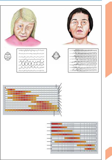
Ictal EEG: Focal activity on

left (frontoprecentral spike Clonus on right side waves)

of face

Clonus in right arm


50 V


Simple partial seizure


1s


Ictal EEG: Bilateral frontotemporal activity Oral automatisms (rhythmic ε waves)

(licking, chewing, lip smacking)

Oral automatisms


(snorting, throat


50 V


clearing, chewing)


1s


Complex partial seizure


Partial seizures (focal epilepsy)


Interictal

Prodromal

Tonic

Clonic


Postictal phase

phase

phase

phase

phase


Normal EEG

Focal or

Ictal ` and _

Rhythmic

Extinction

Irregular and


generalized

spike waves

slowing with

phase

high β and


dysrhythmia


occasional


sub-β wave


and slowing


spikes


activity

Generalized seizures (schematic representation of ictal EEG in grand mal seizure)

Central Nervous System

193

Rohkamm, Color Atlas of Neurology © 2004 Thieme

All rights reserved. Usage subject to terms and conditions of license.

Central Nervous System

Epilepsy: Seizure Types

Site of focus

Seizure type

Symptoms and signs


Frontal lobe

Simple or complex par-


tial seizures with or


without secondary


generalization (hypermo-


tor frontal lobe seizures)

Adversive head movement and other complex motor phenomena (mainly in legs), e. g., pelvic movements, ambulatory automatisms, swimming movements, fencing posture, staring, laughing, outcries, genital fumbling, autonomic dysfunction, speech arrest. Mood changes may also occur. Seizures occur several times a day with an abrupt onset. The unvarying course of the seizures may suggest hysteria. Brief postictal confusion

Temporal lobe

Complex partial seizures

Ascending epigastric sensations (nausea, heat sensa-


with or without second-

tion); olfactory/gustatory hallucinations; compulsive


ary generalization

thoughts, feeling of detachment, déjà-vu, jamais-vu;


oral and other automatisms (psychomotor attacks);


dyspnea, urinary urgency, palpitations; macropsia,


micropsia; postictal confusion

Parietal lobe

Simple partial seizures

Sensory and/or motor phenomena (jacksonian


with or without second-

seizures); pain (rare)


ary generalization


Occipital lobe

Simple partial seizure


with or without second-


ary generalization

(Adapted from Gram, 1990)


! Generalized Seizures

Generalized epilepsy reflects paroxysmal discharges occurring in both hemispheres. The seizures may be either convulsive (e. g., general-

Unformed visual hallucinations (sparks, flashes)

ized tonic-clonic seizure GTCS) or nonconvulsive (myoclonic, tonic, or atonic seizures; absence seizures). Generalized seizures are classified by their clinical features.


Feature

Absence Seizure

Myoclonic

Atonic

Tonic-clonic Seizure


Seizure

(Astatic)


Seizure


Conscious-

Impaired

Unaffected

Impaired

Impaired


ness


Duration

A few (!30) seconds

1–5 seconds

A few sec-

1–3 minutes


onds


Symptoms

Brief absence, vacant

Sudden, bi-

Sudden loss

Initial cry (occasionally); falls


and signs

gaze and blinking fol-

laterally syn-

of muscle

(loss of muscle tone); respira-


lowed by immediate

chronous

tone causing

tory arrest; cyanosis; tonic,


return of mental clar-

jerks in arms

severe falls

then clonic seizures; muscle re-


ity; automatisms (lip

and legs;


laxation followed by deep


smacking, chewing,

often occur in


sleep. Tongue biting, urinary


fiddling, fumbling)

series


and fecal incontinence


may occur


Age group

Children and adoles-

Children and

Infants and

Any age


cents

adolescents

children


Ictal EEG

Bilateral regular

Polyspike

Polyspike

Often obscured by muscle arti-


3 (2–4) Hz spike

waves, spike

waves, flat-

facts

194


waves

waves, or

tening or low-


sharp and

voltage fast


slow waves

activity


(Adapted from Gram, 1990)

Rohkamm, Color Atlas of Neurology © 2004 Thieme

All rights reserved. Usage subject to terms and conditions of license.

Epilepsy: Seizure Types

Fixed stare, blank facial expression


100 V

1s


Generalized 3 Hz


spike-wave activity

Absence

Tonic arm Eyes open, upward gaze position

Mouth open

Leg extension


100 V

1s

Generalized


sharp/slow-wave


activity


Tonic seizure

(in myoclonic/astatic epilepsy)

Body rigid, limbs extended, head back, grimace

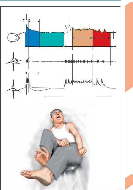
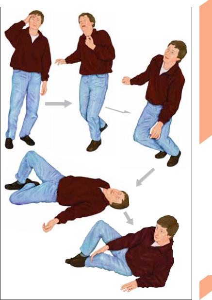
Generalized tonic-clonic seizure

(Grand mal, tonic phase; transition to clonic phase with forceful, rhythmic convulsions)

EEG

EMG (masseter m.)

EMG (biceps brachii m.)

Pupillary diameter

Intravesical pressure

Blood pressure (systolic)

Heart rate

Respiratory rate


Prodromal phase

Ictal phase

Extinction

Recovery phase


(tonic-clonic)

phase


Tonic-clonic grand mal seizure (temporal course)

Central Nervous System

195

Rohkamm, Color Atlas of Neurology © 2004 Thieme

All rights reserved. Usage subject to terms and conditions of license.

Central Nervous System

Epilepsy: Classification

The etiology and prognosis of epilepsy depend on its clinical type. All forms of epilepsy (e. g., absence epilepsy of childhood, juvenile myoclonic epilepsy, temporal lobe epilepsy, frontal lobe epilepsy, reflex epilepsy) are characterized by recurrent paroxysmal attacks; thus classification cannot be based on a single seizure. Epileptic syndromes vary in seizure pattern, cause, age at onset, precipitating factors, EEG changes, and prognosis (e. g., neonatal convulsions, infantile spasms and salaam seizures = West syndrome, Lennox–Gastaut syndrome, temporal lobe seizures). Seizures triggered by fever, substance abuse, alcohol, eclampsia, trauma, tumor, sleep deprivation, or medications are designated as isolated nonrecurring seizures or acute epileptic

reactions. Status epilepticus is a single prolonged seizure or a series of seizures without full recovery in between. Any type of seizure (convulsive or nonconvulsive) may appear under the guise of status epilepticus. In grand mal status epilepticus, patients do not regain consciousness between seizures.

Location-related (focal, partial) epilepsy can be differentiated from generalized epilepsies and epileptic syndromes on the basis of the seizure pattern. Seizures that cannot be classified because of inadequate data on focal or generalized seizure development are called unclassified epilepsy or epileptic syndrome. Other terms used in classification refer to seizure etiology (e. g., idiopathic, cryptogenic, symptomatic).

Type of Epilepsy

Features


Location-related

Partial (focal) seizures

Generalized

Generalized convulsive seizures (GCS)

Idiopathic

No known cause other than genetic predisposition. No manifestations


other than epileptic seizures. Characteristic age of onset

Cryptogenic

Assumed to reflect a CNS disorder of unidentified type. (Once the cause


is identified, epilepsy is classified as symptomatic.)

Symptomatic

Due to an identified CNS disorder or lesion


Type of Epilepsy

Etiology

Epilepsy/Epileptic Syndrome


Location-related

Idiopathic (charac-

Benign epilepsy of childhood with centrotemporal spikes;


(focal, localized,

teristic age of

epilepsy of childhood with occipital paroxysms


partial)

onset)


Cryptogenic or

Variable expression depending on cause and location (e. g., tem-


symptomatic

poral, frontal, parietal, or occipital lobe epilepsy)


Generalized

Idiopathic (charac-

Absence epilepsy of childhood (pyknolepsy); juvenile absence


teristic age of

epilepsy; juvenile myoclonic epilepsy (impulsive petit mal); awak-


onset)

ening grand mal epilepsy (GTCS); epilepsy with specific triggers


(reflex epilepsy)


Cryptogenic or

West syndrome (infantile spasms, salaam seizures); Lennox–


symptomatic

Gastaut syndrome; myoclonic-astatic epilepsy; epilepsy with


myoclonic absence


Symptomatic

Early myoclonic encephalopathy (unspecific etiology); seizures


secondary to various diseases


Unsure whether

Idiopathic or symp-

Neonatal convulsions; acquired epileptic aphasia (Landau–


focal or general-

tomatic

Kleffner syndrome)


ized


196

Variably focal and

Symptomatic

Febrile convulsions; isolated seizure or isolated status epilepti-


generalized

(situation-related

cus; acute metabolic or toxic triggers

seizure)

Rohkamm, Color Atlas of Neurology © 2004 Thieme

All rights reserved. Usage subject to terms and conditions of license.

Epilepsy: Classification

Clonus (left)

Partial seizure, EEG

(right temporoparietal β-wave activity)

Benign neonatal convulsions

1

5

10

15

20


Age of onset


Grand mal epilepsy

Prenatal lesions/disturbances

Metabolic diseases

Congenital anomalies

Encephalitis

Genetic disorders

Head trauma

Brain tumor

Cerebrovascular disorders

Absence

Generalized seizure, EEG

(generalized 3 Hz spike-wave pattern)

Febrile convulsions Lennox-Gastaut syndrome

Epilepsy with myoclonic-astatic seizures

Benign focal epilepsy of childhood Epilepsy with spike waves during sleep Pyknolepsy

Juvenile absence epilepsy Impulsive petit mal Awakening grand mal epilepsy Benign juvenile focal epilepsy

2 3 4 5 10 20 30 50 70

Etiology and age of onset

Age (years)


Central Nervous System

197

Rohkamm, Color Atlas of Neurology © 2004 Thieme

All rights reserved. Usage subject to terms and conditions of license.

Central Nervous System

198

Epilepsy: Pathogenesis and Treatment

Causes. Some patients have a genetic predisposition to epilepsy, particularly those with generalized epilepsies. Some hereditary diseases are associated with epilepsy (e. g., tuberous sclerosis, Sturge–Weber syndrome, mitochondrial encephalopathies, sphingolipidoses). Acquired forms of epilepsy may be focal (possibly with secondary generalization), bilateral, or diffuse (primary generalized epilepsies). The causes include developmental disorders, pyridoxine deficiency, hippocampal sclerosis, brain tumors, head trauma, cerebrovascular disturbances, alcohol, drug abuse, medications, and CNS infections.

Pathophysiology. Seizure activity in the brain is thought to be initiated by a preponderance of excitatory over inhibitory postsynaptic potentials (EPSP, IPSP), resulting in depolarization of nerve cell membranes. Such a depolarization may appear on the EEG as an interictal spike, an initial spike component, or an abrupt depolarization with superimposed high-frequency action potentials (paroxysmal depolarization shift, PDS). The synchronous discharges of large numbers of neurons result in an epileptic seizure. Seizure activity is terminated by active processes such as transmembrane ion transport via sodium–potassium pumps, adenosine release, and the liberation of endogenous opiates, whose combined effect is membrane hyperpolarization, manifested as slow-wave activity in the EEG. Factors favoring the development of seizures include changes in the concentration of electrolytes (Na+, K+, Ca2+), excitatory amino acids (glutamic acid), and inhibitory amino acids (GABA), irregular interneuron connections, and abnormal afferent connections from subcortical structures ( diencephalon, thalamus, brain stem). In focal epilepsy, the epileptic focus is surrounded by an “inhibitory margin”, while the paroxysmal activity of generalized epilepsy is spread throughout the brain.

General treatment measures. Lifestyle changes

(sleep–wake rhythm, avoidance of seizure triggers); chronic use of anticonvulsant medication. Patients with partial seizures preceded by long auras may be able to abort their seizures while still in the aura phase by various concentration techniques (seizure interruption methods).

Antiepileptic drugs (AEDs). AEDs work by a variety of mechanisms, e. g., inhibition of vol-

tage-gated sodium channels (carbamazepine, oxcarbazepine, lamotrigine, phenytoin, valproic acid) or thalamic calcium channels (ethosuximide), or interaction with inhibitory GABA receptors (benzodiazepines, phenobarbital, gabapentin, tiagabine, levetiracetam) or excitatory glutamate receptors (phenobarbital, felbamate, topiramate). Antiepileptic therapy is generally started in patients who have had a single seizure and are thought to be at risk of recurrence, in those with an epileptic syndrome, and in those who have had two or more seizures within 6 months. AEDs used to treat focal, unclassified, and symptomatic tonic-clonic seizures include carbamazepine, gabapentin, lamotrigine, oxcarbazepine, topiramate, levetiracetam, phenytoin, phenobarbital, and primidone; those used to treat generalized seizures include valproic acid, ethosuximide (absences), primidone, phenobarbital (epilepsy associated with myoclonic seizures, tonic-clonic seizures), and lamotrigine. Treatment is always begun with a single drug (monotherapy); if this ineffective, another drug is used instead of or in addition to the first (combination therapy). Antiepileptic therapy can be discontinued in some cases if the risk of seizure recurrence is judged to be low.

Other measures. Surgery (indicated in patients with drug-resistant focal epilepsy and/or resectable lesions, such as brain tumors or unilateral mesial temporal sclerosis). Vagus nerve stimulation by means of an implanted neurocybernetic prosthesis (NCP) is a form of treatment whose efficacy remains controversial.

Prognosis (Table 24, p. 373). Antiepileptic drugs prevent seizure recurrence in roughly 70% of patients, reduce the frequency of seizures in 25%, and are ineffective in 5% (drug resistance), especially those with Lennox–Gastaut syndrome, symptomatic myoclonic epilepsy, and cryptogenic syndromes.

Rohkamm, Color Atlas of Neurology © 2004 Thieme

All rights reserved. Usage subject to terms and conditions of license.


Epilepsy: Pathogenesis and Treatment


Initial spike component


Slow fluctuations

Prodromal phase


Postictal


phase


Interictal


EEG

Silent period

Tonic phase

Clonic


EEG

changes


phase


System


Epileptic seizure (GTCS)


Interictal spikes


500 ms


Nervous


Extracellular


Central

recording


PDS


Membrane hyperpolarization


Intracellular

Neurophysiological changes during epileptic seizure


recording

(data from animal experiments)


Eyes open


Hypersalivation, tongue biting


Symmetric clonic


limb movements


Enuresis, encopresis


Grand mal (GTCS, clonic phase)


199


Rohkamm, Color Atlas of Neurology © 2004 Thieme

All rights reserved. Usage subject to terms and conditions of license.

Central Nervous System

Nonepileptic Seizures

The differentiation of epileptic from nonepileptic seizures is of major prognostic and therapeutic importance. Nonepileptic seizures may or may not involve loss of consciousness. Pseudoseizures resemble epileptic seizures (p. 192 ff), but are of nonepileptic origin. This broad category includes syncope, psychogenic seizures, and simulated seizures. “Pseudoseizure,” in the narrower sense, is a synonym for “psychogenic seizure.”

Nonepileptic seizures are misdiagnosed as epileptic seizures in nearly 20% of patients, roughly 15% of whom are unnecessarily treated with antiepileptic drugs; conversely, some 10% of all epileptic seizures are misdiagnosed as nonepileptic seizures; 20–30% of patients have both epileptic and nonepileptic seizures. In case of doubt, the patient should be referred to a specialist or specialized epilepsy center.

! Syncope

Syncope is defined as a brief loss of consciousness, often involving a fall, due to transient cerebral ischemia or hypoxia (see Table 25, p. 374 for potential causes). In 45% of cases, the cause can be determined from the history and physical examination. Important anamnestic clues include triggers such as excitement or anxiety, precipitating situations (blood drawing, prolonged standing, urination, coughing fits, pain), heart disease, mental illness (generalized anxiety disorder, depression, somatization disorders), and medications. The patient should be evaluated for possible blood pressure abnormalities and for cardiac or neurological disorders (p. 148). EEG yields the diagnosis in only about 2% of cases. Only rare cases of syncope are due to TIA (p. 166). Syncope clinically resembles an epileptic seizure in some ways, but differs in others (see table, below).


Clinical Feature

Syncope

Epileptic Seizure


Triggers

Common

No


Time of day

Mostly diurnal; does not awaken

Day or night; awakens patient from


patient from sleep

sleep


Skin coloration

Pale

Cyanotic or normal


Premonitory symptoms

Tinnitus, visual blurring or blackout,

None or aura


feeling faint, lightheadedness


Type of fall

Collapse or fall over stiffly (often back-

Fall over stiffly


wards)


Duration

Usually ! 30 seconds

1–3 minutes or longer


Abnormal movements

Frequent, arrhythmic, multifocal to

Always generalized, 1–2 minutes


(myoclonus)

generalized, last ! 30 seconds


Eyes

Open

Closed


Urinary incontinence

Occasional

Common


Postictal confusion

Brief or absent

Longer-lasting


Tongue-biting

Occasional

Common


Prolactin, creatine kinase

Normal

Elevated

200


Typical EEG changes

Absent

Common


Focal neurological deficit

Absent

Occasional


Rohkamm, Color Atlas of Neurology © 2004 Thieme

All rights reserved. Usage subject to terms and conditions of license.


Nonepileptic Seizures


Vertigo, light-

Sweating, yawning, tinnitus,

headedness,

unsteadiness, pallor, visual disturbances

malaise

(blurred, gray, black)

Central Nervous System

Warning signs

Fall (by collapsing or falling over stiffly; may cause injury)

Brief unconsciousness (myoclonus possibly accompanied by tonic convulsions)

Brief reorientation phase

201

Syncope

Rohkamm, Color Atlas of Neurology © 2004 Thieme

All rights reserved. Usage subject to terms and conditions of license.

Nonepileptic Seizures


! Psychogenic Seizures


Nonorganic, nonepileptic seizures arising from


psychological factors do not involve loss of con-


sciousness. They are involuntary and uninten-


tional, and thus must be differentiated from


simulated seizures, which are voluntarily, con-


sciously, and intentionally produced events.


Psychogenic seizures may resemble frontal lobe


seizures (p. 194) and are more common in


women than men. About 40% of patients with

System

psychogenic seizures also suffer from true

epileptic seizures. The case history often reveals

characteristic risk factors, which may be bio-


graphical (family difficulties, abuse, divorce,

Nervous

sexual assault in childhood), somatic (genetic

illness), or social (poor living and working con-


predisposition),

psychiatric

(conflicts, stress,


psychosocial gain from illness behavior, mental

Central

ditions). Patients often meet the psychiatric di-

agnostic criteria for a conversion disorder (F44.5


according to the ICD-10). Epileptic seizures in family members, or in the patients themselves, may serve as the prototype for psychogenic seizures.

Premonitory signs. Psychogenic seizures can be induced or terminated by suggestion. They may be preceded by a restless, anxious, or fearful state. They usually occur in the presence of others (an “audience”) and do not occur when the patient is asleep.

Seizure semiology. Psychogenic seizures usually take a dramatic course, with a variable ending. Their semiology is usually of a type more likely to incite sympathy and pity in onlookers than fear or revulsion. Typical features include an abrupt fall or slow collapse, jerking of the limbs, tonic contraction of the body, writhing (arc de cercle), calling out, shouting, rapid twisting of the head and body, and forward pelvic thrusting; the sequence of movements is usually variable. The eyes are usually closed, but sometimes wide open and staring; the patient squeezes the eyes shut when passive opening is attempted. Urinary incontinence or injury (self-mutilation) may also occur. Tongue-bite injuries, if present, are usually at the tip of the tongue (those in true epileptic fits are usually lateral). The patient is

202less responsive than normal to external stimuli, including painful stimuli, but not unconscious

(squeezes eyelids shut when the eyes are

touched, drops arm to the side when it is held over the patient’s face and released). The patient’s skin is not pale or cyanotic during the ictus. Patients who hyperventilate during psychogenic seizures may have carpopedal spasms. Psychogenic seizures often last longer than epileptic seizures.

Postictal phase. No focal neurological deficits can be detected, though there may be a psychogenic postictal stupor. The serum prolactin level is not elevated (which, however, does not rule out a true epileptic seizure). The seizure may be terminated abruptly by suggestion, or by departure of the “audience.” Some patients recall the seizure to some extent, while others emphatically deny memory of it.

! Panic Disorder (ICD-10 F41.0)

Panic disorder is characterized by sudden, unexpected and apparently unprovoked attacks of intense anxiety, which may range in severity from a general feeling of restlessness to a mortal dread. The attacks usually last 5–30 minutes and may awaken the patient from sleep. Accompanying symptoms include feelings of detachment from the environment, i.e., depersonalization (detachment from one’s own body, floating state) and derealization (sensation of being in a dream or nightmare, feeling of unreality); autonomic and other physical symptoms of variable severity, including cardiovascular (tachycardia, palpitations, pallor, chest pain or pressure), gastrointestinal (nausea, dry mouth, dysphagia, diarrhea), respiratory (hyperventilation, dyspnea, smothering sensation), and other manifestations (tremor, twitching of the limbs, dizziness, paresthesia, mydriasis, urinary urgency, sweating). The differential diagnosis includes epilepsy (aura, simple partial seizures), hyperthyroidism, hyperventilation syndrome, pheochromocytoma, heart disease, and hypoglycemia.

Rohkamm, Color Atlas of Neurology © 2004 Thieme

All rights reserved. Usage subject to terms and conditions of license.

Соседние файлы в предмете Нормальная анатомия