

Artificial Organs - Gerald E. Miller
.pdfARTIFICIAL HEART AND CARDIAC ASSIST DEVICES 25
Velocity measurements within ventricular assist devices via flow visualization techniques have been reported by Yamane et al. (2004), Tsukiya et al. (2002), Manning and Miller (2002), Day et al. (2002), Wu et al. (1999), Mulder et al. (1997), Kerrigan et al. (1996), and Miller et al. (1995). Computational fluid dynamics (CFD) techniques have been employed by numerous investigators to analyze various design configurations of circulatory assist devices including Song et al. (2004a,b), Okamoto et al. (2003), Curtas et al. (2002), Anderson et al. (2000), and Pinotti and Rosa (1995), among others.

27
C H A P T E R 3
Cardiac Pacemakers
3.1CARDIAC ELECTROPHYSIOLOGY
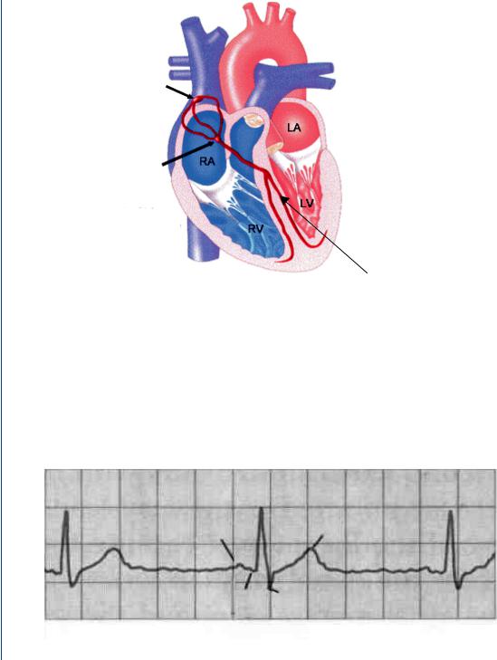
The heart weighs between 7 and 15 ounces (200–425 g) and is a little larger than the size of your fist. By the end of a long life, a person’s heart may have beat (expanded and contracted) more than 3.5 billion times. In fact, each day, the average heart beats 100 000 times, pumping about 2000 gallons (7571 L) of blood. Electrical impulses from your heart muscle (the myocardium) cause your heart to contract, and are created by the movement of ions (principally potassium) across membranes in a fashion called depolarization of cells. This depolarization is propagated along pathways as the ion transport continues. This electrical signal begins in the sinoatrial (SA) node, located at the top of the right atrium. The SA node is sometimes called the heart’s “natural pacemaker” with the average heart beat occurring 72 beats/min. An electrical impulse from this natural pacemaker travels through the muscle fibers of the atria and ventricles, causing them to contract. Although the SA node sends electrical impulses at a certain rate, your heart rate may still change depending on physical demands, stress, or hormonal factors. The initiating electrical signal from the SA node travels down a preferred pathway of specialized conducting myocardial tissue toward the atrioventricular (AV) node, which is the electrical connecting point from the atria to the ventricles. After a slight transmission delay (designed to allow the atria to contract and fill the ventricles before the ventricles contract), the specialized conduction pathway continues into the ventricles. There are two bundles of fibers, called the left and right bundle branches of His, culminating in purkinje fibers, that promote rapid transmission of electrical current through both ventricles. The electrical pathways are shown in Figure 29.
3.2THE ELECTROCARDIOGRAM
The waves of depolarization that spread through the heart during each cardiac cycle generate electrical currents, which in turn spread through the body’s interstitial fluids and eventually up to the body’s surface. Recording electrodes, placed on the surface of the skin on opposite sides of the heart, are used to detect such electrical potentials. These signals are filtered, amplified, and recorded as a measure of the underlying cardiac electrical activity. The record that results from this procedure is termed an electrocardiogram (ECG or EKG). An ECG is an important
28 ARTIFICIAL ORGANS
Sinoatrial (SA) node
Atrioventricular
(AV) node
RA = Right atrium
RV = Right ventricle
LA = Left atrium
LV = Left ventricle
Purkinje fibers
FIGURE 29: Electrical excitatory pathways in the heart.
diagnostic tool to determine whether any cardiac malfunction may have an underlying electrical reason. A typical ECG waveform is seen in Figure 30. The normal ECG consists of three basic features: a P wave, a QRS complex, and a T wave. On some patient waveforms, the QRS complex is seen as three separate waves.
The electrical currents produced as the atrial muscle cells depolarize prior to contraction generate the P wave. This is followed in time by the QRS complex and results from currents
V |
|
R |
|
|
|
o |
P |
T |
l |
||
t |
|
|
a |
|
|
g |
|
|
e |
Q |
S |
|
Time
FIGURE 30: Typical ECG showing P, QRS, and T waves.

CARDIAC PACEMAKERS 29
generated as the ventricles depolarize prior to their contraction. There is a slight time delay between these waves resulting from the delay produced at the AV node, as stated above. Thus, both the P wave and the QRS complex represent depolarization waves. Following ventricular contraction, the ventricle muscle cells repolarize (reverse polarity and reverse ionic transport), and these reversing electrical currents produce the T wave that typically occurs 0.25–0.35 s following ventricular depolarization.
Repolarization of the atria is masked by the QRS complex, and is thus not normally seen on the standard ECG. The intervals between these waves and the width and shape of these waves on the ECG are useful diagnostic measures of cardiac function, as is the overall frequency of the cardiac cycle. The interval between the beginning of the P wave and the beginning of the QRS complex is designated by the PQ interval. Since the Q wave is often absent, it is sometimes termed the PR interval and represents the duration of time (normally about 0.16 s) between the onset of atrial contraction and the onset of ventricular contraction. In patients with certain heart diseases including scarred and/or inflamed tissue, this may lead to a lengthening of the PR interval because more time is required for the depolarization wave to spread through the atrial myocardium and the AV node. The time required for the generation of the QRS complex is termed the QRS duration and represents the amount of time needed for ventricular depolarization. The QT interval extends from the beginning of the QRS complex to the end of the T wave and represents the time required for ventricular contraction and repolarization. Typically, the normal QT interval lasts for approximately 0.35 s. The ST interval represents the time required for the ventricles to repolarize and extends from the S wave to the end of the T wave.
The term tachycardia denotes an overall fast heart rate, typically at more than 100 beats/min. Tachycardia may result from fever, from stimulation of cardiac sympathetic nerves, from certain hormones or drugs, or by weakening of the heart muscle itself. When the myocardium is unable to pump blood effectively, homeostatic reflexes are activated that subsequently increase the heart rate. The term bradycardia denotes a slow heart rate of less than 60 beats/min and is a condition common to athletes, whose enlarged hearts pump a greater stroke volume per heart beat than that of a nonathlete. In addition, bradycardia may also result from decreased body temperature, due to certain drugs, or via stimulation of the heart by its parasympathetic nerve fibers originating from the vagus nerve. Bradycardia may also occur in patients with atherosclerotic lesions of the carotid sinus region of the carotid arteries. The term arrythmia refers to any electrical abnormality in the ECG including rate-related conditions as well as conduction delays or even complete blockages.
The SA node is the natural pacemaker of the heart, providing a stimulating source that initiates the electrical depolarization cycle at approximately 72 beats/min. Should there be a block of the excitation wave between the SA node and the AV node, then the origin of the excitation for the remainder of the heart becomes the AV node, whose normal rhythmic rate is slower than

30 ARTIFICIAL ORGANS
that of the SA node (approximately 55 beats/min). The remainder of the excitatory pathway (AV node to purkinje fibers) remains intact. Should there be a partial or total block between the AV node and the purkinje fibers, then alternate excitatory sources within the ventricular muscle assume the excitatory role, albeit at a much lower rate (30 beats/min). These are readily evident in the ECG waveforms as a reduced heart rate, as widening of the waves within the ECG, and/or as changes in the shape of the waves.
Premature contractions can occur within a normal rhythmic cardiac excitation (called sinus rhythm) due to altered excitatory events within an atrium or a ventricle. Premature atrial contractions (PACs or PABs for premature atrial beats) are atrial beats that occur too early due to an abnormal electrical signal. Often, things such as caffeine, alcohol, medications (especially decongestants), certain medical conditions such as hyperthyroidism, anemia, and hypertension, and stress can trigger PACs. Some people may feel a fluttering in their hearts when experiencing PACs, whereas others have no symptoms. PACs are benign and may require no treatment. Premature ventricular contractions (PVCs or PVBs for premature ventricular beats) are early ventricular contractions that occur when the ventricles contract out of sequence with normal heart rhythm. Though they are generally benign and usually do not require treatment, PVCs may result in more serious arrhythmias in those with heart disease or a history of tachycardia. For these people, antiarrhythmic drugs and an implantable cardioverter defibrillator (ICD) may be prescribed. PVCs most often occur spontaneously; however, like PACs, they can also be triggered by caffeine, alcohol, medications (especially decongestants), certain medical conditions such as hyperthyroidism, anemia, and hypertension, and stress. PACs and PVCs are readily observed as added waves within the ECG. An ectopic or premature atrial beat produces an added P wave within the standard ECG waveform. A PVC produces an added R wave or QRS segment within the normal ECG waveform. Repeated PAC or PCV events may indicate a serious condition that will require intervention.
Fibrillation is caused when the heart muscle begins to quiver, or fibrillate, continually and cannot contract normally. When a heart is in a state of fibrillation, there is no synchronization between the atria and the ventricles, nor any standard heart beat/contraction. This may cause the patient to experience a racing sensation—and sometimes discomfort in the chest—and/or to feel light-headed or faint. Ventricular fibrillation (VF or V Fib) is a life-threatening arrhythmia that necessitates immediate treatment with an external defibrillator, an internal defibrillator (ICD), antiarrhythmic drugs, or VT ablation. Fibrillation is easily recognized on the ECG waveform as a loss of the QRS segment replaced by a noisy, lower amplitude signal. Atrial fibrillation (AF or A Fib) is a very fast, uncontrolled atrial heart rate caused by rapidly fired signals. During an episode of AF, the atrial heart rate may exceed 350 beats/min. Not all of these beats reach the ventricles, so the ventricular rate is not this high. However, the ventricular rate is often higher than normal and can also be erratic, exceeding 100 beats/min. Sometimes an impulse will circle the atria, triggering atrial flutter, which is similar to AF. Alone, AF is rarely serious, but if a

CARDIAC PACEMAKERS 31
patient has high blood pressure, valvular disease, or heart muscle damage, AF can increase the risk of stroke or heart failure.
There are several treatments for AF, including medication, ablation, and an electrical therapy called cardioversion. Electrical cardioversion converts the heart rate back to normal sinus rhythm through the use of a controlled electrical shock that excites all the heart cells at once, allowing the SA node to resume its role as the heart’s natural pacemaker. If medication and electrical cardioversion do not improve the AF, the physician may recommend cardiac ablation to prevent conduction of abnormal electrical impulses from the atria to the ventricles, with implantation of a permanent pacemaker to control heart rate. Ventricular fibrillation (VF) is a chaotic heart rate resulting from multiple areas of the ventricles attempting to control the heart’s rhythm. Ventricular fibrillation can occur spontaneously (generally caused by heart disease) or when ventricular tachycardia has persisted too long. When the ventricles fibrillate, they cannot contract normally, hence, they cannot effectively pump blood. The instant VF begins, effective blood pumping stops. VF quickly becomes more erratic, resulting in sudden cardiac arrest or sudden cardiac death. This arrhythmia must be corrected immediately via a shock from an external defibrillator or an internal device (ICD). The defibrillator can stop the chaotic electrical activity and restores normal rhythm by depolarizing the entire heart with the idea that normal sinus rhythm will be restored if the SA node fires first and initiates the normal excitation process.
3.3CARDIAC PACEMAKER
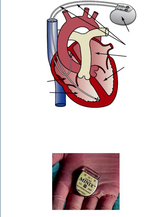
An artificial cardiac pacemaker is an implantable device that produces an excitatory wave at an appropriate site within the heart: a) at the SA node to replace a failing natural SA node (or correct atrial fibrillation) and generate a normal sinus rhythm, b) at the AV node where a malfunctioning AV node exists, or c) within one or both ventricles to continue excitation beyond a partial or total heart block (or to correct ventricular fibrillation). A cardiac pacemaker consists of 1) an electrical device, the pulse generator, which produces the excitatory signal, 2) the wires, called the leads, which connect the pulse generator to the cardiac tissues, 3) a battery system (housed within the pulse generator) to power the pulse generator, and 4) a programmable segment (also housed within the pulse generator) to evaluate the heart excitatory function and send the appropriate excitatory segment from the pulse generator. A schematic diagram of a cardiac pacemaker and the leads is shown in Figure 31.
The batteries within the pulse generator “can” are typically lithium iodide and have a life span of approximately 10–12 years. The pulse generator casing is typically titanium which is welded and hermetically sealed. An epoxy top is attached from which the leads are attached to the pulse generator. This allows for changes in the leads without disrupting the pulse generator case. A typical pulse generator is small in size, often less than an ounce in weight, less than two inches wide, and a quarter-inch thin. Thus, the device is roughly the size of two silver dollars stacked on top of one another. Once implanted in the upper chest, just below the skin
32 ARTIFICIAL ORGANS
Pulse generator
Pacing leads
Left atrium
Left ventricle
Right atrium
Right ventricle
FIGURE 31: Schematic diagram of an implantable cardiac pacemaker.
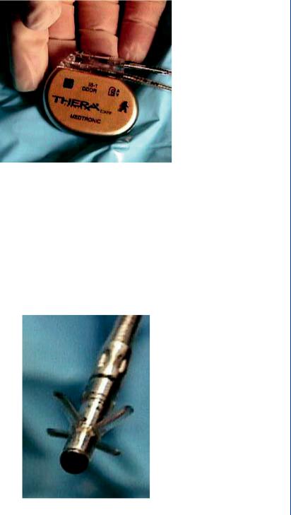
near the collarbone, the pacemaker’s presence is nearly invisible to the eye. When the batteries have become depleted, the entire pulse generator case is replaced. The surgical implantation procedure will be explained in more detail below. A pulse generator is seen in Figure 32, with the leads connected to the “can” seen in Figure 33.
FIGURE 32: Typical pacemaker pulse generator with epoxy top for connection to leads.
CARDIAC PACEMAKERS 33
FIGURE 33: Pulse generator with leads attached.
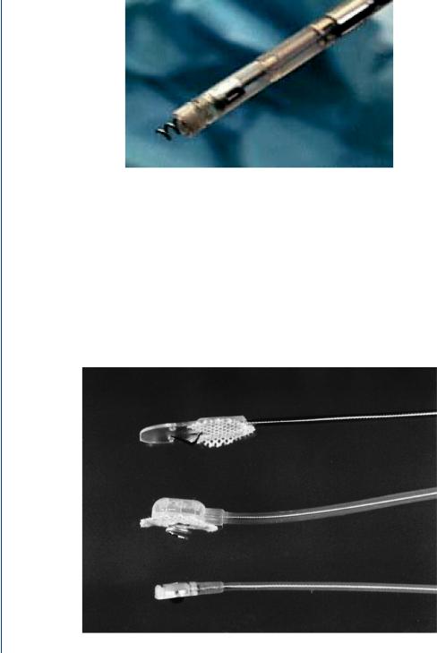
The pacing lead, which connects to the pulse generator header, is a flexible insulated wire with an electrode tip. This tip, which can be inserted through a vein into the heart, carries impulses from the pulse generator to the heart, stimulating the heart to beat. It also carries information from the heart back to the pulse generator, which the physician accesses via a special programmer or into the pulse generator circuit embedded program. Leads that are inserted via a vein into the heart have tines on the end as shown in Figure 34 or have a screw end, which can be threaded into the cardiac muscle as shown in Figure 35.
FIGURE 34: Pacemaker lead with tines on end for embedding into heart tissue.
34 ARTIFICIAL ORGANS
FIGURE 35: Pacemaker lead with screw end for implantation into heart tissue.
The leads shown in Figures 34 and 35 are both designated as endocardial or transvenous leads. The tined lead shown in Figure 34 is a passive fixation device where the tines become lodged in the trabeculae (fibrous meshwork) of the heart. The helix or screw-ended lead shown in Figure 35 is an active fixation device that extends into the endocardial tissue (interior surface of the heart). Such a lead can be positioned anywhere in the heart chamber. The other category of lead is the myocardial or epicardial lead which is secured to the outer surface of the heart as shown in Figure 36. Such leads are connected to cardiac tissue via sutures or screwed into tissue as was the case with the endocardial lead.
FIGURE 36: Myocardial pacemaker leads which are sewn into exterior cardiac tissue via sutures or screwed into the exterior surface.