

McLeod - Swimming Anatomy - 2010
.pdfCHAPTER 1
LEGS
Strong legs are a critical component to reaching your true potential as a swimmer. They are not only the basis for having a powerful and efficient kick but also the key to driving your body off the starting blocks and turn walls. They also play an often overlooked role as a member of the kinetic chain by balancing your stroke mechanics and contributing to a tight streamline.
The lower extremity consists of three major joints-the hip, the knee, and the ankle. Five bones make up the three joints. The pelvis serves as the link between both legs and the torso. Each thigh is composed of a single long bone called the femur. The lower leg contains the tibia and fibula. The talus is the bone that serves as the connecting point between the ankle and lower leg. The hip joint is formed by the bony socket of the pelvis, called the acetabulum, and the head of the femur, which is shaped like a ball. The knee is the junction of the femur and the tibia, and the

ankle is composed of the lower ends of the tibia and fibula and the upper part of the talus.
As a ball-and-socket joint, the hip is capable of a wide range of movements that can be described in three pairs. Flexion involves lifting the thigh upward toward the ceiling as if you are lifting your leg to climb a set of stairs. Extension is movement of the thigh backward. Abduction occurs when the leg is moved to the side away from the midline of the body, and adduction is the movement of bringing the leg back toward the midline of the body. Internal rotation is the process of touching the big toe of each foot together along the midline of the body. External rotation is the opposite and allows you to touch the back end of both heels together.
At the knee, a hinge joint, two primary movements occur. Flexion is the process of pulling the heel to the buttocks, and extension is straightening the knee from a flexed position. Four movements take place at the ankle joint. The process of pointing your toes, as you do in a tight streamline, is plantarflexion. Lifting your toes off the ground and toward your shin is called dorsiflexion. Rolling your ankle inward so that the bottom of your foot faces the midline of your body is inversion. Finally, eversion involves twisting your foot outward as you would before initiating a breaststroke kick.
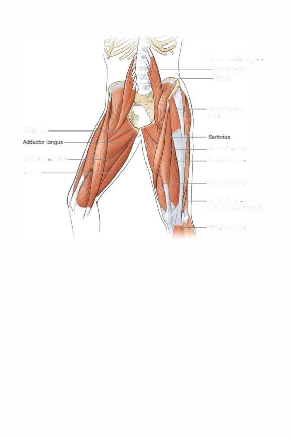
The muscles of the leg can be categorized as those that act on the hip and knee and those that act on the ankle. The thigh and hip muscles can further be categorized into the following groupings: anterior, medial, gluteal, and posterior. Within the anterior grouping are seven muscles. The iliopsoas (figure 7.1 on page 142) is a deep muscle that arises from the anterior aspect of the lumbar vertebrae and the inner aspect of the pelvis and then crosses the hip joint to attach to the proximal femur. The primary movement generated by the iliopsoas is hip flexion. The quadriceps femoris, the largest muscle group in the body, is divided into four separate muscles that are named according to their point of origin. The rectus femoris, the only one to cross both the hip and knee, originates from the anterior aspect of the pelvis. The vastus lateralis arises from the lateral aspect of the femur, the vastus medialis arises from the medial aspect of the femur, and the

vastus intermedius is in the middle. All four muscles have a common insertion on the anterior aspect of the tibia through the patellar tendon and function to extend the knee. Because the rectus femoris crosses the hip joint, it also functions as a hip flexor. The tensor fasciae latae (TFL) runs from the anterior aspect of the pelvis to combine with the iliotibial band (IT band), a thickened band of fascial tissue that runs down the lateral thigh. It then inserts on the lateral aspect of the tibia just below the knee joint. The primary actions of the TFL are hip flexion, abduction, and internal rotation. The final muscle of the anterior group is the sartorius, which is a long straplike muscle that runs diagonally from the anterior pelvis to the medial aspect of the tibia. Its primary actions are to flex, abduct, and externally rotate the hip.
The medial grouping can be divided into the adductor muscle family and two additional muscles that lie in close proximity. The adductor family is composed of three muscles (adductor magnus, adductor longus, and adductor brevis), which all arise from the inferior portion of the pelvis near the midline of the body and attach to the medial aspect of the femur. As the name implies, the primary function of this muscle family is hip adduction. Just superior to the adductors is the pectineus, which also originates from the inferior pelvis near the midline of the body and then inserts along the medial aspect of the femur. Besides assisting the adductors, the pectineus also flexes the hip. The gracilis is the most medial and inferior. It has the same origin as the other muscles but crosses the knee to attach on the medial aspect of the tibia just below the knee joint. Besides adducting the hip, the gracilis is also a secondary flexor of the knee.
Figure 1.1 Muscles of the front of the legs.
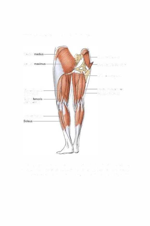

to attach at the medial surface of the superior part of the tibia. Collectively, the muscles extend the hip and flex the knee.
The muscles of the lower leg can be grouped according to their actions at the ankle joint. The gastrocnemius and soleus are the primary plantarflexors and share an insertion into the Achilles tendon. The tibialis anterior and posterior, named according to their attachment location on the tibia, function to invert the foot. The fibularis muscle group (fibularis tertius, fibularis brevis, and fibularis longus), located on the lateral aspect of the ankle joint, originates from the fibula and has the primary function of foot eversion.
For discussion purposes, the muscle recruitment patterns of the freestyle and backstroke flutter kick are described jointly because the patterns are practically identical. The propulsive portion of the flutter kick begins with the torso and core-stabilizing musculature acting as the foundation on which your legs generate their force. The actual kicking movements begin with the hips in a small amount of extension. From this extended position the iliopsoas and rectus femoris are activated to initiate hip flexion. Also acting on the knee joint, the rectus femoris initiates knee extension and is quickly joined by the remainder of the quadriceps group, which helps to increase the force produced during the kick. These muscles remain active throughout the entire propulsive phase of the kick. At the ankle joint, the tibialis anterior and tibialis posterior work in concert to maintain the foot in a position of slight inversion, while contraction of the gastrocnemius and soleus plantarflexes the foot. The hip extension that takes place during the recovery phase is guided by the hamstrings and gluteus maximus. Unlike in flutter kicking, during butterfly and dolphin kicking the torso serves not only as the foundation for the kick but also as a component. The undulating body movements of the torso initiate the kick, and paired movements of the legs follow in a manner identical to the action of the flutter kick. One difference in the paired movement of the legs is that a greater amount of flexion and extension occurs at both the hips and knees. The undulating movement of the torso is guided by the contraction of the abdominal and erector spinae muscles, but the muscles that guide

the movements of the legs are identical to those used in the flutter kick.
The starting point for the propulsive phase of the breaststroke kick is with the feet 8 to 10 inches (20 to 25 cm) apart and the knees and hips flexed. From this position the TFL, gluteus medius, and gluteus minimus internally rotate and abduct the hips, which results in the legs further separating from each other. As the ankles begin to separate, the biceps femoris contracts, pulling on the outer portion of the lower leg, which externally rotates the lower leg and results in further separation of the ankles. At the same time the fibularis muscle group contracts to evert the foot. These combined movements place the legs in the position to begin the whip portion of the kick. From this position the gluteus maximus contracts forcefully to extend the hip, the quadriceps muscle group functions to extend the knee, and the powerful adductor muscles (adductor magnus, adductor longus, adductor brevis, pectineus, and gracilis) pull both legs back toward the midline of the body. At the ankle joint the tibialis posterior, gastrocnemius, and soleus contract to return the ankle to a streamlined plantarflexed position for the glide portion of the stroke. Recovery is accomplished by recruitment of the rectus femoris and iliopsoas, which serve to flex the hip, and recruitment of the hamstrings, which serve to flex the knee.
GtulelJS
GtuteLJiS maximlJs
...c===,
(t:¥-
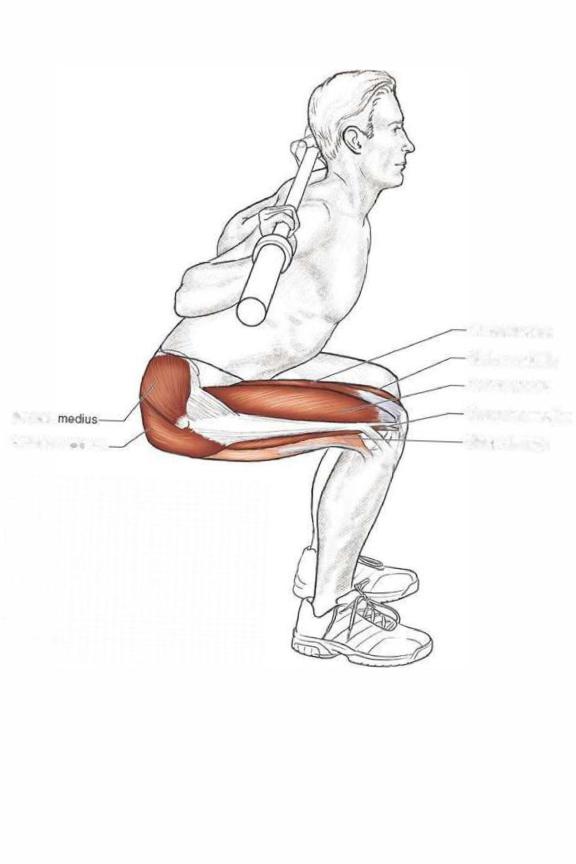
Rectos femoris Vestus m«!ialls \festus lateralis Vastus intel1medhJ5 ---Bioeps femoris

Execution
1 . Rest the barbell across your upper back and position your feet shoulder-width apart.
2.Initiating the movement with your hips, squat down until your thighs are parallel to the ground.
3.Return to the starting position by straightening your legs.