

3 курс / Фармакология / Essential_Psychopharmacology_2nd_edition
.pdfSex-Specific and Sexual Function—Related Psychopharmacology |
549 |
FIGURE 14 — 11. Under normal conditions, when young healthy men are sexually aroused, nitric oxide causes cGMP to accumulate, and cGMP causes smooth muscle relaxation, resulting in a physiological erection, indicated here by an inflated balloon. The erection is sustained long enough for sexual intercourse, and then phosphodiesterase V (PDE V) metabolizes cGMP, reversing the erection, indicated here by a pin ready to prick the balloon.
FIGURE 14 — 12. When a man has diabetes or hypertension, or if he smokes, uses alcohol, takes prescription drugs, or is depressed, there is a good chance that not enough of a signal of sexual desire will be able to get through his peripheral nerves and arteries to produce sufficient amounts of cGMP to cause an erection. This leads to impotence.
injection of the prostaglandin alprostadil produces erections not only in men with organic causes of impotence but also in those with functional causes and even in the common situation of multifactorial causes. Limitations of this somewhat masochistic approach include unacceptability of self-injection, lack of spontaneity, and the possibility of "too much of a good thing," namely a prolonged and painful erection,

550 Essential Psychopharmacology
FIGURE 14-13. Sildenafil, a phosphodiesterase V (PDE V) inhibitor, is able to compensate for faulty signals through the peripheral nerves and arteries that produce insufficient amounts of cGMP to produce or sustain erections. Sildenafil does this by allowing cGMP to build up, since PDE V can no longer destroy cGMP for a few hours. This is indicated by a patch on the balloon in the figure. The result is that normally inadequate nerves and arteries signaling cGMP formation are now sufficient to inflate the balloon, and therefore an erection can occur and sexual intercourse is now possible, until the sildenafil wears off a few hours later.
FIGURE 14—14. Some antidepressants such as serotonin selective reuptake inhibitors (SSRIs) may inhibit nitric oxide synthetase (NOS) and thereby reduce NO and cause erectile dysfunction.
called priapism. Prostaglandin administration will cause an erection whether the man is mentally aroused or not.
Other drugs can affect sexual arousal, including some serotonin selective reuptake inhibitors (SSRIs), which may inhibit NOS directly and can thus cause erectile dysfunction (Fig. 14—14), and some dopaminergic agents, which boost NOS and might some day help erectile dysfunction (Fig. 14—15). Anticholinergic agents can interfere directly with arousal and cause erectile dysfunction. Thus, agents such as

Sex-Specific and Sexual Function—Related Psychopharmacology |
551 |
FIGURE 14—15. Some agents that boost dopamine (perhaps like apomorphine) are promising experimental drugs for enhancing NOS and may be useful to reverse erectile dysfunction.
antipsychotics and tricyclic antidepressants and others with similar properties can cause erectile dysfunction (Fig. 14—16).
Psychopharmacology of Sexual Dysfunction
In summary, numerous agents used in psychopharmacology can facilitate or interfere with each of the three stages of the human sexual response (Fig. 14-16). Understanding the basic mechanisms of neurotransmission for each of these stages (Fig. 14—7), as well as the psychopharmacological mechanisms of action of the various psychotropic drugs that impact these neurotransmitter systems, will facilitate the management of psychotropic drugs in patients with sexual dysfunction.
Estrogen as a Neurotrophic Factor in the Brain
It is well known that ovarian estrogens, especially 17-beta-estradiol, regulate reproductive function and have profound effects on reproductive tissues in women, such as those of the breast and uterus. The long-term positive effects of estrogens outside of the reproductive tissues have also been emphasized, such as estrogen's effects in preserving bone mineralization and in reducing serum cholesterol. Recently there has been growing appreciation for the diversity of effects that estrogen can have on the brain as well, especially in regions of the brain outside of those areas known to be involved in the control of reproductive function and sexual differentiation. These neuronal effects are mediated by the same types of receptors for estrogen that exist in other tissues and have trophic actions on the brain, just as they have on other tissues. Trophic factors have been discussed in Chapter 1 (see Fig. 1 — 19 and Tables 1 — 3 and 1—4). In the brain, estrogen's trophic actions trigger the expression of genes that lead to the formation of synapses.
Estradiol modulates gene expression by binding to estrogen receptors (Fig. 14— 17). Estrogen receptors differ from tissue to tissue and may differ from brain region to brain region. In addition to various forms of estrogen receptors, there are receptors for progesterone and androgens, as well as for other steroids such as glucocorticoids

552 Essential Psychopharmacology
FIGURE 14—16. Psychopharmacological agents can affect all three stages of the human sexual response, both positively and negatively, as summarized here. In stage 1, libido can be enhanced by the norepinephrine and dopamine reuptake inhibitor (NDRI) bupropion, as well as by the dopaminereleasing stimulants amphetamine and methylphenidate. Libido can also be reduced by the dopamine receptor—blocking antipsychotics, some of which also increase prolactin. Stage 2, sexual arousal, can be enhanced by sildenafil, which boosts cGMP action, by prostaglandins, and perhaps by some dopaminergic agents. Sexual arousal can be reduced by some serotonin selective reuptake inhibitors (SSRIs), as well as by agents with anticholinergic properties. Finally, in stage 3, orgasm can be inhibited by SSRIs as well as by beta blockers, which block noradrenergic function.
and mineralocorticoids. Unlike neurotransmitter receptors located on neuronal membranes, receptors for estradiol are located in the neuronal nucleus, so estradiol must penetrate the neuronal membrane and the nuclear membrane to find its receptors, which are therefore located near the genes it wishes to influence. These genes are called estrogen response elements (Fig. 14—17).
The expression of these estrogen response elements within the DNA of the neuron progresses generally in the same manner as the expression of other neuronal genes, which has been discussed in Chapter 2 (see Figs. 2 — 31 to 2—42). The activation of estrogen response elements by estradiol requires "dimerization" (i.e., coupling of two copies of the estrogen receptor) when estrogen binds to the receptor to form an active transcription factor capable of "turning on" the estrogen response element (Fig. 14—18). Formation of transcription factors has also been discussed in Chapter 2 (see Figs. 2 — 33 and 2 — 35 to 2 — 38). Once the estrogen receptors are activated by estradiol into transcription factors, they activate gene expression by the estrogen

Sex-Specific and Sexual Function—Related Psychopharmacology |
553 |
FIGURE 14 — 17. Estrogen modulates gene expression by binding to estrogen receptors. Estrogen receptors differ from tissue to tissue and may differ from brain region to brain region. Unlike neurotransmitter receptors located on neuronal membranes, receptors for estradiol are located in the neuronal cell nucleus, so estradiol must penetrate the neuronal membrane and the nuclear membrane to find its receptors, which are therefore located near the genes that are to be influenced. These genes are called estrogen response elements.
response elements in the neuron's DNA (Fig. 14—19). Gene products that are expressed include direct trophic factors such as nerve growth factor (NGF) and brainderived neurotrophic factor (BDNF), which can facilitate synaptogenesis and prevent apoptosis and neurodegeneration.
Gene products also include neurotransmitter-synthesizing enzymes for the key monoamine neurotransmitter systems that regulate mood and memory (Figs. 14— 20 to 14—22). Thus, the presence of estradiol can be critical to the adequate functioning of the monoamines serotonin (Fig. 14—20) and norepinephrine (Fig. 14— 21) in women. Adult men do not respond to estrogen in this manner. The presence of estradiol in aging women but not in aging men can also be critical to the adequate functioning of acetylcholine in the nucleus basalis of Meynert (Fig. 14— 22). The role of these key cholinergic neurons in the regulation of memory (see Fig. 12 — 11) and in the causation of Alzheimer's disease when they degenerate (see Fig. 12 — 13) have been discussed in Chapter 12. This may explain the emerging role of estrogen in managing memory and Alzheimer's disease in aging women, as discussed below.
Dramatic evidence of estrogen's trophic properties can be observed in hypothalamic and hippocampal neurons in adult female experimental animals within days

554 Essential Psychopharmacology
FIGURE 14—18. The expression of estrogen response elements with the DNA of the neuron must be initiated by estrogen and its receptor. Activation of these genes by estradiol requires "dimerization" (i.e., coupling of two copies of the estrogen receptor) when estrogen binds to the receptor to form an active transcription factor capable of "turning on" the estrogen response element.
and across a single menstrual (estrus) cycle (Figs. 14 — 23 and 14 — 24). During the early phase of the cycle, estradiol levels rise, and this trophic influence induces dendritic spine formation, specifically in the ventromedial hypothalamus and on pyramidal neurons in the hippocampus of female rats. Progesterone administration rapidly potentiates this, so spine formation is at its greatest when both estrogen and progesterone peak, just after the first half of the cycle (Fig. 14—23). However, once estrogen levels fall significantly and progesterone levels continue to rise, the presence of progesterone without estrogen triggers down regulation of these spines and removal of the synapses by the end of the estrus cycle (Fig. 14—23). One hypothesis to explain the mechanism of this cyclical formation and removal of synapses is that estrogen may exert its trophic influence through low levels of glutamate activation (Fig. 14 — 24), leading to spine formation and synaptogenesis: this effect is followed by too much glutamate activation in the absence of estrogen, when progesterone alone leads to excitotoxicity and destruction of these same spines and synapses (Fig. 14 — 24). The hypothesis of how glutamate might mediate excitotoxic synaptic or neuronal toxicity was introduced in Chapter 4 (see Figs. 4— 14 to 4—23) and discussed extensively in Chapter 10 (see Figs. 10 — 26 to 10-33).
Other evidence for the trophic influences of estrogen comes from what happens when the estrogen's effects are blocked with estrogen receptor antagonists. Tamox-

Sex-Specific and Sexual Function—Related Psychopharmacology |
555 |
FIGURE 14—19. Once the estrogen receptors are activated by estradiol into transcription factors, they activate gene expression by the estrogen response elements in the neuron's DNA. Gene products that are expressed include direct trophic factors such as nerve growth factor (NGF) and brainderived neurotrophic factor (BDNF), which can facilitate synaptogenesis and prevent apoptosis and neurodegeneration.
ifen is an estrogen receptor antagonist used for the treatment of breast cancer, especially for breast tumors that themselves express estrogen receptors. Blocking estrogen receptors in breast cancer cells with tamoxifen triggers apoptosis (programmed cell death), presumably due to blocking the trophic effect of estrogen in these tumor cells. Interestingly, tamoxifen is an estrogen receptor antagonist in breast and uterus but is actually a partial agonist in preserving bone mineralization and reducing cholesterol. It is also an estrogen receptor antagonist in brain, since it can induce depression that can be difficult to treat with antidepressants. Thus, individual estrogens such as estradiol and tamoxifen all have tissue-selective estrogen agonist, partial agonist, and antagonist activities. This also extends to the new class of estrogens known as selective estrogen receptor modulators (SERMs), of which raloxifene is the newest available member. Such observations may also explain why some women respond differently to one estrogen preparation than to another, and from a behavioral perspective, why they may have different mood and cognitive responses to one estrogen preparation versus another. Unfortunately, very little work has been done to distinguish the pharmacologic effects of the different available estrogen preparations on estrogen receptor binding in the brain, and the only way

556 Essential Psychopharmacology
FIGURE 14—20. Gene products activated by estradiol interacting with estrogen response elements in the serotoninergic neurons of the midbrain raphe include not only trophic factors, which nourish the growth and synapses of these neurons with nerve growth factor (NGF) and brainderived neuro-trophic factor (BDNF), but also the enzymes and receptors that facilitate serotonergic neurotrans-mission. These receptors may also allow the neuron to have normal mood functions and to be more responsive to antidepressant medications in case of a depressive episode.
to detect these differences at the present time is through trial and error. Nevertheless, the differences in tissue-selective, brain region—selective, or individual-selective estrogen agonist, partial agonist, or antagonist activities can be explained by the fact that there are many different DNA response elements to estrogens, and these may be expressed differently in various tissues, brain regions, and individuals. There may even be state-dependent differences in expression of DNA response elements to estrogens, which vary across the female life cycle or vary depending on the presence or absence of a mood or cognitive disorder. This is the subject of intense current research interest.
Estrogen and Mood Across the Female Life Cycle
Estrogen levels shift rather dramatically across the female life cycle, all in relationship to various types of reproductive events (Fig. 14—25). Thus, levels begin to rise and then cycle during puberty (see also Fig. 14—23). This cycling persists during the childbearing years, except during pregnancy, when a woman's estrogen levels
Sex-Specific and Sexual Function—Related Psychopharmacology |
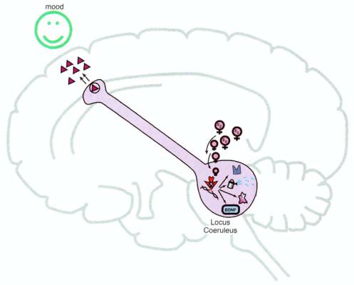
557 |
FIGURE 14-21. Gene products activated by estradiol interacting with estrogen response elements in the noradrenergic neurons of the brainstem locus coeruleus include not only trophic factors that nourish the growth and synapses of these neurons with nerve growth factor (NGF) and brain-derived neurotrophic factor (BDNF), but also the enzymes and receptors that facilitate noradrenergic neu-rotransmission. These receptors may also allow the neuron to have normal mood function and to be more responsive to antidepressant medications in case of a depressive episode.
skyrocket (Fig. 14 — 25). Estrogen levels then plummet precipitously immediately postpartum, and regular menstrual cycles begin again once the mother stops nursing (Fig. 14-25).
Although the median age of menopause, which is the time of complete cessation of menstruation, is 51, women do not begin menopause overnight. The transition period from regular menstrual cycles to complete cessation of menstruation, called perimenopause, can begin 5 to 7 years before menopause and is characterized on onagain off-again cycles and anovulatory cycles, prior to complete cessation of menstrual cycles (Fig. 14—25). Hormone levels can be chaotic and unpredictable during these years. This can be experienced both as a physiological and a psychological stressor. Menopause is the final stage of transition of estrogen in the female life cycle and can be associated with estrogen replacement therapy, which can restore estrogen to its physiological levels during the childbearing years.
There are potential links between these shifts in estrogen levels across the female life cycle and the observation that depression is much more common in women than in men during certain stages of the life cycle. In men, the incidence of depression

558 Essential Psychopharmacology
FIGURE 14—22. Gene products activated by estradiol interacting with estrogen response elements
in the cholinergic neurons of the nucleus basalis of Meynert in the basal forebrain include not only trophic factors that nourish the growth and synapses of these neurons with nerve growth factor (NGF) and brain-derived neurotrophic factor (BDNF), but also the enzymes and receptors that facilitate cholinergic neurotransmission. These receptors may also allow the neuron to function optimally in memory formation, particularly verbal memory in aging women, and to be more responsive to cholinesterase inhibitors in the case of Alzheimer's disease.
rises in puberty and then is essentially constant throughout life, despite a slowly declining testosterone level from age 25 onward (Fig. 14—26). By contrast, in women the incidence of depression mirrors their changes in estrogen across the life cycle (Fig. 14—27). As estrogen levels rise during puberty, the incidence of depression skyrockets, falling again after menopause (Fig. 14—27). Thus, women have the same frequency of depression as men before puberty and after menopause. However, during their childbearing years when estrogen is high and cycling, the incidence of depression in women is two to three times as high as in men (Fig. 14 — 27).
Several other issues are of particular importance to women in terms of assessing their vulnerability to the onset and recurrence of mood disorders across their lifetimes. These are linked to shifts in reproductive hormone status, as outlined in Figure 14—28. First episodes of depression often begin in puberty or early adulthood, when estrogen is first rising; unfortunately these episodes are frequently unrecognized and untreated. Throughout the childbearing years of normal menstrual cycles,