

Biology_of_Turtles
.pdf176 |
Biology of Turtles |
A |
D |
m. spinalis cervico-capitis - cauda hyoidea |
m. spinalis cervico-capitis - cauda cornu branchiale II |
B |
E |
m. spinalis cervico-capitis - cauda squamosi |
m. spinalis cervico-capitis |
C |
F |
m. spinalis cervico-capitis - cauda occipitalis
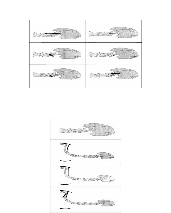
Figure 7.12 Schematic representation of the m. spinalis cervico-capitis complex (A–D), the m. cervicospinalis (E), and the m. articulo-transversalis longus (F) in A. spinifera.
A D
m. articulo-transversalis brevis |
m. transverso-corporis |
B |
E |
m. articulo-cruralis longus |
m. cervico-spinalis lateralis brevis dorsalis |
C |
F |
m. intertransversalis |
m. epistropheo-squamosus ventralis |
Figure 7.13 Schematic representation of the m. articulo-transversalis brevis, the m. articulo-cruralis longus, the m. intertransversalis, the m. transverso-corporis, the m. cervico-spinalis lateralis brevis dorsalis, and the m. epistropheo-squamosus ventralis in A. spinifera.
Cervical Anatomy and Function in Turtles |
177 |
A |
D |
m. epistropheo-squamosus dorsalis |
m. atlanto-exoccipitis |
B |
E |
m. epistropheo-atlantis dorsalis |
m. atlanto-basioccipitis medialis |
C |
F |
m. epistropheo-atlantis ventralis |
m. atlanto-opisthoticus |
Figure 7.14 Schematic representation of the m. epistropheo-squamosus dorsalis, the m. epistropheo-atlan- tis dorsalis and ventralis, the m. atlanto-exoccipitis, the m. atlanto-basioccipitis medialis, and the m. atlantoopisthoticus in A. spinifera.
A
m. epistropheo-odontoideus
B
m. cortico-cervicale I
C
m. cortico-cervicale II
D
m. cortico-cervicale III
Figure 7.15 Schematic representation of the m. epistropheo-odontoideus and the m. cortico-cervicale group in A. spinifera.

178 |
Biology of Turtles |
m.spinalis cervico-capitis (cervico-hyo-capitis, Ogushi, 1913; Figure 7.12). This is a large muscle that consists of four distinct muscle bellies—the cauda hyoidea, the cauda squamosi, the cauda occipitalis, and the cauda cornu branchiale II. The cauda hyoidea (Figure 7.12A) originates at the neural roof of C6, runs anteriorly and merges with the fibers of the m. rectus cervicis. The cauda squamosi (Figure 7.12B) part originates at the neural roof of C5, runs cranially, and inserts by means of a broad aponeurosis at the posterior aspect of the squamosum. The cauda occipitalis (Figure 7.12C) originates at the neural roof of C4, runs cranially, and inserts on the external fascia covering the external adductor. The cauda cornu branchiale II (Figure 7.12D) originates at the neural roof of C3, runs cranially, and inserts at the connective tissue associated with the tip of the second ceratobranchial.
m. cervico-spinalis medialis (Figure 7.12E). This is a segmentally arranged muscle. The muscles originate tendinously at the anterior aspect of the crista mediana ventralis, run anteriorly, and insert tendinously at the posterior aspect of the crista mediana ventralis of the third more cranially positioned vertebra. The muscles originating on the C2-C4 merge and insert jointly at the ventral aspect of the basioccipital by means of a well-developed tendon.
m. cervico-spinalis lateralis longi (Figure 7.12 & Figure 7.13). This consists of three long, superficial muscles that originate at the lateral aspect of the zygapophyses of C5 through C8:
•The m. articulo-transversalis longus (Figure 7.12F) originates on the lateral aspect of the pre-zygapophyses of the last five cranial vertebrae. The muscle runs cranially across the ventral aspect of the two more cranially situated vertebrae and inserts on the transverse process of the following vertebra.
•The m. articulo-transversalis brevis (Figure 7.13A) runs across three vertebrae. It originates at the dorsal aspect of the pre-zygapophyses and inserts jointly with the m. articulotransversalis longus.
•The m. articulo-cruralis longus (Figure 7.13B) originates tendinously at the dorsal aspect of the pre-zygapophyses, runs cranially, and inserts directly at the dorsal aspect of the post-zygapophyses of the third more cranially positioned vertebra.
mm.cervico-spinalis lateralis breves ventrales (Figure 7.13). This consists of two distinct muscle groups, the more laterally positioned mm. intertransversales (Figure 7.13C) and the more medially positioned mm. transverso-corporis (Figure 7.13D). The former originates tendinously at the transverse process, runs cranially, and inserts directly on the posterolateral aspect of the pre-zyg- apophysis and the posterior aspect of the transverse process of the more cranially situated vertebra. The m. transverso-corporis originates on the ventral aspect of the transverse process and the vertebral body, runs cranially, and inserts on the condyle of the more cranially situated vertebra.
mm. cervico-spinalis lateralis brevis dorsales (Figure 7.13E). This is a segmentally arranged muscle running from C7 to C3. Each segment consists of two distinct parts. The medial part originates at the lateral aspect of the crista lateralis of the post-zygapophysis and inserts at the dorsal aspect of the base of the post-zygapophyses on the more cranially positioned vertebra. The lateral part originates tendinously on the connective tissue surrounding the zygapophyseal articulation and inserts at the dorsolateral aspect of the post-zygapophysis of the more cranially situated vertebra.
m. epistropheo-squamosus ventralis (Figure 7.13F). The muscle originates aponeurotically at the posterior aspect of the crista mediana ventralis of the axis. The muscle runs anterodorsally to insert at the processus mastoideus (Ogushi, 1913) of the squamosal.
m. epistropheo-squamosus dorsalis (Figure 7.14A). This originates fleshy at the neural arch and the posterior aspect of the neural roof of the axis. The muscle runs anteriorly and inserts at the processus mastoideus of the squamosal and the posterior edge of the opisthoticum.
m. epistropheo-atlantis dorsalis (Figure 7.14B). This originates at the anterior aspect of the neural arch of the axis. The muscle runs anteriorly and inserts tendinously at the processus articularis transversalis ventralis of the atlas.
Cervical Anatomy and Function in Turtles |
179 |
m.epistropheo-atlantis ventralis (Figure 7.14C). This lies ventral to the previous muscle. It originates fleshy at the lateral aspect of the axis, both dorsal and ventral to the transverse process, and inserts by means of a short aponeurosis at the processus articularis transversalis of the atlas.
m. atlanto-exoccipitis (Figure 7.14D). This originates at the tendinous insertion of the m. rectus lateralis on the atlas and inserts fleshy at the exoccipital.
m. atlanto-basioccipitis medialis (Figure 7.14E). This originates by means of a short tendon at the processus articularis ventralis of the atlas. The muscle runs cranially and diverges toward its origin on the basioccipital.
m. atlanto-opisthoticus (Figure 7.14F). This originates at the dorsolateral aspect of the atlas and inserts at the dorsal surface of the opisthoticum.
m. epistropheo-odontoideus (Figure 7.15A). This originates at the lateral aspect of the atlas, just dorsal to the crista mediana ventralis, and inserts by means of a narrow tendon at the processus odontoideus. m. cortico-cervicale I (Figure 7.15B). This originates fleshy at the anteromedial aspect of the
nuchal plate and inserts musculously at the lateral aspect of the post-zygapophysis of C6.
m. cortico-cervicale II (Figure 7.15C). This originates at the nuchal plate, just posterior to the origin of the m. cortico-cervicale I. The muscle inserts fleshy at the neural roof of C6, just posterior to the insertion of the cauda hyoidea of the m.
cervico-hyo-capitis.
m. cortico-cervicale III (Figure 7.15D). This has a |
|
|
dual origin. The lateral part of the muscle originates at |
|
|
the lateral aspect of the ventral side of the nuchal plate by |
|
|
means of a narrow and long muscular head. The medial |
|
|
head originates posterior to the origin of the m. cortico- |
|
|
cervicale II. Both heads merge toward the origin on the |
|
|
neural roof of C7, lateral to the insertion of the m. cer- |
|
|
vico-spinalis lateralis brevis dorsalis. |
A |
|
m. spinalis dorso-lumbalis. This segmentally arranged |
||
|
||
muscle is positioned in the canalis collateralis vertebralis |
|
|
(Vallois, 1922). The origin is partly on the inner aspect of |
|
|
the canal and partly on the dorsal aspect of the capitulum |
|
|
of the rib. The muscle leaves the canalis collateralis ver- |
|
|
tebralis right posterior to the rib associated with D1 and |
|
|
inserts directly at the post-zygapophysis of C8. |
|
7.3.4Neck Movements
Neck movements in Chelodina are described in detail by Van Damme et al. (1995, 2002), Aerts et al. (2001), Weisgram et al. (1992), and Van Damme and Aerts (1997). Previously unpublished information on neck movements in Apalone and Chelodina are presented here.
7.3.4.1 Neck Retraction and Cervical
Mobility in Apalone ferox
In the extended configuration, joints C9-8, C8-7, and C7- 6 show the largest initial angles. The more cranially positioned joints are all in the extended configuration. The retraction of the neck in Apalone is characterized by relatively small angular changes in the first three (C3-2, C2-1, and S-C1) and the last cervical joint (C9-8). The largest
B
|
C |
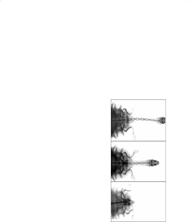
Figure 7.16 Static |
cineradiographs |
recorded in dorso-ventral view, showing the configuration of the cervical vertebrae in (A) fully extended, (B) relaxed, and (C) fully retracted positions of the neck in A. spinifera.
Biology of Turtles
angular changes during retraction occur in the middle region of the neck. Joints C8-7 and C7-6 extend during the course of the retraction. The situation for joints C6-5, C5-4, and C4-3 is more complex. C6-5 initially flexes during the retraction but extends again toward the latter part of neck retraction. The two more cranially situated joints start with an initially extended configuration. During retraction, the angle of these joints gradually increases.
The potential range of movement in the dorso-ventral direction is relatively large in most vertebral joints but largest in joints C8-7, C5- 4, C4-3, and C3-2. However, joint angles during a simulated snorkeling movement are most pronounced at the most posterior cervical joints (D1-C8, C8-7, and C7-6). The anterior joints are kept relatively constant throughout the movements, as is observed during neck retraction (Figure 7.16 and Figure 7.17). Lateral mobility
of the neck in A. spinifera is also surprisingly high, with the more proximal joints being the ones allowing most movement in the horizontal plane. In con-
trast, the last three cervical joints allow almost no lateral A movements.
7.3.4.2 Kinematics of Snorkeling in
C. longicollis
Neck movements during snorkeling in C. longicollis are much slower in comparison with those involved during feeding behavior (Aerts et al., 2001; Van Damme et al., 2002) and escape retraction (Van Damme et al., 1995). The total duration of a typical ventroflexion followed by a dorsiflexion is about 6 s. Each component of the movement takes about 3 s. The representative example of a snorkeling movement in C. longicollis is represented by successive stick diagrams in Figure 7.19. Starting from an extended configuration in which head and neck are slightly depressed, the animal initially balances its neck around this configuration. This configuration is characterized by very small starting angles in all joints (with exception of D1-C8). Minor changes in joint angles occur during the first phase of the movement mainly in the posterior part of the neck (Figure 7.20). During this phase, no conspicuous changes of the elevation of the head and head position are observed. After approximately 3 s, a conspicuous, rather steady increase in the elevation of the head is observed, mainly as a result of changes in the angle of joints C4-3 to C7-6. In patterns observed in C8-7 and D1-C8 in the most posterior part of the neck and S-C1 and C2-1 in the most anterior part of the neck, changes in vertebral joint angle
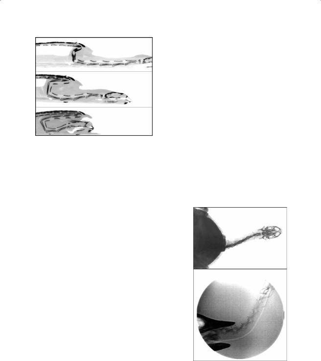
B
Figure 7.18 (A) Static cineradiographs recorded in dorso-ventral view illustrating the position and shape of the cervical vertebrae in C. longicollis. (B) Lateral view of the cervical system in C. longicollis during snorkeling. Note the radio-opaque markers inserted onto the cervical vertebrae to facilitate the analyses of vertebral movements.
Cervical Anatomy and Function in Turtles
appear less conspicuously related to the observed movements, show less overall change, and display a generally more irregular movement pattern. At very high degrees of elevation—at which also head position is also starting to change—changes in joint angle are mainly occurring in joints C5-4, C6-7, and especially C4-3 (more than 30° of dorsiflexion). This is not the case for C6- 5. This joint reaches up to 20° of dorsiflexion and then levels off. Figure 7.21 illustrates the absolute range of movement (expressed in degrees of flexion) of each joint during the whole sequence. Notice the relatively minor joint rotations in the anterior (S-C1, C2-1, and C3-2) and posterior parts (C8-7, D1-C8) of the neck. As mentioned previously, the most conspicuous changes in joint angle are observed in the middle part of the neck, especially in joints C4-3, C5-4, and C7-6.
In summary, the range of mobility in the vertical plane of the cervical joints is limited, especially in comparison with the changes of joint angle in the horizontal plane. Dorsiflexion of the joints is more conspicuous than ventroflexion. Ventroflexion in the joints is nearly non-existent. The dominant changes in joint angle are observed in the middle part of the neck.
Breathing
Horizontal extension
Ventroflexion
C
7.4 |
Discussion |
|
|
|
|
7.4.1 |
Vertebral Structure |
A |
|
|
|
|
|
|
|
||
The nature of retraction of the head-neck system in turtles (in the |
B |
ΔT = 0.25 sec. |
|||
vertical plane as in Apalone or in the horizontal plan as in Chelo- |
|
|
|
|
|
dina) has profound implications on the morphology of the cervi- |
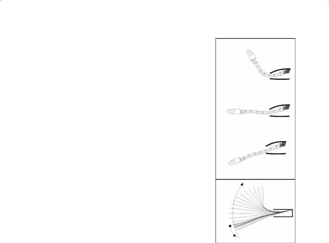
Figure 7.19 (A) |
Schematic |
|||
cal system. Although in both species the cervical vertebrae are |
illustration of the neck configu- |
||||
markedly elongated, in Chelodina the vertebrae are rather narrow |
ration in C. longicollis. (B) Stick |
||||
and tall. However, in Apalone the situation is reversed and the |
diagram representing |
an |
actual |
||
vertebrae are wide and rather shallow. Another marked difference |
snorkeling |
movement |
in C. |
||
is the presence of a pronounced ventral vertebral crest in Chelo- |
longicollis. |
|
|
dina. In Apalone, these crests are only present in the most anterior |
|
vertebrae. The transverse processes that serve as insertion sites |
|
for the muscles of the longissimus are well developed in Chelodina and are positioned laterally at the border between the neural roof and the vertebral centrum. Because the longissimus system is absent in Apalone, the transverse processes are poorly developed and positioned more cranially. Also, the structure of the posterior zygapophyses is markedly different between the two species. Whereas in Chelodina there is only a single basal element carrying the two horizontally oriented articular facets, in Apalone two separate post-zygapophyses are present. Additionally, the articular facets are much more curved in Apalone compared to those in Chelodina. However, in both species the structure of the zygapophyses limits ventroflexion considerably.
7.4.2Cervical Musculature
The differences in retraction mode in the two groups are also reflected in the structure and differentiation of the cervical musculature. As mentioned previously, for example, the longissimus system appears to be completely absent in Apalone. As head-neck movements typically occur in the vertical plane in these animals, this is not unexpected. Alternatively, in Chelodina the longissimus system is strongly developed. Notable in the cryptodires is the cortico-cervical muscular system that
182 |
Biology of Turtles |
Degrees
80 |
|
Head position |
|
|
8 |
40 |
C5−4 |
|
|
|
|
|||
40 |
|
|
|
4 |
0 |
|
|
|
|
|
|
|||
0 |
|
Elevation |
|
|
|
|
|
|
|
|
|
|||
|
|
|
|
–40 |
|
|
|
|
|
|
||||
–40 |
|
|
|
|
0 |
|
|
|
|
|
|
|||
|
|
|
|
|
|
|
|
|
|
|
|
|||
80 |
|
|
|
|
|
|
|
|
|
|
|
|
|
|
S−C1 |
|
|
|
|
40 |
40 |
C6−5 |
|
|
|
|
|||
40 |
|
|
|
|
20 |
|
|
|
|
|||||
|
|
|
|
|
|
0 |
|
|
|
|
|
|
||
0 |
|
|
|
|
|
|
|
|
|
|
|
|
||
|
|
|
|
|
|
0 |
–40 |
|
|
|
|
|
|
|
–40 |
|
|
|
|
|
|
|
|
|
|
|
|
||
80 |
C2−1 |
|
|
|
|
40 |
40 |
C7−6 |
|
|
|
|
||
40 |
|
|
|
|
20 |
|
|
|
|
|||||
|
|
|
|
|
|
0 |
|
|
|
|
|
|
||
0 |
|
|
|
|
|
|
|
|
|
|
|
|
||
|
|
|
|
|
|
|
|
|
|
|
|
|
||
|
|
|
|
|
|
0 |
–40 |
|
|
|
|
|
|
|
–40 |
|
|
|
|
|
|
|
|
|
|
|
|
||
80 |
C3−2 |
|
|
|
|
40 |
0 |
C8−7 |
|
|
|
|
||
40 |
|
|
|
|
|
|
|
|
|
|||||
|
|
|
|
|
|
20 |
–20 |
|
|
|
|
|
|
|
0 |
|
|
|
|
|
|
0 |
–40 |
|
|
|
|
|
|
–40 |
|
|
|
|
|
|
|
|
|
|
|
|
||
|
|
|
|
|
|
–60 |
|
|
|
|
|
|
||
|
|
|
|
|
|
|
|
|
|
|
|
|
||
80 |
|
|
|
|
|
|
40 |
|
|
|
|
|
|
|
C4−3 |
|
|
|
|
40 |
D1−C8 |
|
|
|
|
||||
40 |
|
|
|
|
|
|
20 |
0 |
|
|
|
|
|
|
0 |
|
|
|
|
|
|
|
|
|
|
|
|
||
|
|
|
|
|
|
0 |
–40 |
|
|
|
|
|
|
|
–40 |
|
|
|
|
|
|
|
|
|
|
|
|
||
|
0.0 |
1.0 |
2.0 |
3.0 |
4.0 |
5.0 |
6.0 |
|
0.0 |
1.0 |
2.0 |
3.0 |
4.0 |
5.0 |
|
|
|
Time (s) |
|
|
|
|
|
Time (s) |
|
||||
20
0
20 |
|
|
0 |
|
|
20 |
(°) |
|
Angle |
||
0 |
||
Joint |
||
10 |
||
|
||
0 |
|
–10
20
10
0
–10
6.0
Figure 7.20 Kinematic plots illustrating time profiles of joint angle (dashed curves, right vertical axis) and elevation angle with respect to the horizontal of the more distal of the segments constituting the joint (solid curve, left vertical axis) during a snorkeling movement in C. longicollis. The first panel represents elevation of the head segment and the rectilinear distance (right axis in cm) between head and carapace.
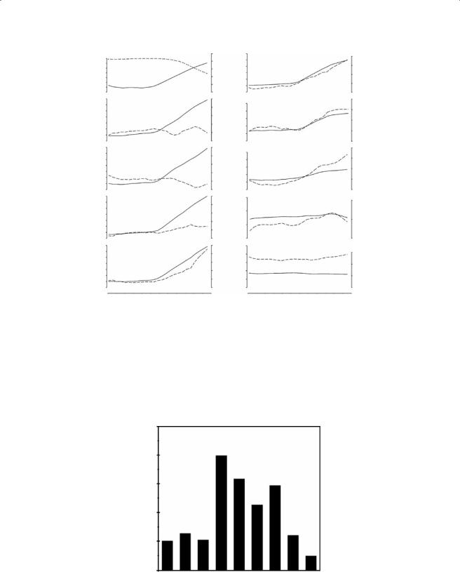
Degrees
Absolute Range of Movement
50
40
30
20
10
0 S-C1 C2-1 C3-2 C4-3 C5-4 C6-5 C7-6 C8-7 D1-C8
Joint
Figure 7.21 Movement range of the cervical joints in C. longicollis in the vertical plane.

Cervical Anatomy and Function in Turtles |
183 |
presumably functions to protract and retract the neck. Preliminary electromyographic data for the cryptodire T. scripta elegans (Van Damme, unpublished) support this hypothesis. In pleurodires like Chelodina, this system is absent. Rather unexpectedly, the m. sphincter colli is poorly developed in Apalone. In Chelodina and most other pleurodires, this muscle appears much better developed and may actually play a role in aligning the cervical muscle bundles along the cervical column during neck movements. However, this remains speculative and must be tested by electromyography. Also, the dominant head retractor muscle, the m. retrahens capitis et collique in Chelodina (Shah, 1963) and the m. carapace-basioccipitis in Apalone (Ogushi, 1913), is notably different in the two species. In Apalone, this muscle consists of a paired massive muscle bundle running from the first caudal vertebra to the back of the skull. In Chelodina, this muscle is more complex and consists of several discrete bundles that insert on specific sites along the cervical column.
However, a striking similarity among the two groups is the extreme length of the head retractor muscle. Presumably, elongation of the retractor muscle allows for more sarcomeres to be placed in series, which may considerably increase the contraction speed of the muscle (Josephson, 1975). Moreover, by increasing the number of sarcomeres in series, each individual sarcomere has to contract less and may thus potentially operate continuously on the plateau of its length-tension relationship. However, for an extremely elongated muscle to contract efficiently, all parts must contract simultaneously. The polyneural, polysegmental innervation of the m. carapaco-basioccipitis in turtles (Guthe, 1981) may allow for this. Unexpectedly, the m. carapace-basioccipitis consists not only of fast twitch fibers but also has a considerable population of tonic fibers (Guthe, 1981), which may help control the position of the head and neck during slow movements or near-stationary behaviors such as snorkeling.
7.4.3Movement Patterns
The cranio-cervical system in turtles can be characterized as an open kinematic chain of eight vertebrae and nine joints. Because each joint theoretically has three rotational degrees of freedom (and to a limited extent also three translational degrees of freedom), and because the positioning of the vertebrae must be achieved by the combined actions of roughly 50 symmetrical muscle bundles, the control of the position of the head in space obviously presents a challenging control problem (Aerts et al., 2001). However, the control of the system is largely simplified by a number of morphological constraints limiting the mobility at each joint. For example, in Chelodina the horizontal nature of the zygapophyseal articular facets and the vertical orientation of the articular facets of the condyle and the vertebral centrum will facilitate movements in the horizontal plane. Additional simplification of the control of the neck in Chelodina is achieved by the presence of distinct areas of rotation along the neck (Van Damme et al., 1995). For instance, the joint between cervical vertebrae 7 and 8 and the joints between cervical vertebrae 6-5 and 5-4 typically show the greatest range of angular change. Moreover, the resting position of the neck is characterized by a distinct bend at these locations, facilitating the correct retraction of the neck using only a simple activation of the m. retrahens capitis et collique (Aerts et al., 2001). Dorso-ventral mobility is greatest at the joints C3-2 through C6-5. The joints surrounding the bi-convex articular centrum of C5 show the greatest angular change during dorsiflexion.
The cervical system in Apalone is notably divergent and the kinematic patterns are also markedly different from those observed in Chelodina. Upon retraction of the neck, the more caudally positioned vertebrae are the first to start their rotation around a transverse axis. The angular changes in all vertebrae are negative during retraction, suggesting that the neck is retracted as a safetylinked bicycle chain. Cervical vertebrae 5, 6, and 7 undergo an angular change of nearly 180°, which implies that these vertebrae end up with their ventral aspect alongside the ventral aspect of the immobile dorsal vertebrae. This is made possible by the nearly complete reduction of the ventral crest of these vertebrae. It is also striking that C8 remains in a near-vertical position at the moment of full protraction. The more anterior vertebrae (C1 to C4) do not contribute to neck retraction at

184 |
Biology of Turtles |
all. The bicycle-chain-like retraction pattern observed for Apalone is in contrast to the retraction patterns suggested in other cryptodires such as Testudo or Trachemys (Scanlon, 1982; Weisgram & Splechtna, 1990). In the latter species, as well as in Chelodina, distinct rotation centers are present where the majority of the angular rotation takes place. Thus, the extremely elongated neck and associated morphology of Apalone may not be representative for the cryptodiran condition in general but provide an excellent example of a highly specialized cervical system that can be compared to the condition in Chelodina. Clearly, further investigation into the morphology and function of the cervical system in turtles is needed to increase our understanding of the evolution of the cervical system and its control in turtles. Especially insightful would be studies exploring cervical structure and function in a broader sample of turtles including short-necked representatives of both cryptodires and pleurodires. Functional approaches including electromyography of the cervical muscles, albeit challenging, are essential to further our understanding of the evolution of the cervical system in turtles.
Acknowledgments
We thank A. De Schepper (University Hospital Antwerp) for allowing us to use the cineradiography machine and CT scanner; the Antwerp Zoo for providing us with the specimens of Chelodina longicollis; G.A. Wood (Wood, 1982) for providing us with a software package for data smoothing and differentiation; and A. Cuylits for making the drawings of the cervical vertebrae in C. longicollis. This study was supported by IWONL-grant 910091 (JV) and FKFO-grant 2.9005.90 (FDV). AH is a postdoctoral fellow of the fund for scientific research, Flanders, Belgium (FWO-Vl).
References
Aerts, P., Van Damme, J., and Herrel, A., Intrinsic mechanics and control of fast craniocervical movements in aquatic feeding turtles, Am. Zool., 41(6), 129–1310. 2001.
Bojanus, L.H., Anatome Testudinis Europaeae, Vol. 1., Vilnae, Luthuania, republished in 1970, Facsimile reprints in Herpetology, no. 26, Society for the Study of Amphibians and Reptiles, Ohio, 1819.
Bojanus, L.H., Anatome Testudinis Europaeae, Vol. 2., Vilnae, Luthuania, republished in 1970, Facsimile reprints in Herpetology, no. 26, Society for the Study of Amphibians and Reptiles, Ohio, 1821.
Dalrymple, G.H., Intraspecific variation in the cranial feeding mechanism of turtles of the genus Trionyx, J. Herpetol., 11, 255–285, 1977.
Deban, S.M., Wake, D.B., and Roth, G., Salamander with a ballistic tongue, Nature, 389, 27–28. 1997. Ernst, C.H., and Barbour, R.W., Turtles of the World, Washington, DC: Smithsonian Institution Press, 1989. Gans, C., Why develop a neck?, in The Head-Neck Sensory Motor System, A. Berthoz, P. Vidal, and W. Graf
(eds.), New York: Oxford University Press, 1992, 17–21.
George, J.C., and Shah, R.V., The myology of the head and neck of the common Indian pond turtle, Lissemys punctata granosa Schoepff, J. Anim. Morph., 1, 1–12, 1954.
George, J.C., and Shah, R.V., The myology of the head and neck of the Indian tortoise, Testudo elegans, J. Anim. Morph. Physiol., 2, 1–13, 1955.
Guthe, K.F., Reptilian muscle: Fine structure and physiological parameters, in Biology of the Reptilia, Vol 1, C. Gans and T. Parsons (eds.), New York: Academic Press, 1981, 265–354.
Heidweiller, J., Van Der Leeuw, A.H.J., and Zweers, G.A., Cervical kinematics during drinking in developing chickens, J. Exp. Zool., 262, 135–153, 1991.
Herrel, A., Meyers, J.J., Nishikawa, K.C., and Aerts, P., The mechanics of prey prehension in chameleons, J. Exp. Biol., 203, 3255–3263, 2000.
Hofstetter, R., and Gasc, J.P., Vertebrae and ribs of modern reptiles, in Biology of Reptilia, Vol. 1, C. Gans and T. Parsons (ed.), New York: Academic Press, 1969, 201–231.
Irschick, D.J., and Garland, T. Jr., Integrating function and ecology in studies of adaptation: Investigations of locomotor capacity as a model system, Ann. Rev. Ecol. System., 32, 367–396, 2001.
Josephson, R.K., Extensive and intensive factors determining the performance of striated muscle, J. Exp. Biol., 194, 135–154, 1975.
King, G., Reptiles and Herbivory, London: Chapman & Hall, 1996, 160.

Cervical Anatomy and Function in Turtles |
185 |
Ogushi, K., Anatomische Studien an der japanischen dreikralligen Lippenschildkröte (Trionyx japonicus): Muskel und peripheres nervensystem, Morph. Jb., 46, 299–562, 1913.
Pritchard, P.C.H., Piscivory in turtles, and evolution of the long necked Chelidae, Symp. Zool. Soc. Lond., 52, 87–110, 1984.
Romer, A.S., and Parsons, T.S., The Vertebrate Body, Philadelphia: W.B. Saunders, 1977, 161–167.
Scanlon, T.C., Anatomy of the neck of the Western Painted turtle (Chrysemys picta belli Gray; Reptilia, Testudinata), unpublished diss., University of Michigan, University Microfilms International, 1982.
Shah, R.V., The neck musculature of a Cryptodire (Deirochelys) and a Pleurodire (Chelodina) compared, Bull. Mus. Comp. Zool., 129, 343–368, 1963.
Vaillant, M.L., Mémoire sur la disposition des vertebres cervicales chez les chéloniens, Ann. Sci. Nat. Zool. Paleont., Ser. 6, 10, 1–106, 1881.
Vallois, H.V., Les transformations de la musculature de l’épisome chez les vertébrés, Arch. Morph. Gen. Exp., 13, 180–217, 1922.
Van Damme, J., and Aerts, P., Kinematics and functional morphology in aquatic feeding in Australian snakenecked turtles (Pleurodira; Chelodina), J. Morph., 233 , 113–125, 1997.
Van Damme, J., Aerts, P., and De Vree, F., Kinematics of the escape head retraction in the common snake-necked turtle, Chelodina longicollis (Testudines: Pleurodira: Chelidae), Belg. J. Zool., 125, 215–235, 1995.
Van Damme, J., and Aerts, P., Cervical movements during prey capture in Australian snake necked turtles, genus Chelodina (Pleurodira, Chelidae), in Topics in Functional and Ecological Vertebrate Morphology, A Tribute to Frits De Vree, P. Aerts, K. D’Août, A. Herrel, and R. Van Damme (eds.), Shaker Publishing BV, 2002, 77–94.
Weisgram, J., and Splechtna, H., Intervertebral mobility in the neck of two turtle species (Testudo hermanni hermanni, Pelomedusa subrufa), Zool. Jb. Anat., 120, 425–431, 1990.
Weisgram, J., and Splechtna, H., Cervical movements during feeding in Chelodina novaeguinaeae (Chelonia, Pleurodira). Zool. Jb. Anat., 122, 331–337, 1992.
Williams, E.E., Variation and selection in the cervical centre articulations of living turtles, Bull. Amer. Mus. Nat. Hist., 94(9), 505–562, 1950.
Wood, G.A., Data smoothing and differentiation procedures in biomechanics, in Exercise and Sports Sciences Reviews, 10, 308–362, 1982.
Wren, K., Claussen, D.L., and Kurz, M., The effects of body size and extrinsic mass on the locomotion of the ornate box turtle, Terrapene ornata, J. Herpetol., 32, 144–150, 1998.
Zani, P.A., Gottschall, J.S., and Kram, R., Giant tortoises walk without inverted pendulum mechanical-energy exchange, J. Exp. Biol., 208, 1489–1494, 2005.