

Paluch et al. BMC Biology (2015) 13:47 |
Page 8 of 14 |
several days [72]. Another recent study used this quantitative order parameter analysis to determine the effect of substrate elasticity on differentiating myoblasts [73], indicating that mechanical stimuli and respective change of cytoskeleton structure do play a role in differentiation of more than one cell type.
Such significant differences in macroscopic or global structures are most likely induced by distinct molecular compositions of the actomyosin stress fibers on the nano-scale, a hypothesis that might be confirmed in the near future by super-resolution microscopy methods now available. It is important to note that this type of analysis is now done with fixed cells at distinct times, just like one would do for protein or transcription analysis. To get a better insight into the complex and also highly dynamic processes it will be of paramount importance to follow the temporal evolution of stress-fiber structure and organization followed by live cell imaging and quantitatively extract parameters of the kinetics.
Analyzing transcription profiles and biochemical signaling cascades is essential to finally elucidate the complex processes underlying mechanosensory phenomena. However, the microand nano-structural properties of those mechanically active structures contain valuable information on the route from outside mechanical signals to inside biochemical regulation. In the same way that cell biology relied for a long time on descriptive phenotyping by analyzing global cell shape, we should use the additional information of the morphology of stress fibers to complete our picture of mechano-guided differentiation of hMSCs.
Reversible adhesion in climbing animals - is it similar to cell adhesion?
Walter Federle
Not only cells but also many climbing animals such as insects, spiders and geckos are able to move around in their environment, yet have to resist detachment forces by forming adhesive contacts. These animals possess special attachment structures on their feet that allow them to cling to substrates. When climbing on trees they may be challenged by the forces of gravity and wind, or by predators trying to dislodge them [74]. Cells, in turn, can be exposed to shear flows in blood vessels or to tensile stresses within tissues. Many animals can climb in a three-dimensional environment such as the forest canopy by repeatedly attaching and detaching their feet, whereas cells can migrate on substrates and within tissues by forming new adhesive contacts at their front and releasing them again at their trailing edge [75]. By way of these functional similarities, it is perhaps natural to compare cell adhesion with the adhesion of climbing animals. Is cell adhesion similar to the adhesion of insects, spiders and geckos?
Comparison of adhesive strength
Starting with the physical mechanism of adhesion, both cells and climbing animals take advantage of van der Waals forces [76, 77]. These forces only become significant when two objects are in intimate contact with separation distances less than approximately 10 nm [78]. The contacts formed by cell adhesion molecules such as integrin or cadherin and those of gecko adhesive hairs are likely within this range [79, 80]. In addition to van der Waals forces, cell adhesion is strongly dependent on electrostatic forces comprising hydrogen bonds, double layer forces and forces between charged domains of interacting proteins [79]. An important role of electrostatic forces via contact electrification has also been proposed recently for the adhesion of geckos [81], and similar mechanisms are possible for insects [82]. Although more evidence is needed to confirm the extent to which contact electrification contributes to animal adhesion, both cells and climbing animals are probably affected by electrostatic interactions. In addition, the adhesion of many climbing animals involves capillary forces, arising from tiny amounts of fluid secreted into the adhesive contact zone [83–87]. As cells live in a watery environment, this adhesive mechanism is usually absent in cells.
Given that both cells and whole animals adhere by van der Waals forces and probably electrostatic forces, one might expect their adhesive stresses to be of similar magnitude. Let us briefly consider the consequences if this were indeed the case: depending on the geometry, adhesion forces scale with the length or the area of adhesive contacts [88]. For example, while the pull-off force for a suction cup is proportional to its area, the force needed to peel off a piece of Scotch tape depends on its width. For isometric organisms, weight increases with the cube of linear dimensions, and therefore faster than areaor length-specific adhesion. As a consequence, adhesion per body weight is expected to decrease for larger animals. Despite their relatively large body size, however, geckos can easily hang from a single toe, and weaver ants can carry more than 100 times their own body weight whilst walking upside down on a smooth surface (Fig. 3a, b). Clearly, these animals use only a small fraction of their body surface area (that is, the adhesive organs on their feet) for attachment, and at least the gecko does not seem to employ any specific adhesion molecules to achieve high levels of forces.
Cells have an average mass of approximately 1 ng [89] and are thus 5 to 11 orders of magnitude lighter than geckos and insects. Their weight-specific adhesion should thus be enormous in comparison to animals. Moreover, about half of a cell’s surface is in relatively close contact with the substrate, a much larger proportion than for a climbing animal. Thus, one might expect cells to adhere
Paluch et al. BMC Biology (2015) 13:47 |
Page 9 of 14 |
||
|
|
|
|
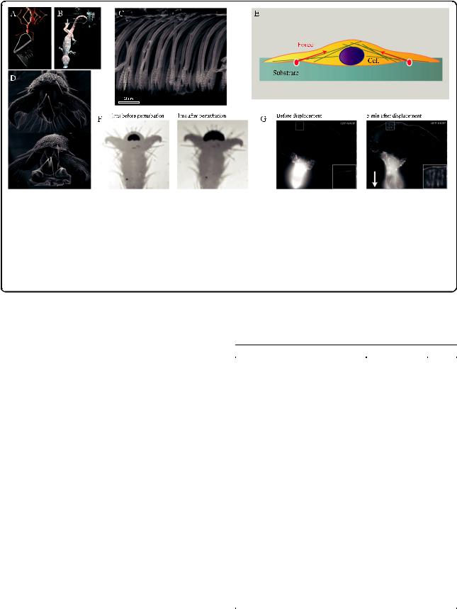
Fig. 3. Surface adhesion in climbing animals and cells. a Weaver ant (Oecophylla smaragdina) carrying more than 100 times its body weight upside-down on a smooth surface (photo: Thomas Endlein). b Tokay gecko (Gekko gecko) attached by a single toe to a tilted glass surface. Reproduced from [130] with permission from the Journal of Experimental Biology. c Lateral view of adhesive setae in a longhorn beetle (Clytus arietis) showing non-adhesive orientation of seta tips and anti-adhesive corrugations on the dorsal side. Reproduced from [131] with permission from the Journal of Experimental Biology. d Weaver ant adhesive pad in the retracted (top) and the extended position (bottom). Reproduced from [114]. e Adherent cell on a deformable substrate. Inward forces are transmitted via the cytoskeleton and the focal adhesions to the substrate. Adapted from [75]. f Rapid increase in adhesive contact area in stick insects (Carausius morosus) in response to a rapid displacement of the substrate. Adapted from [121]. g B16 melanoma cell (expressing fluorescent marker for focal adhesions) before and 5 minutes after displacement of cell body by a microneedle (direction shown by arrow), showing growth of peripheral focal contacts in the region opposite the cell body (enlarged in insets), stimulated by tension. Reproduced from [123] with permission from the Journal of Cell Science
very firmly to any substrate and to one another, even without any specific adhesion molecules, potentially limiting or preventing locomotion. Are the adhesive stresses of cells and climbing animals indeed comparable, and how do they compare with typical levels for van der Waals forces?
van der Waals forces are considered weak intermolecular forces, but they still can produce maximum contact strengths of the order of 100 MPa, sufficient for a 1 cm2 contact to support the weight of a small family car [76]. Table 1 shows that the stresses measured for animal adhesives are at least two orders of magnitude below these theoretical strength levels, probably a result of stress concentrations and surface contamination. However, adhesive and shear stresses of cells are much smaller still, by several orders of magnitude. Why are stresses so much smaller for cells, although they use similar molecular forces for attachment?
Firstly, cells live in a watery environment. Water not only provides viscous ‘squeeze-out’ resistance that makes it harder for objects to come into close contact, but it also shields surface charges, and intervening layers of water reduce van der Waals forces [76]. However, there are many examples of marine organisms that achieve high adhesion strengths under water (e.g., barnacles and mussels) [90–92], so that submersion alone may not fully explain the low stress levels of cells.
Table 1 Shear and adhesive strength of animal adhesive pads in comparison with single cells
|
Strength (kPa) |
Source |
Shear forces |
|
|
Gecko seta: real contact area |
53,300 |
[124, 125] |
Gecko seta: projected contact area |
2,880 |
|
Beetle pad: real contact area |
681 |
[112] |
Beetle pad: projected contact area |
259 |
|
Weaver ants |
405 |
[126] |
Stick insects |
299 |
[112] |
Barnacles |
10-300 |
[127] |
Fibroblast cells (whole) |
0.048 |
[128] |
Fibroblast cells (focal contacts) |
5.5 |
[103] |
Adhesion |
|
|
Gecko seta: real contact area |
10,700 |
[124, 125] |
Gecko seta: projected contact area |
576 |
|
Beetle pad: real contact area |
86.9 |
[112] |
Beetle pad: projected contact area |
35.5 |
|
Stick insect pad |
44.6 |
[112] |
Barnacles |
100-1000 |
[90] |
Ants |
~50 |
[126] |
Endothelial cells |
0.56-1.1 |
[129] |
|
|
|
Paluch et al. BMC Biology (2015) 13:47 |
Page 10 of 14 |
A second key factor that reduces cell adhesion is longrange repulsion by the glycocalyx, a layer of glycoproteins and glycolipids on the outer cell surface [93, 94]. These surface molecules carry negative charges that inhibit adhesion between cells or negatively charged surfaces. The electrostatic repulsion by the glycocalyx is further enhanced by steric repulsion and osmotic effects [93]. Treatments reducing the negative charge on substrates were found to increase cell adhesion dramatically [95], and it is likely that excessive non-specific adhesion would lead to cell clustering and render cells unable to move. Cell locomotion is indeed enhanced on more negatively charged substrates [96]. Thus, while sufficient adhesion to diverse substrates via non-specific adhesion is essential for climbing animals to prevent detachment, the priority for cells may be exactly the opposite. Cells have to reduce or prevent non-specific adhesive interactions to maintain motility and to allow the controlled formation and release of specific adhesive contacts. Prevention of unwanted adhesion is also a common theme in the larger world of climbing animals. Many plant surfaces prevent insect adhesion with the help of epicuticular wax crystals or surface textures that trap lubricating water films [97]. In many insects, fields of microtrichia occur in regions immediately outside adhesive contact zones; it is likely that these surface structures are nonadhesive and facilitate detachment when the foot pad is rolled off, or in some systems prevent the self-matting of adhesive hairs (Fig. 3c) [98].
One consequence of the interplay of short-range attraction between cell adhesion molecules and long-range repulsion by the glycocalyx is that adhesion is concentrated locally in small adhesion domains such as focal contacts or desmosomes, which are connected intracellularly to filaments of the cytoskeleton [99]. This arrangement is functionally similar to adaptations in animal adhesive structures. The division of one large contact into many sub-contacts (contact splitting [100]) is one design principle of ‘hairy’ adhesive systems found in insects, spiders and geckos, which consist of dense arrays of microscopic hairs. Contact splitting can increase adhesion if the forces of individual contacts scale with their width or perimeter (comparing a smooth and a hairy toe pad of the same size, the hairy pad has a much smaller total contact area, but a much larger total contact perimeter), and if different contacts are loaded simultaneously so that a condition of ‘equal load sharing’ is achieved [101]. Cells are able to control the pulling forces on focal contacts by contractile stress fibers and remodeling of the actin fiber network [75, 102, 103]. Climbing animals can actively distribute loads between different legs or pads; but within each individual pad, the stress distribution depends mainly on the pad’s stiffness and mechanical design. The adhesion forces of many animals scale with contact area [88], suggesting
that their pads are designed in a way that allows them to achieve uniform load distribution, but the underlying mechanisms are still unclear. Separate subcontacts also help for attachment on uneven substrates. Although gecko and insect adhesive hairs are made of relatively stiff keratin and cuticle, respectively, arrays of adhesive hairs are compliant and can maintain adhesion even on substrates with significant surface roughness [104, 105].
Adhesion and locomotion
One of the most apparent parallels between cell adhesion and whole-animal adhesion is that, in both systems, the contacts are reversible and dynamic to allow locomotion. When adhesive pads of climbing animals are sheared (pulled) towards the body, they adhere firmly, but they detach when pushed. A pull maximizes adhesion by increasing not only the adhesive contact area, but also the force per contact area [106].
Cells and animals are able to switch between different types of adhesive bonds, depending on whether weak temporary or strong permanent adhesion is required. Leucocytes provide a well-known example of an active change from weak/transient to stronger/more permanent adhesion [107]. Leucocytes moving from the circulatory system into infected tissues undergo states of weak adhesion, in which they 'roll' on the wall of venules, followed by firm adhesion to endothelial cells and transmigration through the endothelium, each involving different adhesion molecules (selectins and integrins) with varying binding affinities. The different steps of this cascade are triggered by the release of cytokines from infected tissues which trigger the expression of selectin molecules on the inner vessel wall, and by the release of chemokines which activate integrins on the leucocytes. While the shear force exerted by the blood flow is sufficient to maintain dissociation of selectins to induce cell rolling, it is too small to dislodge cells in the firm adhesion state [108]. There are some analogous cases of animals switching between different adhesive mechanisms for temporary and permanent adhesion. For example, limpets use a glycoprotein glue and achieve high adhesive strengths when stationary at low tide, but suction (achieving lower adhesive strengths) when moving around underwater at high tide [109]. However, most animals do not control adhesion by varying the chemistry of their adhesive bonds (this would probably be too slow for climbing animals), but by changing the geometry of the adhesive contact.
When climbing rapidly, geckos, spiders and insects are able to attach and detach their feet within tens of milliseconds. Although foot detachment can be very fast, it occurs without any measurable force peaks in geckos and insects [110]; [T. Endlein & W. Federle, unpubl. results]. The key principle underlying this impressive performance is the