

Paluch et al. BMC Biology (2015) 13:47 |
Page 7 of 14 |
Mechano-guided differentiation of human mesenchymal stem cells: actomyosin stress fibers as collective mechanosensors
Carina Wollnik, Galina Kudryasheva and Florian Rehfeldt
It is nowadays well acknowledged that mechanical stimuli can be as important for cells as traditional biochemical cues [62]. They also impact the efficacy of drugs [63] and can influence the morphology and growth phase of cellular aggregates [64]. Especially striking are the experiments by Engler et al., demonstrating that substrate elasticity can direct differentiation of human mesenchymal stem cells (hMSCs) towards various linages (neural, muscle, bone) [13]. Here, it is particularly interesting that mimicking Young’s modulus of the in vivo environment drives naïve adult stem cells towards the respective cell type. While the initial cue (elastic properties of the matrix) and the overall outcome (changes in transcription) are well-defined, the integration of the mechanical stimuli into biochemical signaling pathways is still not fully understood. Recently, there is mounting evidence that direct mechanical coupling to and perturbation of the nuclear envelope and the nucleus might be an alternative or additional route to alter gene regulation [65].
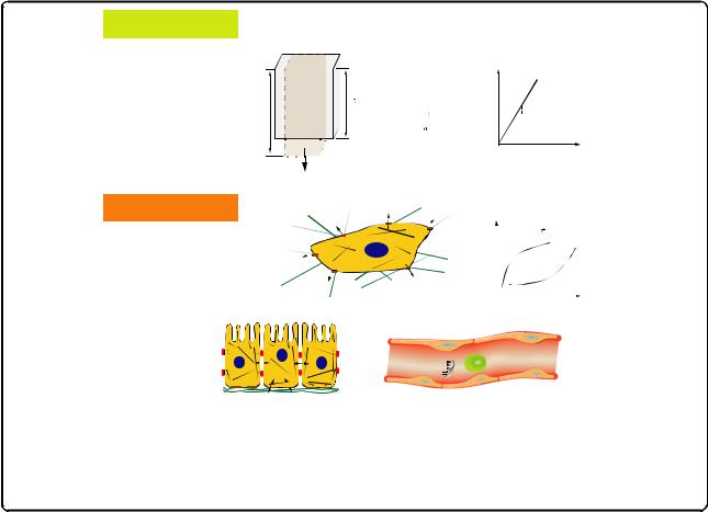
To explain such a mechanical pathway, it is essential to understand how forces can be transmitted to the nucleus. Actomyosin stress fibers are key players in cell adhesion and cell-matrix interactions as they anchor at focal adhesion sites and create cellular contractility (Fig. 2a) [66, 67]. Because they also connect directly to the nuclear lamina [68, 69], stress fibers are able to pass on stress and strain and deform the nucleus [65]. A closer look at the actomyosin filaments in hMSCs revealed that quantification of their structure and organization by means of an order parameter S showed significant differences with respect to the substrate elasticity at an early stage of mechano-induced differentiation (24 hours) [70, 71]. On soft (1 kPa) and rigid (34 kPa) substrates the stress fibers are organized more isotropically (S = 0.08 and 0.58, respectively), while on 11 kPa substrates (an intermediate elasticity and matching the in vivo stiffness of relaxed muscle) the actomyosin bundles were parallel aligned and showed high anisotropy as indicated by an order parameter S = 0.63 (Fig. 2b). This early morphological marker can be understood in terms of a collective mechanosensor and is experimentally observable long before lineagespecific genes are upregulated, a process that usually takes
engineering materials
material model assumptions
homogeneous
L
isotropic linear elastic
small deformations
stress(σ) = |
F |
(force) |
|
|||
A |
(area) |
|
(Pa) |
|||
|
|
|||||
L |
|
|
||||
|
|
|
||||
strain(ε) = |
|
L -L ( length) |
σ |
|||
|
|
|
|
|||
|
|
L |
(length) |
|
||
F
Hooke’s Law
σ =E ε

 E... elastic modulus
ε (%)
proteins, cells, tissues
material model assumptions
heterogeneous anisotropic, hierarchical viscoelastic
large deformations
epithelium
|
|
|
|
E const. |
|
|
|
σ (Pa) |
|
tissue cells in |
|
|
||
|
ε (%) |
|||
fibrous 3D extracellular matrix |
|
|||
|
|
|||
shear stress |
|
|
circulating cells |
|
|
|
|
endothelium |
|
|
|
|||
|
|
|
||
|
|
|
|
|
Fig. 2. Acto-myosin stress fibers are key mechanical regulators in cell-matrix mechanosensing. a Sketch of a cell adhering to a substrate of elasticity E. Actomyosin stress fibers (magnified in the inset zoom) are connected via focal adhesions and extracellular matrix proteins to the micro-environment and generate contractile forces that enable the cell to sense the mechanical properties of the substrate. The cytoskeleton is also connected to the nuclear lamina, thus providing a direct mechanical route to gene regulation. Adapted from [67] with permission from The Royal Society of Chemistry. b Non-monotonic dependence of stress fiber structure quantified by an order parameter S of hMSCs grown on substrates of different elasticity E can be used as early morphological marker for mechano-guided differentiation. Scale bar is 50 μm. Adapted from [70] with permission from the Nature Publishing Group