

Paluch et al. BMC Biology (2015) 13:47 |
Page 4 of 14 |
can guide differentiation of stem cells (see also the pieces by Ben Fabry and Carina Wollnik et al. in this forum). Furthermore, strong attachment forces may be required for cells migrating against a flow, such as in blood vessels. Another key question is the molecular basis of friction. It is unclear whether it depends on the chemistry of the cell surface and the substrate only, or if geometric features such as substrate rugosity and geometry might also play a role. Finally, it will be exciting to elucidate in what physiological contexts friction-based migration occurs in vivo.
Acto-myosin cycling kinetics and focal adhesion reinforcement drives cellular durotaxis
Ben Fabry
Durotaxis describes the movement of cells along a stiffness gradient of the substrate. Similar to chemotaxis where cells migrate towards a concentration gradient of chemokines, durotaxis is thought to be important for tissue formation during embryogenesis, or the migration of cells during wound healing, inflammation, and metastasis. Obviously, durotaxis cannot be fully understood without a basic understanding of cell migration. Migration, in turn, cannot be understood without some understanding of the underlying fundamental processes: cell adhesion (and deadhesion), spreading, and contraction. The canonical picture of how cells crawl on a planar substrate is that of a sequential four-step process [35]. First, cells form protrusions at the leading edge, driven by actin polymerization. Second, these protrusions are attached to the substrate through the formation of focal adhesions. Third, these focal adhesions are connected to actin stress fibers that are tensed through the contractile activity of myosin motors. Finally, the focal adhesions at the rear end of the cells de-adhere under the influence of time and contractile force. Each of these processes can contribute to durotaxis.
Focal adhesions generate friction between the cell and the substrate. Stronger and longer-lasting adhesions give rise to higher friction and thus result in a lower speed of cell migration. As discussed in more detail below, cells form stronger adhesions and consequently migrate more slowly on stiffer substrates [36]. If we consider that cell migration on a mechanically isotropic substrate is a directionally random process, it follows that cells spend less time in regions with low stiffness, and thus more time in regions with higher stiffness. The net effect is durotaxis, and to understand this, we need to understand why adhesions become stronger on stiffer substrates.
For reasons that are still not fully known, adhesions are reinforced (they become larger and stronger) under mechanical load [37, 38]. The mechanical load equals the internal contractile stress of the cytoskeleton and at the same time the external substrate tractions. Cells usually generate higher tractions on stiffer substrates [39];
hence, adhesions also become stronger on stiffer substrates [36]. Thus, we next need to understand why tractions, and cell contractility, are stiffness-dependent.
As cells contract, the resulting traction forces deform the underlying substrate. The softer the substrate is, the more it deforms. Large deformations, however, pose a problem for the cell for two reasons, both of which were first discovered in muscle tissue. First, the force-generating contractile apparatus of the cells has to shorten, which reduces the force it can generate because the overlap between actin and myosin filaments becomes suboptimal [40]. Second, larger deformations require a larger speed of contraction, and more and more of the myosin-generated forces are wasted to overcome the internal friction with actin [41]. Thus, cells cannot keep up large traction forces on soft substrates, with the consequence that focal adhesions become instable, which allows the cell to migrate faster until it reaches a region of higher substrate stiffness.
This physical picture of durotaxis as presented here is of course highly simplified and neglects important biological details. For example, myosin activation is not constant in a cell but is actively controlled by force-dependent signaling cascades that originate at focal adhesions [42]. Nonetheless, key aspects of durotaxis can be explained without such complex biochemical signaling events. All that is needed are two force-sensitive processes. One appears to be the dependence of myosin-generated cytoskeletal forces on the sliding speed, which is governed by acto-myosin crossbridge cycling kinetics [41], and the other appears to be the stress-dependent reinforcement of focal adhesions, which may also be governed by a simple physical mechanism, namely the catch-bond kinetics reported for focal adhesion proteins [43]. Given the fundamental importance of durotaxis for essential cell behavior in the living organism, it may be sensible that it relies not on complex signaling cascades that can be easily deregulated, but on robust physical principles.
May the force (deformation) be with you
Jens Moeller and Beth L Pruitt
Do cells sense and respond to forces or deformations?
Cells within tissues are subjected to exogenous, physiological forces, including fluid shear stress or mechanical load, while at the same time cells exert acto-myosin- generated contractile forces to the extracellular matrix (ECM) and to neighboring cells via cell-ECM and cellcell adhesions [44]. Hooke’s Law and Newton’s Laws of equilibrium readily relate the linear extension of a ‘spring’ to forces, and using appropriate material models we can further relate forces to stresses (force/area). All mechanical measurements revolve around exquisitely precise displacement measurements, yet these displacement data must be converted to estimate force via a set of material deformation models [45]. By necessity, these models are
Paluch et al. BMC Biology (2015) 13:47 |
Page 5 of 14 |
oversimplified because proteins, cells, and tissues present highly anisotropic, heterogeneous, nonlinear mechanical properties that vary widely and depend on the composition, architecture, and environmental conditions, as well as the direction, nature and rate of load application [46]. But do not despair, for while ‘essentially, all models are wrong, some are useful’ [47]. Although we need a material model to estimate forces and stresses, we can directly observe substrate displacements and calculate strains (changes in length/original length) or three-dimensional deformation fields (Fig. 1). To unravel how cells convert these mechanical cues into biochemical signals
(mechanotransduction), we must also consider how the structures of proteins and protein networks are altered upon mechanical load. The cellular microenvironment consists of protein networks of varying biochemical and physical properties, including matrix composition, dimensionality and stiffness, all of which have been shown to coregulate cell function, differentiation, tissue homeostasis and organ development [48, 49].
ECM remodeling provides a force-feedback loop
Not only do cell mechanical properties exhibit ratedependent behaviors such as non-linear viscoelasticity or
A T, pH,c, ...
 -myosin stress fibers
substr |
E |
|
B E= 1kPa |
11kPa |
34 kPa |
24 hours
S = 0.08 |
0.63 |
0.58 |
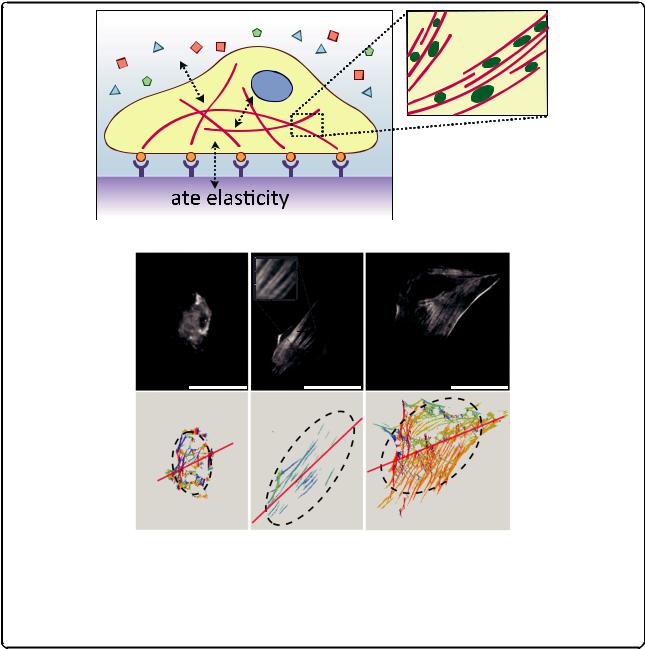
Fig. 1. Hooke's law for linear elastic engineering materials compared to complex material models for biological specimens. The ratio between applied stress σ (force/area) and resulting strain ε (deformation) is described by the elastic modulus E for homogenous, isotropic, linear elastic materials. For biological specimens, the material model assumptions are more difficult and depend on the specific system. Proteins, cells and tissues consist of multiple heterogeneous, anisotropic building blocks of various length scales that are hierarchically organized and exhibit rate-dependent, non-linear, viscoelastic stress–strain responses. Comparison of mechanical properties across systems and among different testing methods requires careful assessment of testing conditions and calibration schemes, which are not yet standardized
Paluch et al. BMC Biology (2015) 13:47 |
Page 6 of 14 |
thermodynamic instabilities, but the substrate itself also changes with time. Cells secrete and remodel the ECM that comprises their tissue microenvironment in a load history-dependent manner. For example, bone resorption by cell-secreted proteases as in microgravity results in more porous, weaker ECM networks while bone growth by cell-secreted and remodeled ECM reinforcement occurs with weight-bearing exercise [50]. Variations in ECM biophysical properties within or across a tissue are not only graded in their composition, crosslinking, dimensionality and stiffness by the cells that created them, but these properties also feedback on cell responses via mechanically mediated cell signaling pathways and enable longrange signaling by cells through the ECM. The biochemical and physical properties of cell-generated ECM protein networks (for example, collagens and laminins in basement membranes or collagens, fibronectin and elastins in blood vessels) co-regulate cell functions such as motility, proliferation, apoptosis [51], stem cell differentiation [13] and organ development [48]. Different cell types show preferences for substrates of different rigidity, which in turn can elicit different cell-ECM traction forces [52]. Unlike inert materials with fixed properties, living cells remodel themselves and their environment under chronic loading and also actively generate mechanical forces through actomyosin-generated tension in the cytoskeleton.
How do molecular mechanisms integrate to cellular mechanotransduction?
Even though we can relate the deformations of the cells and the cellular microenvironment to forces, mechanically mediated signaling at the molecular level is also fundamentally governed by deformation and rate sensitivity. Binding kinetics, protein crosslinking and availability of binding sites depend on both the protein sequence and protein conformation, a function of thermodynamic energy. Changing protein conformations can encode distinct functional states with different ligand binding affinities or binding availability by exposing cryptic binding sites for other binding partners. For example, the rod domain of talin, a focal adhesion protein linking integrins to the actin cytoskeleton, undergoes large conformational changes under acto-myosin-generated forces and exposes cryptic binding sites for vinculin [53]. Vinculin recruitment in turn contributes to a reinforcement of the focal adhesion complex to transduce higher load between the cells and the ECM [54]. Similarly, actomyosin-generated tension facilitates vinculin binding to a cryptic binding site in α- catenin in cell-cell adherens junctions to regulate tissue organization [55]. Within the fibrous ECM, fibronectin unraveling is controlled by Rho-mediated cell contractility to expose cryptic self-assembly sites and binding sites for
other proteins and growth factors [56, 57]. All those mechanosensitive proteins consist of multiple domains with a range of threshold unfolding loads. At appropriate force thresholds, stiff protein domains (β-sheets, α-helices, barrels) first reorient in the direction of loading as flexible linker chains connecting them (turns, loops, hairpins) stretch and rotate; the individual domains unfold in the order of their stiffness and thus contribute to highly nonlinear force-displacement behavior as proteins can unfold up to >10 times their equilibrium length. Most of the protein force spectroscopy studies quantified force thresholds for these phenomena through a complex set of assumptions both about the measurement tools and the sample to arrive at forces at the scale of picoNewtons in such load-bearing proteins as cadherin [6] and vinculin [5]. While pN molecular forces may be sufficient for mechanically switching individual protein functions or binding affinities, aggregated forces measured at the cellular level are much higher and result in deformation on the surrounding ECM on the order of several cell lengths. Indeed, fibrous scaffolds transmit tension over long distances through their rope-like interconnections [58].
How can we measure deformations (forces)?
Applying or measuring displacements (and inferring forces from these measurements) across the length scales of proteins, cells, and tissues requires a range of techniques and several biochemical sensors and microfabricated devices have been developed for this purpose. Optical and magnetic tweezers, Förster Resonance Energy Transfer (FRET) molecular tensions sensors, and atomic force microscopy (AFM) are widely used to study conformational changes of individual mechanosensitive proteins under mechanical load [59], while optical stretchers, micropipette aspiration, AFM and microelectromechanical systems (MEMS) enable single cell mechanobiological studies (see [60] for a review). Meanwhile, traction force microscopy of fiducial markers embedded in compliant substrates and microfabricated post arrays are commonly used to measure the displacement fields of single cells and microtissues [61]. Given the dynamic nature of the state of cells, ECM and proteins, estimates of small forces or heterogeneous mechanical properties are not easily compared between labs using the same method, let alone across methods, and standardized calibration and measurement schemes are needed. Nevertheless, the variability of life is perhaps greater still, and thus mechanobiologists can learn a great deal from appropriately designed experiments and controls to look for relative changes in a consistent framework of measurements and models.