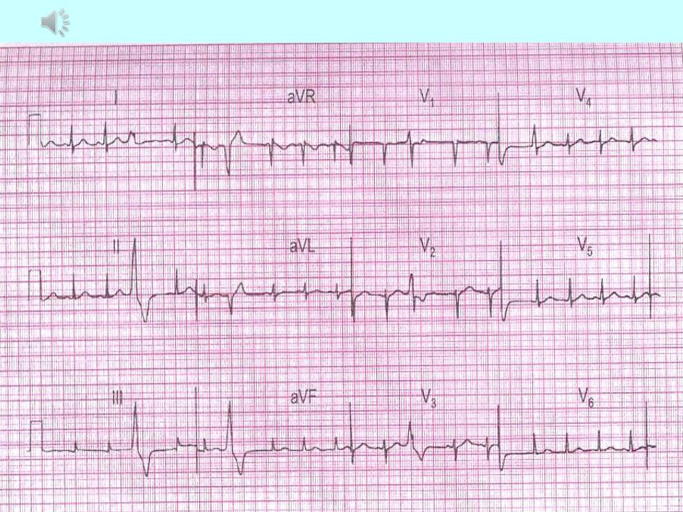
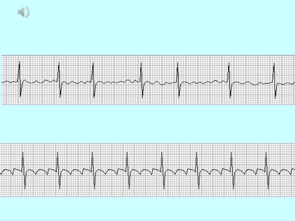
Supraventricular extrasystole:

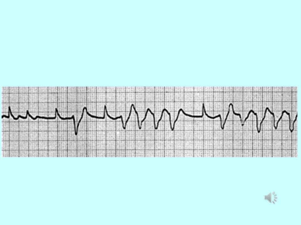
Ventricular extrasystole:
This is a premature excitatory impulse occurs in various parts of the conducting system ventricles. The main ECG signs:
Premature contraction
1.Absence of the P wave in the extrasystolic complex.
2.Significant expansion (more about, 1 s) and deformation
ventricular complex (splitting, bifurcation teeth, serration, large amplitude compared to normal complexes).
3.Discordant displacement of the R (S) -T segment and the T wave (asymmetrical two-phase or negative) on in relation to the main tooth of the QRS complex.
4.Usually full compensatory pausе.

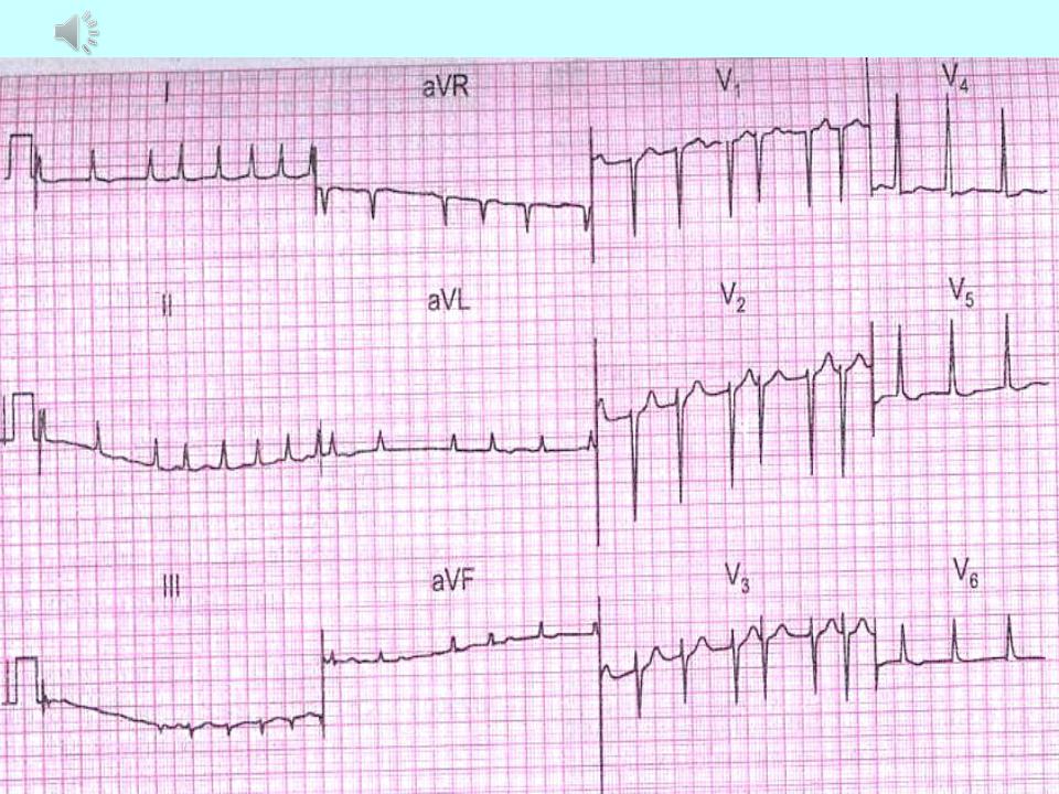
Ventricular extrasystole:
ECG can determine the place of occurrence ventricular premature beats: from the left ventricle, from
right ventricle, from the base of the ventricles and apex
heart, and most accurately on the chest leads. With left ventricular extrasystole in V1, V2, as well as III
aVF derivations QRS complex of extrasystoles is presented
the main R wave, and in leads V5, V6, I and aVL - a wave
S.
With right ventricular extrasystole, on the contrary: in
leads V1, V2, III, a VF QRS complex is directed downward, and
Ventricular extrasystole:

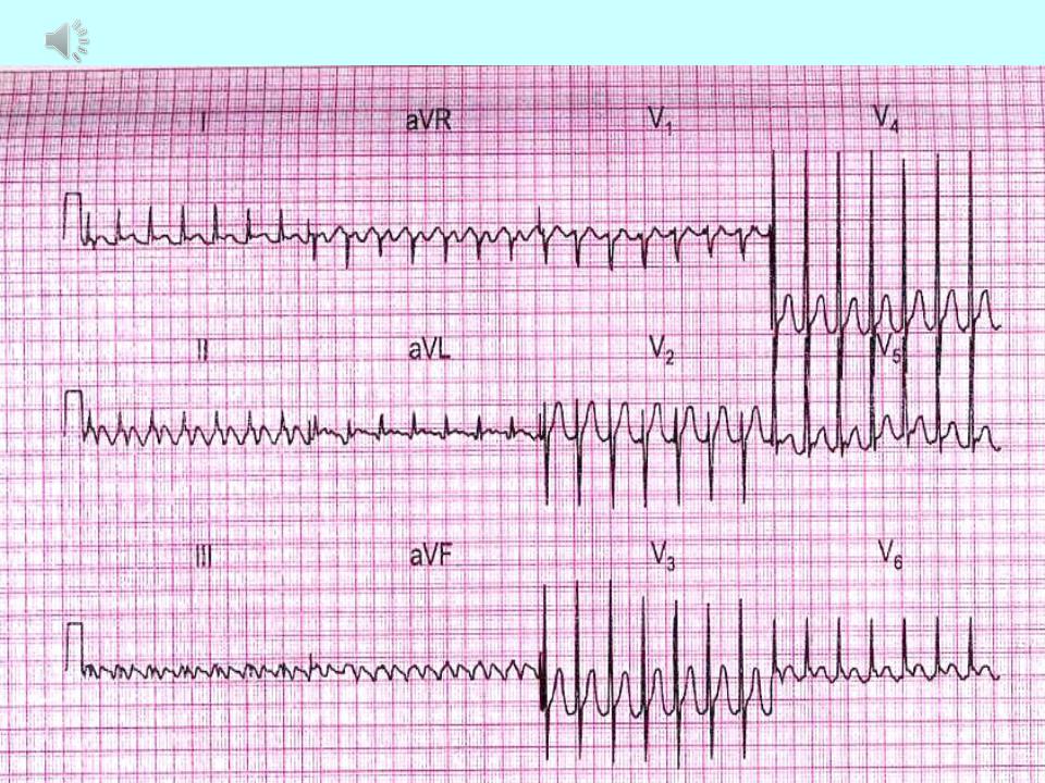
Paroxysmal tachycardia:
It starts suddenly and also suddenly ending attack of increased heart rate contractions from 140-160 to 250-260 in 1 min while maintaining correct rhythm. Distinguish between atrial, atrioventricular (from the AV junction) and ventricular form.
The main ECG signs of supraventricular paroxysmal tachycardia:
1.Altered (reduced, deformed, biphasic or negative) P wave in front of complex QRS or its absence.
2.Usually, normal, unchanged QRS complexes.
2.Possible lengthening of the interval P-Q (R) or loss
individual QRS complexes (development
atrioventricular block I or I degree).
Paroxysmal ventricular tachycardia:
1.Paroxysmal ventricular tachycardia ≤ 120 bpm.
2.P-waves are absent.
3.QRS-complexes are wide and irregular.
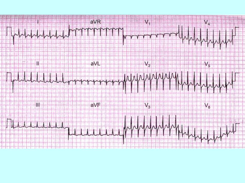
Atrial fibrillation and flutter:
1.Atrial fibrillation - more than 400 P-waves per min, QRS-frequency of 150-180 bpm, f-
2.Atrial flutter atrial frequency is about 300
Atrial fibrillation: