

Cardiology / Пропедевтика внутренних болезней (кардиология, часть 2) Ослопова 2006 года
.pdf38
The lipid hypothesis postulates that an elevation in plasma low density lipoproteins (LDL) levels results in penetration of LDL into the arterial wall, leading to lipid accumulation in smooth muscle cells and in macrophages (foam cells). LDL also augments smooth muscle cell hyperplasia and migration into the subintimal and intimal region in response to growth factors. LDL is modified or oxidized in this environment and is rendered more atherogenic. Small dense LDL cholesterol particles are also more susceptible to modification and oxidation. The modified or oxidized LDL is chemotactic to monocytes, promoting their migration into the intima, their early appearance in the fatty streak, and their transformation and retention in the subintimal compartment as macrophages. Scavenger receptors on the surface of macrophages facilitate the entry of oxidized LDL into these cells, transferring them into lipid-laden macrophages and foam cells. Oxidized LDL is also cytotoxic to endothelial cells and may be responsible for their dysfunction or loss from the more advanced lesion.
Proliferating smooth muscle cells also accumulate lipid. As the fatty streak and fibrous plaque enlarge and bulge into the lumen, the subendothelium becomes exposed to the blood at sites of endothelial retraction or tear, and platelet aggregates and mural thrombi form. Release of growth factors from the aggregated platelets may increase smooth muscle proliferation in the intima. Alternatively, organization and incorporation of the thrombus into the atherosclerotic plaque may contribute to its growth.
The chronic endothelial injury hypothesis postulates that endothelial injury by various mechanisms produces loss of endothelium, adhesion of platelets to subendothelium, aggregation of platelets, chemotaxis of monocytes and T-cell lymphocytes, and release of platelet-derived and monocyte-derived growth factors that induce migration of smooth muscle cells from the media into the intima, where they replicate, synthesize connective tissue and proteoglycans, and form a fibrous plaque. Other cells (eg, macrophages, endothelial cells, arterial smooth muscle cells) also produce growth factors that can contribute to smooth muscle hyperplasia and extracellular matrix production.
These two hypotheses are closely linked and not mutually exclusive. Modified LDL is cytotoxic to cultured endothelial cells and may induce endothelial injury, attract monocytes and macrophages, and stimulate
39
smooth muscle growth. Modified LDL also inhibits macrophage mobility, so that once macrophages transform into foam cells in the subendothelial space they may become trapped. In addition, regenerating endothelial cells (after injury) are functionally impaired and increase the uptake of LDL from plasma.
The atherosclerotic plaque may grow slowly and over several decades may produce a severe stenosis or may progress to total arterial occlusion. With time, the plaque becomes calcified. Some plaques are stable, but others, especially those rich in lipids and inflammatory cells (eg, macrophages) and covered by a thin fibrous cap, may undergo spontaneous fissure or rupture, exposing the plaque contents to flowing blood. These plaques are deemed to be unstable or vulnerable and are more closely associated to the onset of an acute ischemic event. The ruptured plaque stimulates thrombosis; the thrombi may embolize, rapidly occlude the lumen to precipitate a heart attack or an acute ischemic syndrome, or gradually become incorporated into the plaque, contributing to its stepwise growth.
Risk Factors
Major nonreversible risk factors for atherosclerosis include age, male sex, and family history of premature atherosclerosis. Major reversible risk factors are discussed below. Evidence also strongly suggests that physical inactivity is associated with an increased risk of coronary artery disease (CAD). Although personality type has been proposed as a risk factor, its role is controversial.
Abnormal serum lipid levels: Elevated levels of low density lipoprotein (LDL) and reduced levels of high density lipoprotein (HDL) predispose to atherosclerosis. The association of total serum cholesterol and LDL cholesterol levels with the risk of CAD is direct and continuous. HDL levels are inversely correlated with CAD risk. The main causes of reduced HDL are cigarette smoking, obesity, and physical inactivity. Low HDL is also associated with the use of androgenic and related steroids (including anabolic steroids), β-blockers, hypertriglyceridemia, and genetic factors.
Cholesterol level and CAD prevalence are influenced by genetic and environmental factors (including diet). Persons with low serum cholesterol levels who move from a country with a low CAD prevalence to a country with a high CAD prevalence and who tend to alter their
40
eating habits accordingly develop higher serum cholesterol levels and an increased risk of CAD.
Hypertension: High diastolic or systolic BP is a risk factor for stroke, MI, and cardiac and renal failure. The risk associated with hypertension is lower in societies with low average cholesterol levels.
Cigarette smoking: Smoking increases the risk of peripheral artery disease, CAD, cerebrovascular disease, and graft occlusion after reconstructive arterial surgery. Smoking is particularly hazardous in persons at increased cardiovascular risk. There is a dose relationship between the risk of CAD and the number of cigarettes smoked daily. Passive smoking may also increase the risk of CAD. Men and women are both susceptible, but the risk for women may be greater. Nicotine and other tobacco-derived chemicals are toxic to vascular endothelium.
Cigarette smoking increases LDL and decreases HDL levels, raises blood carbon monoxide (and could thereby produce endothelial hypoxia), and promotes vasoconstriction of arteries already narrowed by atherosclerosis. It also increases platelet reactivity, which may favor platelet thrombus formation, and increases plasma fibrinogen concentration and Hct, resulting in increased blood viscosity.
Diabetes mellitus: Both insulin-dependent and non-insulin- dependent diabetes mellitus are associated with earlier and more extensive development of atherosclerosis as part of widespread metabolic derangement that includes dyslipidemia and glycosylation of connective tissue. Hyperinsulinemia damages vascular endothelium. Diabetes is a particularly strong risk factor in women and significantly negates the protective effect of female hormones.
Obesity: Some studies have found that obesity, particularly truncal obesity in men, is an independent risk factor for CAD. Hypertriglyceridemia is commonly associated with obesity, diabetes mellitus, and insulin resistance and appears to be an important independent risk factor in persons with lower LDL or HDL levels and in the nonelderly. Not all triglyceride elevations are likely to be atherogenic. Smaller, denser very low density lipoprotein particles may carry greater risk.
Physical inactivity: Several studies have associated a sedentary lifestyle with increased CAD risk, and others have shown that regular exercise may be protective.
41
Hyperhomocysteinemia: Elevated blood homocysteine due to a genetically determined decrease in its metabolism may cause vascular endothelial injury, which predisposes the vessels to atherosclerosis.
Chlamydia pneumoniae infection: Chlamydia pneumoniae infection or viral infection may play a role in endothelial damage and chronic vascular inflammation that may lead to atherosclerosis.
Symptoms and Signs
Atherosclerosis is characteristically silent until critical stenosis, thrombosis, aneurysm, or embolus supervenes. Initially, symptoms and signs reflect an inability of blood flow to the affected tissue to increase with demand (eg, angina on exertion, intermittent claudication).
Symptoms and signs commonly develop gradually as the atheroma slowly encroaches on the vessel lumen. However, when a major artery is acutely occluded, the symptoms and signs may be dramatic.
Diagnosis
Atherosclerosis is suspected based on the risk factors and on its symptoms and signs, of which there may be few. Atheromatous obstruction is commonly confirmed by arteriography or Doppler ultrasonography.
Hyperlipidemia commonly presents with symptoms and signs of premature obliterative atherosclerosis affecting the brain (cerebral transient ischemic attacks or stroke), heart (angina pectoris or MI), intestine, and lower extremities (intermittent claudication). Xanthomas (in the creases of hands and elbows and along tendon sheaths) and xanthelasmas are sometimes associated with hyperlipidemia, particularly of the familial type. Recurrent attacks of acute pancreatitis, with or without alcoholism, suggest hypertriglyceridemia. A family history of hyperlipidemia or onset of cardiovascular disease before age 60 is further reason to look for premature atherosclerosis.
Prevention
The most effective way to prevent the cardiovascular and cerebrovascular complications of atherosclerosis and the associated arterial thrombosis is to prevent atherosclerosis itself. Reversible risk factors for atherosclerosis are
abnormal serum lipid levels,
hypertension,
cigarette smoking,
42
diabetes mellitus,
obesity,
physical inactivity,
hyperhomocysteinemia, and possibly
C. pneumoniae infection.
Increased understanding of these risk factors and their role in the etiology, pathogenesis, and course of atherosclerosis will lead to more focused intervention for preclinical or overt atherosclerotic disease and will thereby contribute to further declines in morbidity and mortality.
Treatment
Treatment of established atherosclerosis is directed at its complications (eg, angina pectoris, MI, arrhythmias, heart failure, kidney failure, ischemic stroke, and peripheral arterial occlusion).
CORONARY ARTERY DISEASE
Most coronary artery disease (CAD) is due to subintimal deposition of atheromas in the large and medium-sized arteries serving the heart. Less often, CAD is due to coronary spasm, which is usually idiopathic (with or without associated atheroma) or may be due to drugs such as cocaine. Rare causes include an embolus to the coronary artery, Kawasaki syndrome, and vasculitis.
Coronary atherosclerosis is characteristically insidious in onset, is often irregularly distributed in different vessels, and can abruptly interfere with blood flow to segments of the myocardium, most often due to rupture of an eccentric atheromatous plaque with consequent intraluminal thrombosis.
The major complications of CAD are angina pectoris, unstable angina, MI, and sudden cardiac death due to arrhythmias. In the USA, CAD is the leading cause of death in both sexes, accounting for about one third of deaths each year.
Although the precise pathogenesis of CAD is unclear, the risk factors are well known: high blood levels of low density lipoprotein cholesterol (LDL-C) and lipoprotein a, low blood levels of high density lipoprotein cholesterol (HDL-C) and serum vitamin E, and poor physical fitness.
43
High blood levels of triglycerides and insulin reflecting insulin resistance may be risk factors, but the data are less clear. CAD risk is increased by tobacco use; diets high in fat and calories and low in phytochemicals (found in fruits and vegetables), fiber, and vitamin E and C or, at least in some persons, diets with relatively low levels of omega-3 polyunsaturated fatty acids (PUFAs); poor stress management; and inactivity. Several systemic diseases (eg, hypertension, diabetes, hypothyroidism) are also associated with increased CAD risk.
Homocysteine has recently been identified as a risk factor for coronary, peripheral, and cerebral vascular disease. Patients with homocystinuria, a rare recessive disease, have plasma homocysteine levels 10 to 20 times above normal (hyperhomocysteinemia) and accelerated, premature vascular disease. Homocysteine has a direct toxic effect on endothelium and promotes thrombosis and oxidation of LDL.
Normal values range from about 4 to 17 µmol/L. Modest elevations of total plasma homocysteine have multiple causes, including low levels of folic acid, vitamins B6 and B12, renal insufficiency, certain drugs, and
44
genetically controlled variations in homocysteine metabolic enzymes. Patients with homocysteine values in the top 5% have a 3.4 greater risk of MI or cardiac death than those in the lower 90% after adjustment for other risk factors. Increased homocysteine levels are associated with increased risk regardless of etiology. Recent studies suggest a graded risk even in normal-range homocysteine; thus, reduction of normal plasma levels may be advantageous. The most simple and effective way to reduce plasma homocysteine is administration of folic acid 1 to 2 mg/day, which has essentially no side effects except in untreated vitamin B12 deficiency. Many authorities recommend that patients with CAD be screened for plasma homocysteine levels and, unless the values are in the lower normal range, treatment be initiated with folic acid.
Patients with CAD undergoing atherectomy have biologic markers suggesting coronary artery localization of Chlamydia infection. The role of this and other putative infectious agents in the genesis of CAD is being investigated
Ischemic heart disease (IHD; also known as coronary heart disease
— CHD) is usually caused by structural disorder of the coronary arteries (coronary artery disease — CAD), although disorders of small coronary vessels may occasionally lead to similar symptomatology.
Ischemic heart disease produces five clinical syndromes:
1.angina pectoris — stable or unstable, and variant;
2.myocardial infarction.
3.постинфарктный кардиосклероз
4.heart failure
5.arrhythmias
6.sudden cardiac death.
ANGINA PECTORIS
A clinical syndrome due to myocardial ischemia characterized by precordial discomfort or painflul constricting sensation of pressure, which may radiate to the arms, the throat, back and epigastrium. It is usually provoked by activity that increases heart rate and blood pressure, thereby increasing myocardial oxygen demand, for example exercise, emotion, stress, fear or sexual intercourse. The pain or tightness of ‗stable‘ angina typically starts while walking and is relieved in a few minutes by rest or sublingual nitroglycerin.
45
Etiology
The cause is usually critical coronary artery obstruction due to atherosclerosis. Spasm (idiopathic or due to cocaine) or, rarely, a coronary embolism may be causative. Disease other than atherosclerosis (eg, calcific aortic stenosis, aortic regurgitation, hypertrophic subaortic stenosis) can cause angina directly (by increasing cardiac work) or in combination with CAD.
Pathology and Pathogenesis
Usually, patients with long-standing angina are found at autopsy to have extensive coronary atherosclerosis and patchy myocardial fibrosis. There may be gross or microscopic evidence of old MI.
Angina pectoris occurs when cardiac work and myocardial O2 demand exceed the ability of the coronary arteries to supply oxygenated blood. Heart rate, systolic tension or arterial pressure, and contractility are the major determinants of myocardial O2 demand. An increase in any of these factors in a setting of reduced coronary blood flow may induce angina. Thus, exercise in the patient with a critical degree of coronary stenosis induces angina relieved by rest.
As the myocardium becomes ischemic, coronary sinus blood pH falls, cellular K loss occurs, lactate production replaces lactate use, ECG abnormalities appear, and ventricular performance deteriorates. Left ventricular (LV) diastolic pressure frequently rises during angina, at times to levels inducing pulmonary congestion and dyspnea. The discomfort of angina pectoris is believed to be a direct manifestation of myocardial ischemia and the resultant accumulation of hypoxic metabolites.
Symptoms and Signs The discomfort of angina pectoris is not usually perceived as pain. It may be a vague, barely troublesome ache, or it may rapidly become a severe, intense precordial crushing sensation. It has a variable location but is most commonly felt beneath the sternum. It may radiate to the left shoulder and down the inside of the left arm, even to the fingers; straight through to the back, into the throat, jaws, and teeth; and occasionally down the inside of the right arm. It may also be felt in the upper abdomen. Because discomfort seldom occurs in the region of the cardiac apex, the patient who points to this precise area or describes fleeting, sharp, or hot sensations usually does not have angina.

46
Between and even during attacks of angina, signs of heart disease may be absent. However, during the attack, heart rate may increase modestly, BP is often elevated, heart sounds become more distant, and the apical impulse is more diffuse. Palpation of the precordium may reveal localized systolic bulging or paradoxical movement, reflecting segmental myocardial ischemia and regional dyskinesia. The second heart sound may become paradoxical because of more prolonged LV ejection during the ischemic episode. A fourth heart sound is common. A midsystolic or late-systolic apical murmur--shrill but not especially loud due to localized papillary muscle dysfunction secondary to ischemia-- may occur.
Angina pectoris is typically triggered by physical activity and usually persists no more than a few minutes, subsiding with rest. Response to exertion is usually predictable, but in some persons a given exercise that is tolerated one day may precipitate angina the next. Angina is worsened when exertion follows a meal. Also, symptoms are exaggerated in cold weather: walking into the wind or first contact with cold air on leaving a warm room may precipitate an attack.
Angina may occur at night (nocturnal angina) preceded by a dream that is accompanied by striking changes in respiration, pulse rate, and BP. Nocturnal angina may also be a sign of recurrent LV failure, an equivalent of nocturnal dyspnea. Attacks may vary from several days to occasional episodes with symptom-free intervals of weeks, months, or years. They may increase in frequency (crescendo angina) to a fatal outcome or may gradually decrease or disappear if adequate collateral coronary circulation develops, if the ischemic area becomes infarcted, or if heart failure or intermittent claudication supervenes and limits activity.
Angina may occur spontaneously at rest (angina decubitus), usually accompanied by modest increases in heart rate and a rise in BP that may be marked. If the angina is not relieved, the higher BP and fast heart rate increase unmet myocardial O2 need and make MI more likely.
Because the characteristics of angina are usually constant for a given patient, any deterioration in the pattern of symptoms-- increased intensity, decreased threshold of stimulus, longer duration, occurrence when the patient is sedentary or waking from sleep--should be considered serious. Such changes are termed unstable angina.
47
Classification.
Canadian functional сlassification (1976) of stable angina is used in clinical practice.
I functional class — Ordinary physical activity does not cause symptoms. Angina does not occur on walking or stair climbing. Attacks appear during hard, fast or long-term exertion.
II functional class — Comfortable at rest, ordinary physical activity causes symptoms. Angina appears on walking or fast stair climbing, going uphill, after meals, in cold weather, against the wind, emotional exertion. Walking on distance more than 100—200 m on flat place or stair climbing more than 1 stair-well at a normal pace and in normal conditions.
III functional class — Comfortable at rest, less than ordinary activity causes symptoms. Walking on flat place or stair climbing more than 1 stair-well at a normal pace and in normal conditions provoke angina attack appearance.
IV functional class — Symptoms present at rest, inability of any kind of physical ability without discomfort. Angina attack appearance is possible at rest.
Diagnosis
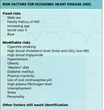
Diagnosis is based on a characteristic complaint of chest discomfort brought on by exertion and relieved by rest. Diagnosis may be confirmed if reversible ischemic ECG changes are seen during a spontaneous attack (Fig.19).
48
Fig.19. Angina pectoris associated with ECG changes. During anginal pain, there are usually ST-segment changes on the ECG. This ECG was taken during an episode of exercise-induced angina, and it shows ST-segment depression (4 mm) in leads V4—6, standard leads II and Ill and lead aVF.
A wide variety of changes may appear: ST segment depression (typically), ST segment elevation, decreased R-wave height, intraventricular or bundle branch conduction disturbances, and arrhythmia (usually ventricular extrasystoles). Between attacks, the ECG (and usually LV function) at rest is normal in about 30% of patients with a typical history of angina pectoris, even with extensive three-vessel CAD (an abnormal resting ECG alone does not establish or refute the diagnosis). Alternatively, diagnosis can be confirmed by a test dose of sublingual nitroglycerin, which characteristically should relieve the discomfort in 1.5 to 3 min.
Exercise stress ECG testing: Because the diagnosis of angina is usually primarily based on the patient's history, exercise testing in a patient with typical symptoms is generally used to determine functional and ECG response to graded stress.
The patient exercises to a predetermined goal (eg, 80 to 90% of maximal heart rate, which can be approximated as 220 less the age in
49
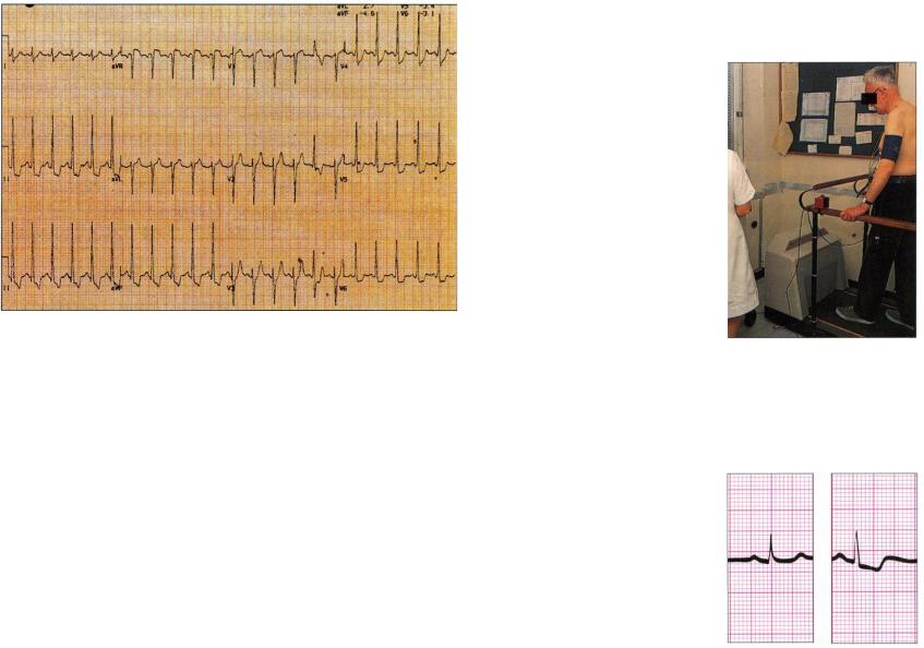
years), unless distressing cardiovascular symptoms (dyspnea, reduced endurance, fatigue, hypotension, or chest pain) supervene (Fig.20).
Fig.20. The exercise treadmill test may reveal signs of ischemia on the ECG when the resting trace is normal.
The ischemic ECG response during or after exercise is characterized by a flat or downward-sloping ST segment depression >= 0.1 millivolts (1 mm on the ECG when properly calibrated) lasting >= 0.08 sec (Fig.21).
Fig.21 A positive exercise test as shown in lead II. The trace taken before exercise is normal, but the second trace, recorded 2 minutes after the end of exercise, shows ST segment depression with T-wave inversion. Analysis of the full trace may show further evidence of ischemia
50
Interpretation of exercise testing is further complicated by the increased incidence of CAD with age; tests are falsely positive in >= 20% patients under age 40 but in < 10% over age 60. The frequency of true-positive tests increases with the number of coronary arteries obstructed, and greater degrees of ST segment depression generally correlate with more extensive disease.
Exercise testing is most predictive of CAD in men with chest discomfort suggestive of angina (specificity, 70%; sensitivity, 90%).
Exercise tests are more difficult to interpret in women aged < 55; a high incidence of false-positive responses, probably related in part to a lower pretest incidence of the disease in the younger population, reduces the specificity. However, women are more likely than men to have an abnormal ECG in the presence of disease (32 vs. 23%). The falsenegative rate in women is comparable to that in men, suggesting that a negative test is a reliable indicator of absence of disease.
In patients with atypical symptoms, a negative exercise test generally rules out angina pectoris and CAD. A positive test may indicate exercise-induced ischemia but may not explain atypical symptoms, suggesting the need for further investigation.
Patients with unstable angina or those in whom recent MI is suspected should not undergo exercise testing. However, with proper indications and close monitoring, an exercise test in an ischemic patient carries a low risk. The patient's response provides valuable prognostic information and helps to evaluate the need for angiography and possible bypass surgery in those on maximal medical therapy. A complete life support system, including emergency drugs, airway, and defibrillator, should be immediately available for any patient undergoing exercise testing.
Coronary angiography documents the extent of anatomic coronary artery obstruction. Coronary angiography findings parallel postmortem findings, but the extent and severity of disease are usually underestimated. Vessels as small as 1 mm may be visualized with highquality imaging. CAD is recognized by narrowing, beading, or occlusion of the vessels. Obstruction is assumed to be physiologically significant when the luminal diameter is reduced > 70%, which correlates well with the presence of angina pectoris; lesser degrees of obstruction are unlikely to result in ischemia, unless spasm or thrombosis is superimposed.
51
Evaluation of wall motion by LV angiography is important if not contraindicated by potential adverse effects of contrast agent on renal or ventricular function.
Echocardiography can be used for anatomic and functional myocardial analysis. Valve anatomy is well depicted, and PA pressure can be reliably estimated. Patients with poor ventricular function, evidence of reduced contractility, have a decreased life expectancy. Yet, if poor function is due to CAD, these patients benefit most from coronary artery bypass grafting, if they survive the operation.
Radionuclide images provide information about cardiac anatomy, cardiac function, myocardial perfusion, and metabolism.
Differential Diagnosis
Many conditions must be considered in the differential diagnosis (eg, abnormalities of the cervicothoracic spine, costochondral separation, nonspecific chest wall pain). However, few truly mimic angina, which is generally so characteristic that errors in diagnosis usually result from careless history taking (Table 6)
|
|
|
Table 6 |
|
Major causes of chest pain which is not IHD |
||
|
|
|
|
Cause |
|
Features |
Further |
|
|
|
investigations |
Esophageal/gall |
|
Associated with dyspepsia, |
Endoscopy |
bladder/peptic |
|
waterbrash, related to food, not |
Ultrasound of gall |
ulcer |
|
related to exertion, relieved by |
bladder |
|
|
antacids |
|
Lung/pulmonary |
|
Pain is pleuritic, worse on |
Chest X-ray |
embolism |
|
breathing, coughing and |
Ventilation/perfusion |
|
|
sneezing. May have cough, or |
scan |
|
|
infected sputum, or blood in |
|
|
|
sputum. There may be a friction |
|
|
|
rub. |
|
Other cardiac |
|
Dissecting aneurysm – very |
Chest X-ray |
causes |
|
severe pain especially in back. |
CT scan |
|
|
Patient hypotensive and may |
|
|
|
collapse. |
|
|
|
|
|
|
52 |
|
|
|
|
Pericarditis – sharp pain worse |
|
Echocardiogram |
|
|
|
|
||
|
on breathing and lying flat – |
|
|
|
|
better it upright. Pericardial rub |
|
|
|
|
may be heard. |
|
|
|
|
Mitral valve prolapse – pain is |
|
Echocardiogram |
|
|
often vague central chest – |
|
|
|
|
comes on after exercise – often |
|
|
|
|
present in young women. |
|
|
|
Musculoskeletal |
Sharp pain related to position |
|
Chest X-ray |
|
|
and movement. May be |
|
Spine and rib X-ray |
|
|
localized to chest wall – |
|
|
|
|
perhaps a history of injury |
|
|
|
|
(includes costochondritis - |
|
|
|
|
Tietze's syndrome). |
|
|
|
Functional |
Present in anxious young |
|
Rebreathing from |
|
|
women and men – no apparent |
|
bag if |
|
|
cause is found. May be |
|
hyperventilation. |
|
|
associated with hyperventilation |
|
Otherwise diagnose |
|
|
(Da Costa's syndrome). |
|
by exclusion. |
|
GI disorders: Diagnostic difficulties arise when the patient has atypical anginal symptoms, especially GI symptoms (eg, bloating; belching, which may give relief; abdominal distress) that are often ascribed to indigestion. Peptic ulcer, hiatus hernia, and gallbladder disease may cause symptoms similar to angina pectoris or may precipitate attacks in persons with preexisting CAD. Nonspecific changes in the T waves and ST segments have been reported in esophagitis, peptic ulcer disease, and cholecystitis, which can further complicate diagnosis.
Dyspnea: Angina may be confused with dyspnea, partly because of the sharp and reversible rise in LV filling pressure that often accompanies the ischemic attack. The patient's description may be imprecise, and whether the problem is angina, dyspnea, or both may be difficult to determine. Recurrent breathlessness on mild exertion may reflect increased LV filling pressure secondary to ischemia, with or without pain.
53
Silent ischemia: Twenty-four hour Holter monitoring has revealed a surprising incidence (up to 70% of episodes) of T-wave and ST segment abnormalities in the absence of pain in patients with CAD. Such changes are rare in persons without CAD. Radionuclide studies have documented myocardial ischemia in some persons during mental stress (eg, mental arithmetic) and during spontaneous ECG change. Silent ischemia and angina pectoris may coexist. In silent ischemia, the prognosis is defined by the severity of CAD. Revascularization may improve prognosis by reducing the incidence of subsequent MI or sudden death.
Prognosis
The major adverse outcomes are unstable angina, MI, recurrent MI, and sudden death due to arrhythmias. Annual mortality is about 1.4% in men with angina and no history of MI, a normal resting ECG, and normal BP. The rate rises to about 7.5% if systolic hypertension is present, to 8.4% when the ECG is abnormal, and to 12% if both risk factors are present.
Lesions of the left main coronary artery or in the proximal anterior descending vessel indicate particularly high risk. Although outcome correlates with number and severity of coronary vessels involved, in stable patients the prognosis is surprisingly good, even with three-vessel disease, if ventricular function is normal.
Reduced ventricular function, often measured by analysis of ejection fraction, adversely influences prognosis, especially in patients with three-vessel disease.
Prognosis also correlates with symptoms; it is better in patients with mild or moderate angina (class I or II) than in those with severe exerciseinduced angina (class III).
Age is a major risk factor in the elderly.
Treatment
The major tenet of treatment is to prevent or reduce ischemia and minimize symptoms. The underlying disease, usually atherosclerosis, must be delineated and the primary risk factors reduced as much as possible. Smokers should quit: Discontinuing smoking for >= 2 yr reduces the risk of MI to the level of those who never smoked. Hypertension should be treated diligently because even mild hypertension increases cardiac work. Angina sometimes improves markedly with treatment of mild LV failure. Paradoxically, digitalis
54
occasionally intensifies angina, presumably because increased myocardial contractility raises O2 demand in the presence of fixed coronary blood flow. Aggressive reduction of total and LDL cholesterol (with dietary treatment supplemented by drugs as necessary) in patients at risk retards progression of CAD and may cause some lesions to regress. An exercise program emphasizing walking often improves the sense of well-being, reduces risk, and improves exercise tolerance.
Three classes of drugs are usually effective, alone or in combination, in relieving symptoms: nitrates, β-blockers, and Ca blockers.
Nitroglycerin is a potent smooth-muscle relaxer and vasodilator. Its major sites of action are in the peripheral vascular tree, especially in the venous or capacitance system and on the coronary blood vessels. Even severely atherosclerotic vessels may dilate in areas without atheroma. Nitroglycerin lowers systolic BP and dilates systemic veins, thus reducing myocardial wall tension, a major determinant of myocardial O2 need. Overall, the drug helps balance myocardial O2 supply and demand.
Sublingual nitroglycerin 0.3 to 0.6 mg is the most effective drug for the acute episode or for prophylaxis before exertion. Dramatic relief is usual within 1.5 to 3 min, is complete by about 5 min, and lasts up to 30 min. The dose may be repeated after 4 to 5 min three times if initial relief is incomplete. Patients should carry nitroglycerin tablets or aerosol spray with them at all times to use promptly at the onset of an angina attack.
β-Blockers completely block sympathetic stimulation of the heart and reduce systolic pressure, heart rate, contractility, and cardiac output, thus decreasing myocardial O2 demand and increasing exercise tolerance. Additionally, they increase the threshold for ventricular fibrillation. Because tissue O2 requirements are met by greater O2 extraction from capillary blood, systemic arteriovenous O2 difference is widened. These drugs are extremely useful in reducing symptoms and are well tolerated by most patients.
Ca blockers are the important third arm in the approach to angina pectoris and CAD. These vasodilators are useful in the treatment of angina with hypertension and counter coronary spasm if present. They are often highly effective in variant angina (see below), but their effectiveness may be limited by negative chronotropic and inotropic effects (diltiazem, verapamil).
55
Antiplatelet drugs are important in opposing platelet aggregation, which is pivotal in the genesis of MI and unstable angina. Aspirin, which binds irreversibly to platelets and inhibits cyclooxygenase and platelet aggregation in vitro, has been shown in epidemiologic studies to reduce coronary events (MI, sudden death) in CAD patients.
Angioplasty involves insertion of a balloon-tipped catheter into an artery at the site of a partially obstructive atherosclerotic lesion. Inflation of the balloon can rupture the intima and media and dramatically dilate the obstruction. About 20 to 30% of obstructions reocclude in a few days or weeks, but most can be redilated successfully. Use of stents significantly reduces the reocclusion rate, which continues to decline with application of newer techniques. Repeat angiography 1 yr later reveals an apparently normal lumen in about 30% of vessels undergoing the procedure. Angioplasty is an alternative to bypass surgery in a patient with suitable anatomic lesions. The risk is comparable with that of surgery: Mortality is 1 to 3%; MI rate is 3 to 5%; emergency bypass for intimal dissection with recurrent obstruction is required in < 3%; and the initial success rate is 85 to 93% in experienced hands. Results continue to improve with advances in technique, catheter and balloon mechanics, and pharmacotherapy to maintain postangioplasty patency.
Coronary arterial bypass surgery is highly effective in selected patients with angina.
UNSTABLE ANGINA
(Acute Coronary Insufficiency; Preinfarction Angina; Crescendo Angina; Intermediate Syndrome)
Angina characterized by a progressive increase in anginal symptoms, new onset of rest or nocturnal angina, or onset of prolonged angina.
Unstable angina is precipitated by an acute increase in coronary obstruction due to rupture of the fibrous plaque covering an atheroma with consequent platelet adhesion. In unstable angina, >= 1/3 of patients studied angiographically have partially occluding thrombi in the vessel subtending the recurrent ischemic area. Because recognition of a thrombus on angiography may be difficult, the incidence is probably underreported.
56
Compared with stable angina, the pain of unstable angina is generally more intense, lasts longer, is brought on by less effort, occurs spontaneously at rest (angina decubitus), is progressive (crescendo) in nature, or involves any combination of these changes.
About 30% of patients with unstable angina will probably suffer an MI within 3 months of onset; sudden death is less common. Presence of marked ECG changes with chest pain is an important marker for subsequent MI or death.
Unstable angina is a medical emergency to be treated in a cardiac care unit (CCU). Both heparin and aspirin reduce the incidence of subsequent MI. To reduce intracoronary clotting, aspirin 325 mg po and IV heparin should be instituted immediately. If aspirin cannot be tolerated or is contraindicated, ticlopidine 250 mg bid or clopidogrel 75 mg/day is a possible alternative. Ticlopidine requires monitoring of WBC at regular intervals because of the risk of neutropenia.
Cardiac work should be reduced by slowing heart rate and lowering BP with β-blockers and IV nitroglycerin, thus restoring the balance between cardiac O2 demand and coronary blood flow. Contributing disorders (eg, hypertension, anemia) should be vigorously treated. Bed rest, nasal O2, and nitrates are useful. Ca blockers may be useful for patients with hypertension and possible coronary artery spasm. Thrombolytic drugs are not useful and may be harmful. Use of the antiplatelet glycoprotein IIb/IIIa receptor antagonist, the humanized chimeric Fab fragment abciximab, has been shown to improve outcome in a randomized trial in patients with refractory unstable angina. Tirofiban has been shown to prevent cardiac ischemic events in unstable angina and non-Q-wave infarction. Other IIb/IIIa receptor antagonists are being evaluated in acute ischemic syndromes.
The patient's symptoms should be brought under control within a few hours of intensive treatment. After 24 to 48 h, if therapy is not effective, more aggressive treatment may be required. The intra-aortic counterpulsating balloon reduces systolic afterload and increases diastolic pressure, the driving force for coronary blood flow. It frequently relieves continuous anginal pain and may be used to support the circulation during diagnostic cardiac catheterization prior to revascularization with coronary bypass surgery or angioplasty. Angiography may be indicated in a patient poorly responsive to medical
57
therapy in order to identify the culprit lesion and evaluate the extent of CAD and LV function, with a plan for percutaneous transluminal coronary angioplasty or coronary artery bypass grafting if technically feasible.
VARIANT ANGINA
(Prinzmetal's Angina)
Angina pectoris that is usually secondary to large vessel spasm and is characterized by discomfort at rest and by ST segment elevation during the attack.
Most patients have significant fixed proximal obstruction of at least one major coronary vessel. Spasm usually occurs within 1 cm of the obstruction (often accompanied by ventricular arrhythmia). Between anginal attacks, which tend to occur with regularity at certain times of day, the ECG may be normal or may present a stable abnormal pattern. Ergonovine IV has been used as a provocative test to induce spasm, but this should be done only by experienced personnel in an angiographic laboratory. Although the average survival at 5 yr is 89 to 97%, patients with variant angina and severe coronary artery obstruction are at greater risk. Relief of variant angina is usually prompt after sublingual nitroglycerin; Ca blockers appear to be highly effective.
MYOCARDIAL INFARCTION
Ischemic myocardial necrosis usually resulting from abrupt reduction in coronary blood flow to a segment of myocardium.
Etiology and Pathogenesis
In > 90% of patients with acute MI, an acute thrombus, often associated with plaque rupture, occludes the artery (previously partially obstructed by an atherosclerotic plaque) that supplies the damaged area. Altered platelet function induced by endothelial change in the atherosclerotic plaque presumably contributes to thrombogenesis. Spontaneous thrombolysis occurs in about 2/3 of patients so that, 24 h later, thrombotic occlusion is found in only about 30%.
MI is rarely caused by arterial embolization (eg, in mitral or aortic stenosis, infective endocarditis, and marantic endocarditis). MI has been reported in patients with coronary spasm and otherwise normal coronary arteries (Fig.22). Cocaine causes intense coronary arterial spasm, and users may present with cocaine-induced angina or MI.