

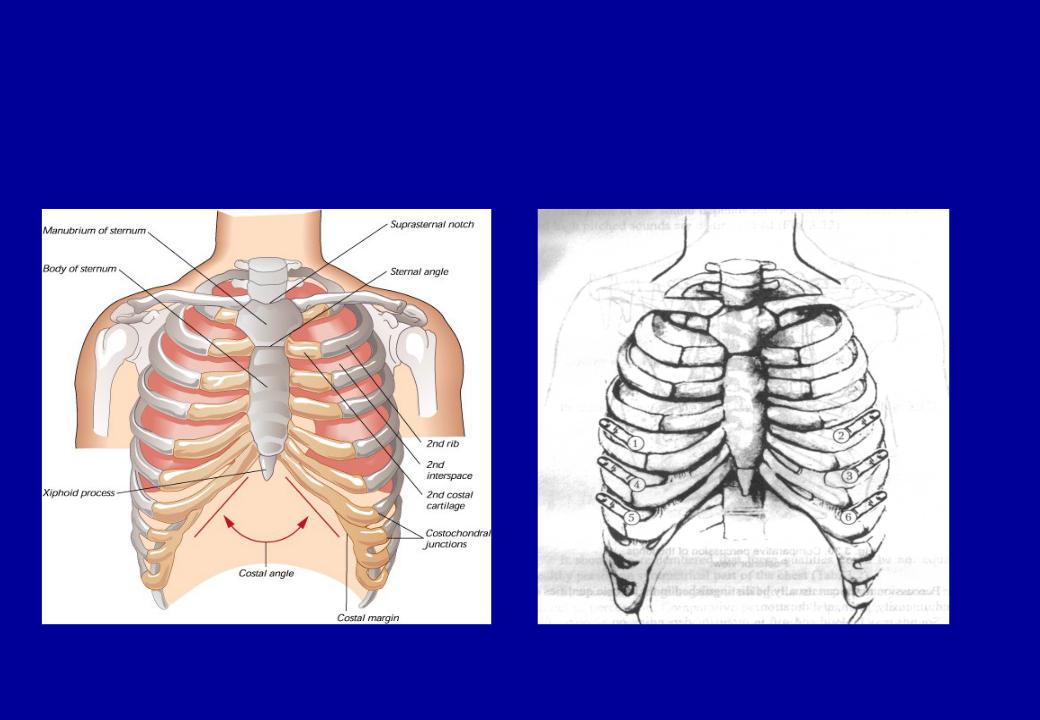
Topographic regions and lines of the chest

Percussion
Press The last 2 phalanges of your left middle finger firmly on on the area to be percussed and raise the second and fourth fingers off the chest surface; otherwise, both sound and tactile vibrations will be blunted

Use a two quick, sharp wrist motion
The best percussion site is between the proximal and distal interphalangeal joints.

Percussion of the chest
This is the objective method of examination based on evaluation of sound types during the knocking of the thorax
Comparative – revealing of percussion sound features
on symmetrical areas of the chest:
SupraclavicularisClavicularisSubclavicularisAxillarisSuprascapularisInterscapularisSubscapularis
Topographic - aimed to determining :
lower borders of the lungsupper borders of the lungsthe width of Crenig’s area
active and passive mobility of lower borders of the lungs
width of Traube’s area
Comparative percussion

Comparative percussion
Resonant - Clear
pulmonary
Intermediate -
pulmonary sound becomes duller
Dull
Hyperresonant –
Tympanic
Bandbox sound - over
the hyper inflated lungs of emphysema

The main symptoms based on comparative percussion
Percussion sound on the symmetric areas :
Clear pulmonary (in healthy persons) Dullness (dulling)
Infiltration of lung tissue (tuberculosis, pneumonia, pneumosclerosis, lung cancer, abscess, lung gangrene)
Accumulation of liquid in pleural cavity Stony dull – large pleural effusion
pleural thickening
Tympanic
Increasing the air capacity of lung tissue (bronchial asthma, lung emphysema)
Formation the cavity with air in lung parenchyma (released form contents caverns, abscess, bronchoectasis)
Accumulation of air in pleural cavity (pneumothorax)

The main symptoms based on topographic percussion
1.Lower borders:
Removal down (lung emphysema, bronchial asthma, lower standing of diaphragm)
Removal upper (athelectasis, surgical ablation the part of lung, higher standing of diaphragm, subdiaphragmal abscess)
2.Upper borders:
Removal down (tuberculosis of lung apexes, pneumosclerosis, athelectasis of lung apexes)
Removal upper (lung emphysema, bronchial asthma)
3. Width of Traube’s area:
Increasing more than 6 sm - lung emphysema, bronchial asthma Decreasing less than 4 sm - tuberculosis of lung apexes, pneumosclerosis, athelectasis of lung apexes

Topographic percussion
Topographic percussion of the lungs. Anterior view.

Topographic percussion
Topographic percussion of the |
Topographic percussion. |
lungs. Lateral view. |
Posterior view. |