

Pulmonology / Topic_8_resp_syst
.pdfMinistry of Health of Ukraine
Kharkiv National Medical University
AUSCULTATION OF THE LUNGS: ADDITIONAL RESPIRATORY SOUNDS (RALES, CREPITATION, PLEURAL FRICTION SOUND). LABORATORY SPUTUM AND PLEURAL FLUID ANALYSIS. INSTRUMENTAL METHODS OF RESPIRATORY ORGANS EXAMINATION
Methodical instructions for students
Рекомендовано Ученым советом ХНМУ Протокол №__от_______2017 г.
Kharkiv
KhNMU
2017
Auscultation of the lungs: additional respiratory sounds (rales, crepitation, pleural friction sound). Laboratory sputum and pleural fluid analysis. Instrumental methods of respiratory organs examination: Меthod. instr. for students / Authors. Т.V. Ashcheulova, O.M. Kovalyova, G.V. Demydenko. – Kharkiv: KhNMU, 2017. – 28 p.
Authors: Т.V. Ashcheulova
O. M. Kovalyova
G.V. Demydenko
AUSCULTATION OF THE LUNGS
Adventitious (added) sounds
Three types of adventitious sounds can be heard in pulmonary pathology: rales, crepitation, and pleural friction
sound.
Rales are generated in bronchi and bronchioles. Dry and moist rales are distinguished.
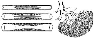
Dry rales can be caused by narrowing of airways, or by presence of viscous sputum in them (Fig. 1).
2

Fig. 1. Mechanism and site of dry rales generation
Dry rales are continuous musical sounds, persist throughout the respiratory cycle, and vary greatly in their character, pitch, and intensity. Depending on character and pitch, dry rales are divided into sibilant and sonorous rales.
Sibilant rales (wheezes) are relatively high pitched, whistling sounds (Fig. 2).
Fig. 2. Sibilant rales.
Sibilant rales signify obstruction in small bronchi in:
bronchial asthma (total bronchospasm during attack);
bronchitis (non-uniform swelling of the bronchial mucosa due to inflammation, or viscous sputum narrows the lumen of bronchi);
tuberculosis or tumor of bronchus (localized constriction of the bronchus. Limited dry rales over apex of the lung can suggest early symptom of tuberculosis).
Sonorous rales (rhonchi) are relatively low pitched, sonoring sounds (Fig. 3).
Fig. 3. Sonorous rales.
Sonorous rales are generated by vibration of the viscous secretions or in widespread obstruction of medium and large bronchus. The most common cause of sonorous rales is bronchitis. They may be also heard in bronchial asthma, tuberculosis, and bronchocarcinoma.
Intensity and transmission of the dry rales depends on the size and depth of the affected bronchi. In localized affection of medium and large bronchus insignificant amount of low pitched and soft rales is heard. Widespread bronchi inflammation or bronchospasm in asthma attack both sibilant and sonorous rales of different tone and intensity are heard. Such rales can be heard at a distance during expiration. If dry rales are caused by accumulation of the viscous secretions in the lumen of bronchi, they can be altered by coughing or deep inspiration to shift mucus.
Moist rales (clackles) are generated in bronchi and cavities in the lungs in the presence of liquid secretions (sputum, congestive fluid, blood).
Airflow in liquid-containing bronchi causes formation of air bubbles, which break to produce specific cracking sound. Similar sound can be heard when bubbling air through the water using small tube. Such sounds are called bubbling or moist rales.
Moist rales are discontininuous sounds, intermittent, nonmusical, and brief. Moist rales are heard throughout the respiratory cycle, but as speed of airflow in inspiration is higher, rales are somewhat louder during inspiration.
Moist rales are subdivided into fine, medium, and coarse bubbling rales depend on the caliber of bronchi where they are originated.
Fine bubbling rales ( . . . . . . . ) generate in small bronchi and bronchioles. These rales are soft, high-pitched, and
very brief. |
|
|
|
|
|
|
|
|
|
|
|
|
|
Medium bubbling rales ( |
|
|
|
|
|
|
|
) |
|
originate in bronchi of medium caliber. They are somewhat louder, and not |
|||
so brief. |
|
|
|
|
|
|
|
|
|
|
|
|
|
Coarse bubbling rales ( |
|
|
|
|
|
|
|
|
|
|
) |
produce in bronchi of large caliber, large bronchiectasis, and also in fluid- |
|
|
|
|
|
|
|
|
|
|
|
||||
containing lung cavity communicated with large bronchus (Fig. 4). Coarse bubbling rales are loud, low-pitched, and longer.
3
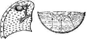
Fig. 4. Mechanism of the coarse bubbling rales generation over the cavity.
Moist rales are classified into consonating and non-consonating rales.
Consonating rales are heard when liquid-containing bronchi or cavity are surrounded by solid lung tissue. The cavity itself act as resonator to intensify loudness of rales.
Non-consonating rales are heard in bronchitis or acute pulmonary edema caused by left ventricular failure, when intensity of rales produced are dampened by air-containing lung tissue.
The most common causes of the moist rales include:
acute and chronic bronchitis (bilateral, symmetrical, of various caliber, non-consonating rales);
bronchopneumonia (consonating rales);
bronchiectasis (of various caliber, over limited area, non-consonating rales);
cavity in the lungs (coarse bubbling, over limited area, as rule over lung apices, consonating rales);
pulmonary edema due to the left ventricular failure (bilateral symmetrical, of different caliber, nonconsonating rales).
Crepitation is generated in alveoli, when they contain small amount of liquid secretion. During expiration alveoli stick together as a result of fluid presence. During inspiration alveolar walls separate with difficulty only at the end of inspiration to produce late inspiratory slight cracking sound (Fig. 5). Crepitation somewhat resembles sound produced by rubbing a lock of hair near the ear.
 Fig. 5. Mechanism of crepitation.
Temporary crepitation in first deep inspiration can be heard in the patients with grave cardiovascular and infectious diseases, in aged persons, especially so if the patient was in lying posture before auscultation.
Relatively constant crepitation can be due to:
acute lobar pneumonia at the initial and final stages (insignificant amount of exudates at the initial stage causes so-called indux crepitation – quit, remote sound. During next stage of disease alveoli are overfilled with inflammatory fluid and crepitation therefore disappears. At the final stage due to resolution of exudates, loud, as near the ear, crackling sound – redux crepitation is heard again);
pulmonary tuberculosis (in small amount of inflammatory fluid in alveoli);
lung infarction (in small amount of blood in alveoli);
4

congestive heart failure (in small amount of congestive fluid in alveoli);
compressive atelectasis (alveoli are compressed by pleural air or fluid, and separate therefore with difficulty).
Pleural friction sound (pleural rub, friction rub) is diagnostic added sound of pleurisy.
The smooth surfaces of visceral and parietal pleura lubricated by pleural fluid, allow pleura to move easily and noiseless during breathing.
Adventitious sound known as pleural friction sound, generates as a result of decreased amount of pleural fluid in dry pleurisy due to dehydrotation:
intestinal infections (cholera, dysentery);
profuse bleeding;
profuse diarrhea;
profuse vomiting;
or cicatrices, commissures, bands between pleural layers at the focus of inflammation, or when fibrin deposits on inflamed pleura to make it surface rough in:
pleuropneumonia;
rheumatic pleurisy;
pleural tuberculosis;
tumor;
effusive pleurisy at the period of rapid resorption of exudates.
Pleural friction rub is heard throughout inspiration and expiration, and is differentiated by intensity, location and duration.
A soft friction rub in early dry pleurisy may be mistaken for crepitation or fine bubbling rales but is not altered by coughing as rales; it can be louder by pressure with stethoscope. During rapid resorption of pleural effusion, pleural rub becomes louder, and more intense. Such sound is so rough that can be felt even during palpation.
Pleural friction sound is best heard at the lung bases due to better respiratory mobility, and rarely at the lung apices (tuberculosis with involvement of the pleura, for example).
Duration of the friction rub varies in different diseases. Periodic pleural friction sound is typical to rheumatic pleurisy. It is heard a few hours, temporary disappears, and then appears again. In dry pleurisy of tuberculosis etiology and pleurisy with effusion at resorption stage, pleural rub is heard for a week and over. Longstanding, for years after pleurisy, friction rub can be sometimes heard due to significant roughness of the pleural surfaces.
Characteristics of adventitious sounds are summarized in Tab. 1.
Tab. 1. Differential diagnosis of adventitious sounds.
|
|
|
|
Pleural |
Signs |
Dry rales |
Moist rales |
Crepitation |
friction sound |
Relation to |
Best heard |
Best heard |
Heard at the |
Heard |
the |
during |
during |
end of |
throughout |
respiratory |
expiration |
inspiration |
inspiration |
respiratory |
phases |
|
|
|
cycle |
Change |
Decrease or |
Decrease or |
Without |
Without |
during cough |
change |
disappears |
changes |
changes |
|
character |
|
|
|
Pressure with |
Without |
Without |
Without |
|
the |
changes |
changes |
changes |
Increase |
stethoscope |
|
|
|
|
Breathing |
|
|
|
|
movement |
|
|
|
Only this |
with close |
Absent |
Absent |
Absent |
sound is |
nose and |
|
|
|
heard |
mouth |
|
|
|
|
INSRUMENTAL AND LABORATORY METHODS
Diagnostic procedures for assessing the patients with suspected or known respiratory system disease include imagine studies, technique for obtaining biological specimens, and method used to characterize the functional changes developing as a result of disease.
Imagine studies
5
Imagine studies used to examine the patients with disorders of the respiratory system include:
Roentgenoscopy
Roentgenography (radiography)
Fluorography
Computed tomography
Magnetic resonance imaging
Scintigraphic imaging
Bronchography
Pulmonary angiography
Ultrasound examination
Roentgenoscopy is the most common method for assessing relative lungs translucency, and for the diagnostic evaluation of disease involving the pulmonary parenchyma (consolidation of the pulmonary tissue, pneumosclerosis, tumor), the pleura (pleural fluid or air, pleural adhesions), and, to a lesser extent, the airways. Presence of the cavity in the lungs can also be determined roentgenoscopy.
Roentgenography (radiography, x-rays). Routine chest radiography generally includes both posteroanterior and lateral views, and used for film recording – radiograph. The detail that can be seen on radiograph allows better recognition of parenchymal and airway diseases (indistinct focal consolidations, bronchovascular pattern, etc.).
Fluorography – a variant of radiography, is a convenient method for screening the population. The image in fluorography is made on a role film of a small size.
Computed tomography is cross-sectional scanning of the chest. This technique is more sensitive than plain radiography in detecting respiratory abnormalities. Computed tomography makes possible to distinguish more accurate tumors, small indurations, cavities and caverns in the lungs. This method is far better than radiographic studies at characterizing tissue density, distinguishing subtle differences in density between adjacent structures, and providing accurate size assessment of lesions. The use of computed tomographic scanning of the chest is very useful as a means of gathering quantitative information about specific radiographic findings.
Magnetic resonance imaging provides a less detailed view of the pulmonary parenchyma as well as poor spatial resolution. However, magnetic resonance imaging offers several advantages over computed tomography in certain clinical settings: for imaging abnormalities near the lung apex, the spine, and the thoracoabdominal junction. Vascular structures can be distinguished from nonvascular without the need of contrast.
Scintigraphic imaging. Administered radioactive isotopes allow the lungs to be imaged with a gamma camera. The most common use of such method is ventilation-perfusion lung scanning performed for detection of pulmonary embolism. Radioactive isotopes can be injected intravenously; albumin macroaggregates labeled with technetium 99m is used for this purposes, or inhaled – radiolabeled xenon gas. When injected intravenously, the distribution of the trapped radioisotope follows the distribution of blood flow. When inhaled, radioisotopes can be used to demonstrate the distribution of ventilation.
Bronchography is an integral part of the diagnosis evaluation of diseases of bronchi. The standard technique requires the injection of contrast medium, usually iodolipol, into the bronchi lumen. This may be done through a catheter passed via the nose or mouth through the anaesthetized larynx. Then radiographs are taken, that give a distinct patterns of the bronchial tree. This procedure is of particular importance to the evaluation of bronchiectasis, abscesses, caverns in the lungs, and compression of the bronchi by tumor.
Pulmonary angiography. The technique of the pulmonary angiography requires the injection of radiopaque contrast medium into the pulmonary artery through a previously threaded catheter. Radiographs are taken, on which the pulmonary arterial system can be visualized. Pulmonary angiography in pulmonary embolism demonstrates the consequences of an intravascular clot (a defect in the lumen of a vessel, or abrupt termination of the vessels). Suspected pulmonary arteriovenous malformation can be also visualized by this method.
Ultrasound examination generally is not useful for evaluation of parenchyma of the lungs due to physical properties of the ultrasound waves: ultrasound energy is rapidly dissipated in air-containing pulmonary tissue. However, it is helpful in the detection and localization of pleural fluid and therefore is often used as a guide to placement of a needle for sampling of the liquid in thoracentesis.
Techniques for obtaining biologic specimens
Techniques for obtaining biologic specimens, some of which involve direct visualization of the part of the respiratory system, include
Collection of the sputum
Thoracentesis
Bronchoscopy
Collection of the sputum
6
Sputum is pathological secretion expectorated from the respiratory tract. Sputum should be collected after thorough mouth and throat rinsing in the morning hours before breakfast. To collect sputum for more than 12 hours is not expedience because long-standing storage leads to rapid flora multiplying and autolysis of the formed elements.
SPUTUM ANALYSIS
Clinical sputum analysis includes: macroscopic, microscopic, and bacterioscopic studies.
Macroscopic study
In macroscopic study amount, character, color, consistence, and admixture in the sputum are assessed.
Amount of the sputum
Daily amount and amount of separate portions of the sputum depends on the character of the diseases from one side, and from the patient ability to expectorate from other one.
Scarce amount of sputum observes in the patients with inflammation of the respiratory tract: in laryngitis, trachitis, at initial stage of acute bronchitis, bronchial asthma out of attack, and in bronchopneumonia.
Ample amount of sputum (from 0.5 to 2 liters) secrete from the cavity in the lungs, in bronchus (bronchiectasis, pulmonary abscess), or in pulmonary edema due to significant transudate in bronchi.
Significant amount of purulent sputum may forms layers on standing. Two-layers (pus and plasma) sputum is typical to pulmonary abscess, three-layers (pus, plasma, and upward mucus) – to bronchiectasis, pulmonary tuberculosis (in cavern presence).
Character of the sputum
Character of the sputum is determined by its composition: mucus, pus, blood, and serous fluid.
Mucous sputum consists of mucus – product of mucous glands. Such sputum is produced in acute bronchitis, at the peak of bronchial asthma attack.
Mucopurulent sputum is mixture of mucus and pus, moreover mucus is predominant part, and pus in a form of traces or small bundles is observed. Mucopurulent sputum can be obtained in chronic bronchitis, trachitis, bronchopneumonia, and tuberculosis.
Puromucous sputum contains pus and mucus; pus is predominant part of the sample. Such sputum arises in chronic bronchitis, bronchiectasis, pulmonary abscess, etc.
Purulent sputum without mucus admixture appears in opened to the bronchus pulmonary abscess, in rupture of the pleural empyema to the bronchus lumen.
Mucous-bloody sputum consists mainly of mucus with streaks of blood, and can be produced in inflammation of upper respiratory ducts, pneumonia, lung infarction, congestion in the pulmonary circulation, and bronchogenic tumor.
Mucopurulent bloody sputum contains uniform mixed mucus, blood and pus. Such sputum arises in tuberculosis, bronchiectasis, actinomycosis of the lungs, and bronchogenic tumor.
Bloody sputum observes in pulmonary hemorrhage: tuberculosis, wounds of the lungs, actinomycosis, and bronchogenic tumor).
Serous sputum is plasma of the blood that passes to the bronchi in edema of the lungs.
Serous bloodstained foamy sputum is characteristic of pulmonary edema, when not only plasma, but also erythrocytes penetrate from pulmonary alveoli to the bronchi.
Color of the sputum
Color of the sputum depends on its character, and also by inspirited particles. Predominance of one of substrates gives sputum corresponding hue.
Mucous sputum is usually colorless, transparent, and glass-like.
Mucopurulent sputum is glass-like with yellow tint as its main component is mucus, on the background of which pus traces is observed.
Puromucous sputum is yellow-greenish due to predominance of pus.
Purulent sputum is greenish-yellow due to the pus.
Mucous-bloody sputum is glass-like (due to predominance of mucus) with pink or rusty tint (due to the presence of changed or unchanged blood pigment - hematin). Rusty sputum is characteristic of acute lobar pneumonia, when blood is not expectorated immediately from the respiratory tract and stays there for sometimes. The hemoglobin converts into hemosiderin to give a rusty hue to the sputum.
Mucopurulent bloody sputum is glass-like (predominance of mucus), with yellow traces (pus), with red color streaks (fresh blood) or rusty hue (changed blood pigment).
Bloody sputum is of red color. Peculiarity of the pulmonary hemorrhage is the presence foamy secretions due to the air bubbles.
Serous sputum is transparent-yellow (color of penetrated blood plasma), and foamy.
Sputum containing foreign admixtures has color of these admixtures: white in millers, black – in miners, blue in inspiration of ultramarine paint, etc.
Consistency of the sputum
Consistency tightly connected with sputum character and may be tenacious, thick, and liquid.
7

Tenacity of the sputum depends on the presence of mucus and amount of it. For example, in bronchial asthma, acute and chronic bronchitis, bronchopneumonia consistency of the sputum is tenacious.
Thickness of the sputum is caused by the presence of the large amount of the formed elements – leucocytes, various epithelium cells (bronchiectasis, chronic bronchitis, pulmonary abscess, and tuberculosis).
Liquid sputum can be in large it amount, when the plasma is significant composing component (pulmonary hemorrhage, pulmonary edema).
Odor of the sputum
Fresh sputum is usually odorless. Unpleasant smell can appears in protracted conservation of the sputum. Foul odor of freshly expectorated sputum can be caused by it retaining in bronchi and cavities in the lungs due to putrefactive decomposition of proteins. Unpleasant odor sputum can be had in chronic bronchitis with bad bronchi drainage, strong smell – in bronchiectasis, pulmonary abscess, sometimes in tuberculosis, in malignant tumor with necrosis, fetid (putrid) odor is characteristic of tissue decomposition – gangrene.
Admixture
The following elements can be seen in the sputum by an unaided eye:
Curschmann spirals – has diagnostic significance in bronchial asthma;
Fibrin clots –has significance in fibrinous bronchitis, and rarely in lobar pneumonia;
Lentil or rice-like bodies (Koch’s lens) – observe in sputum in cavernous tuberculosis;
Purulent plugs (Dittrich’s plugs) - occurring in bronchiectasis, gangrene, chronic abscess, and fetid bronchitis.
Diphtherias films;
Necrotic pieces of the lungs – observes in pulmonary gangrene and abscess;
Pieces of the pulmonary tumor;
Actinomycete;
Lime grains – in decomposition of old tubercular foci;
Echinococcus bubbles – observe in sputum in rupture of echinococcus cyst in the lung and expectoration of plentiful amount of colorless transparent fluid;
Foreign bodies.
Physical properties of the sputum revealed in macroscopic examination are summarized in Tab. 2.
|
|
|
Tab. 2. Physical properties of the sputum |
|||
Characte |
Consisten |
Color |
Odor |
Layer |
Disease |
|
r |
-cy |
|
|
ness |
|
|
Mucous |
Tenacious |
Glass- |
Odorless |
Absent |
Acute |
|
|
|
like |
|
|
bronchitis, |
|
|
|
|
|
|
at the peak |
|
|
|
|
|
|
of |
the |
|
|
|
|
|
bronchial |
|
|
|
|
|
|
asthma |
|
|
|
|
|
|
attack |
|
Muco- |
Tenacious |
Glass- |
Odorless |
Absent |
Chronic |
|
purulent |
thick |
like with |
|
|
bronchitis, |
|
|
|
yellow |
|
|
trachitis, |
|
|
|
traces |
|
|
broncho- |
|
|
|
|
|
|
pneumonia |
|
|
|
|
|
|
, |
|
|
|
|
|
|
tuberculosi |
|
|
|
|
|
|
s |
|
Puro- |
Thick, |
Yellow- |
Un- |
Three |
Chronic |
|
mucous |
tenacious |
greenish |
pleasant |
layers |
bronchitis, |
|
|
|
|
|
|
bronchiect |
|
|
|
|
|
|
asis, |
|
8

|
|
|
|
|
pulmonary |
||
|
|
|
|
|
abscess |
|
|
Purulent |
Thick |
Greenish |
Sharp un- |
Two |
Pulmonary |
||
|
|
-yellow |
pleasant |
layers |
abscess, |
|
|
|
|
|
|
|
gangrene |
||
|
|
|
|
|
of |
|
the |
|
|
|
|
|
lungs |
|
|
Mucous |
Viscous, |
Rusty, |
Odorless |
Absent |
Inflammati |
||
bloody |
tenacious |
glass- |
or |
|
on |
of |
the |
|
|
like, |
without |
|
upper |
|
|
|
|
reddish |
un- |
|
respiratory |
||
|
|
|
pleasant |
|
ducts, |
|
|
|
|
|
odor |
|
pneumonia |
||
|
|
|
|
|
, |
|
|
|
|
|
|
|
bronchoge |
||
|
|
|
|
|
nic |
tumor, |
|
|
|
|
|
|
lung |
|
|
|
|
|
|
|
infarction, |
||
|
|
|
|
|
conges- |
|
|
|
|
|
|
|
tion |
in |
the |
|
|
|
|
|
lesser |
|
|
|
|
|
|
|
circulation |
||
Muco- |
Tenacious |
Glass- |
Un- |
In |
Tuberculos |
||
purulent |
thick |
like, |
pleasant, |
large |
is, |
|
|
bloody |
|
reddish |
putre- |
amount |
bronchiect |
||
|
|
with |
factive |
three |
asis, |
|
|
|
|
purulent |
|
layers |
bronchoge |
||
|
|
traces |
|
|
nic tumor |
||
Bloody |
Liquid, |
Red |
Odorless |
Absent |
Pulmonary |
||
|
foamy |
|
|
|
hemorrhag |
||
|
|
|
|
|
e: in |
|
|
|
|
|
|
|
tuberculosi |
||
|
|
|
|
|
s, wounds, |
||
|
|
|
|
|
lung tumor |
||
Serous |
Foamy, |
Transpa- |
Odorless |
Absent |
Pulmonary |
||
|
liquid, |
rent |
|
|
edema |
|
|
|
sticky |
yellowish |
|
|
|
|
|
Microscopic study
Sputum elements revealed in microscopic study can be divided into three main groups: cellular, fibrous, and crystal formations (Tab. 3).
Tab.3. Sputum elements in microscopic study
|
Cellular elements |
|
Fibrous elements |
|
Crystal elements |
|
Epithelium cells |
|
Curschmann spirals |
|
Charcot-Leyden crystals |
|
(squamous, columnar) |
|
Elastic fibbers |
|
Crystals of hematoidin |
Macrophages |
|
Fibrin fibers |
|
Crystals of cholesterol |
|
|
Leucocytes, |
|
|
Crystals of fatty acid |
|
|
(eosinophils) |
|
|
|
|
Erythrocytes
Tumor cells
Cellular elements
9

Squamous epithelium – is epithelium of mucous membrane of mouth cavity, nasopharynx, larynx, and vocal chords. Single cells of squamous epithelium are always observed in sputum, and have no diagnostic significance.
Columnar ciliated epithelium – is epithelium of bronchi and trachea mucous membrane. It is contained in small quantity in any sputum, but its large amount is found in acute bronchitis, in bronchial asthma attack, and in acute infections of upper respiratory tract.
Alveolar macrophages. Insignificant quantities of alveolar macrophages are present in any sputum, large amount – in various inflammatory processes of bronchi and pulmonary tissue: pneumonia, bronchitis, and professional diseases of the lungs. Siderophages arise in the sputum of the patients with congestion in the pulmonary circulation, especially in mitral stenosis; in lung infarction, acute lobar pneumonia.
Leucocytes observe in any sputum; in mucous – single, and in purulent – all microscope vision area. Their large amount is characteristic of inflammatory and especially purulent process. Sometimes among leucocytes eosinophils can be identified. Eosinophils are the large leucocytes with uniform large lustrous grains. Eosinophils presence in the sputum suggest bronchial asthma or chronic bronchitis with asthma component.
Erythrocytes Single erythrocytes can be visible at any sputum; in large quantity observed in bloody sputum: pulmonary hemorrhage, lung infarction, congestion in the pulmonary circulation, etc.
Malignant tumor cells. Sputum with such cells is underwent then special cytological study. Tumor cells are found in the sputum especially when tumor degrades or growth endobronchially.
Fibrous elements
Curschmann spirals – are found in the sputum of patients with respiratory pathology accompanied by bronchospasm: bronchial asthma, bronchitis with asthmatic component, bronchial tumor.
Elastic fibers presence in the sputum indicates degradation of the pulmonary tissue: in tuberculosis, pulmonary abscess, and tumor.
Fibrin fibers - are found in fibrinous bronchitis, tuberculosis, actinomycosis, and lobar pneumonia.
Crystal elements
Charcot-Leyden crystals. Presence of Charcot-Leyden crystals in the sputum is characteristic of the bronchial asthma even not in attack, and between attacks period. Less frequently they can be observed in the sputum of patients with eosinophilic bronchitis, lobar pneumonia, and bronchitis.
Hematoidin crystals. These crystals are the product of hemoglobin degradation, and are formed in hemorrhage, and necrosis tissue.
Cholesterol crystals – observed in the sputum of the patients with tuberculosis, tumor, pulmonary abscess, etc. Fatty acid crystals – are frequently found in purulent sputum (Dittrich’s plugs), produced in sputum congestion
in the cavity (abscess, bronchiectasis).
Bacterioscopic study
Tuberculosis mycobacteria presence in the sputum indicates tuberculosis.
Pneumococcus, streptococcus, staphylococcus, Pfeiffer’s bacillus – all these microorganisms occur in small amount in the sputum of the respiratory ducts of healthy persons and only become pathogenic under the certain unfavorable condition to cause pneumonia, lung abscess, bronchitis.
Microbes, their virulence and drug-resistance can be identifying by bacterioscopic study. Sputum analysis in selected respiratory pathology is represented in Tab. 4.
Tab. 4. Sputum analysis in selected respiratory pathology
Disease |
Sputum |
Sputum |
Macroscop |
Microscopic |
|
amount |
character |
ic study |
study |
Acute |
Scarce, in |
Mucous, |
|
Columnar |
bronchitis |
later stages |
mucous- |
--- |
epithelium, |
|
– large |
purulent |
|
leucocytes- |
|
amount |
|
|
moderate |
|
|
|
|
amount, in long- |
|
|
|
|
standing course |
|
|
|
|
- macrophages |
|
|
Mucous- |
|
Leucocytes – |
Chronic |
Various |
purulent, |
|
large amount; |
bronchitis |
|
mucous- |
--- |
erythrocytes, |
|
|
purulent |
|
macrophages |
|
|
bloody |
|
|
|
|
Puro- |
Dittrich’s |
Leucocytes – |
Bronchi- |
Ample |
mucous |
plugs |
many; fatty |
ectasis |
(morning) |
three- |
|
acids, |
|
|
layers |
|
hematoidin, |
|
|
|
|
cholesterol |
10