

Pulmonology / Topic_8_resp_syst
.pdf
|
|
|
|
crystals |
Bronchial |
Scare |
Mucous |
Curschma |
Columnar |
asthma |
|
|
nn spirals |
epithelium, |
|
|
|
|
Charcot-Leyden |
|
|
|
|
crystals, |
|
|
|
|
eosinophils, |
|
Scare |
Sticky, |
Fibrin |
Macrophages, |
Lobar |
initially, |
rusty |
clots, |
leucocytes, |
pneumoni |
ample – |
initially, |
changed |
erythrocytes, |
a |
later |
later |
blood |
hematoidin |
|
|
mucous- |
|
crystals, |
|
|
purulent |
|
pneumococcus |
|
Ample in |
Purulent |
Necrotic |
Leucocytes, |
Pulmonar |
secrete to |
with foul |
pieces of |
elastic fibers, |
y abscess |
bronchus |
odor |
the lung |
fatty acid, |
|
|
|
tissue |
hematoidin, |
|
|
|
|
cholesterol, |
|
|
|
|
crystals, |
|
|
Mucous- |
“Koch’s |
Tuberculosis |
Pulmonar |
Various |
purulent, |
lens” in |
mycobacteria; |
y |
|
sometimes |
cavern |
elastic fibers, |
tuberculos |
|
with blood |
presence |
and various |
is |
|
|
|
crystals |
|
|
Mucous- |
Tissue |
Atypical cells |
Broncho- |
Various |
bloody, |
pieces in |
|
pulmonary |
|
mucous- |
ample |
|
tumor |
|
purulent |
sputum in |
|
|
|
bloody |
tumor |
|
|
|
|
degradatio |
|
|
|
|
n |
|
Thoracentesis (pleurocentesis) is performed to sampling of pleural fluid for diagnostic purposes; in the case of a large effusion to remove fluid from the pleural cavity; and, whenever necessary, to administer drugs.
Technique. The patient should sit facing the chair back with arms crossed on the chest. The puncture is done in posterior axillary line at the preliminary determined by percussion point of maximum dullness – usually 7th or 8th interspaces at the upper edge of the underlying rib (at the lower edge intercostals vessels are located). Previous the place of the puncture is treated with alcohol iodine and then anesthetized. Sampling is obtained by 10 ml syringe with a thick and long needle. For diagnostic purposes 50-150 ml of fluid is taken, and then puncture site after needle removing is treated with a 5% iodine solution.
STUDY OF THE PLEURAL FLUID
Diagnostic sampling allows the collection of liquid for macroscopic, chemical, microscopic, and bacteriologic
studies.
Macroscopic study
In macroscopic study character, color, consistency, and relative density of the pleural fluid are assessed. Character. The pleural fluid is divided into two large groups transudates and exudates.
Transudates – are non-inflammatory fluid that occurs in disorders of lymph and blood circulation in the lungs (for example in heart failure).
Exudates – are of inflammatory character, and occur in inflammatory affection of the pleura. Exudates can be:
Serous and serofibrinous in exudative pleurisy, rheumatic pleurisy;
Seropurulent and purulent in bacterial pleurisy;
Hemorrhagic more frequent in traumatic pleura affection, less frequently in infarction of the lungs, and tuberculosis;
Chylous in congestion of lymph or destruction of the thoracic duct by a tumor or an injury;
Chylous-like in chronic inflammation of serous membrane as a result of cellular degradation with fatty degeneration;
Putrefactive in wounds associated with putrefactive flora.
Transparency of pleural fluid depends on its character. Transudates and serous exudates are transparent and slightly opalescent. Another exudates in most cases are turbid that can be caused by abundance of leucocytes (seropurulent, purulent), erythrocytes (hemorrhagic), fat drops (chylous), and cellular detritus (chylous-like).
11
Color of the pleural fluid is also depends on its character. Transudates have pale yellow color, serous exudates
– from pale to golden yellow, in jaundice – deep yellow. Purulent and putrefactive effusions are of grayish-white or greenish-yellow color; in blood admixture they can be reddish or more frequent – grayish-brown. The color of hemorrhagic exudates varies from pink to dark red or even brown depending on amount of blood in the fluid, and also on the time of its retention in pleural cavity. Chylous exudates resemble thin milk.
Consistency of pleural fluid in transudates and exudates is usually liquid. Only purulent exudates are thick and cream-like. In old encapsulated empyema the pus can be of puree consistency with grains and fibrin flakes.
Odor. The pleural fluid is a rule odorless. Only putrefactive exudates have unpleasant, offensive smell due to decomposition of protein by anaerobic enzymes.
Relative density of the pleural fluid is determined by urometer. Relative density of transudates is less than of exudates. Relative density of transudates varies from 1005 to 1015 g/cm2; relative density of exudates is usually higher than 1015 g/cm2 (1018-1022).
Chemical study
Protein level in the pleural fluid is assessed by refractometer. The relative density and protein contents are the main criteria that allow the effusion to be classified as either exudative or transudative. Protein content in transudates is 5-25 g/l (0.5-2.5%), in exudates – more than 30 g/l (3-8%). Qualitative protein content is also of great diagnostic significance for differentiation between transudates and exudates.
Correlation of protein fractions of exudates is about the same as of blood serum; albumin-globulin ratio is 0.5:2; the fibrinogen contents is lower than that of blood (0.05-0.1%) but its quantity is sufficient to clot spontaneously.
In transudates albumin-globulin ratio is 2.5:4; albumin prevail while fibrinogen is absent or almost absent (therefore transudates does not clot).
Rivalta’s reaction was proposed for differentiation between transudates and exudates. In a cylinder filled with 100-150 ml of distillated water and 2-3 drops of acetic acid, 1-2 drops of the punctate are added. Exudates drop cause turbidity in a form of white cloud (or like cigarette smoke), which sinks to the bottom of a cylinder (positive reaction). Transudates drops or do not leave a cloudy trace, or it can be insignificant and quickly disappears (negative reaction).
Lucaerini test. To 2 ml of 3% hydrogen peroxide solution placed on a watch glass (against a black background) one drop of punctate is added. Exudates drop leaves opalescence turbidity (positive reaction); transudates drop cause no turbidity (negative reaction). In both reactions the cause of turbidity is the presence of seromucin – mucopolysaccharide complex in exudates. In transudates seromucin is absent.
Microscopic study
Microscopy allows study cellular composition of the pleural precipitate obtained by centrifuging. A native preparation before staining is recommended to study.
Native preparation
Study of the native preparation allows assessing quantity of cellular elements, qualitative content of precipitate, presence of suspected atypical cells, etc. In native preparation the following elements can be revealed.
Erythrocytes in small quantity can be present in any pleural fluid because of puncturing of the tissues. In transudates and serous exudates insignificant amount of erythrocytes is detected; in hemorrhagic exudates in patients with tumor, infarction of the lung, injuries, hemorrhagic diathesis they usually covered all vision area.
Leucocytes in a small quantity (to 15 in vision field) are revealed in transudates and in a large amount – in fluid of inflammatory genesis (especially in purulent exudates). Qualitative content of leucocytes are assessed in stained preparations.
Mesothelium cells are recognized by their large size (to 50 mcm). Transudates contain significant amount of mesothelium cells. They also can be determined in exudates in canceromatosis, and sometimes in tuberculosis.
Tumor cells. Exudates sometimes contain cells suspected for tumor according to absence of distinct cellular borders, polymorphism of their size and shape. The nature of tumor cells is difficult to assess in native preparation.
Stained preparation
Cytological picture of the pleural fluid is different and depend on character, etiology and duration of liquid presence. In stained preparation the following cellular elements are differentiated.
Neutrophils are present in exudates of any etiology. In serous exudates of tubercular or rheumatic etiology they are found in significant amount at initial stage of exudates development (approximately during first 10 days), and then their amount gradually decreases – replaced by lymphocytes. Long-standing neutrophilia indicates grave course of disease; appearance of predominant amount of neutrophils is a sign of transition of serous exudates to purulent. In purulent exudates neutrophils are prevalent cells.
Lymphocytes are obligatory elements of any exudates. They are predominant in cytological picture of serous exudates at a peak of clinical manifestation (80-90% of all leucocytes).
Eosinophils are contained sometimes in serous and hemorrhagic exudates of various etiology: in rheumatic, tubercular, tumor exudates, composing 20-80% of all cellular elements.
Macrophages resemble morphologically monocytes, but differ from they by the presence in the cytoplasm of phagocytosis products.
12
Mesothelial cells are always present in transudates, at initial stage and at the period of reparation of exudates, in significant amount in canceromatosis of serous membrane. In long-standing and sometimes in acute pleural affections and also in transudates coarse vacuolized mesothelial cells acquire many properties of blastoma cells that can be lead to mistakes.
Malignant cells. It is very difficult to differentiate between tumor and mesothelial cells. Luminescent microscopy helps in this situation: when stained with rhodamine, acridine orange or some other fluorochromes, tumor cells luminescence differently than normal cells.
Bacterioscopic study
Transudates as a rule are sterile in microbiological studies, but they can be infected during repeated thoracentesis.
Exudates may be sterile, for example in rheumatic pneumonia, tumor of the lung, and lymphosarcoma. Bacterioscopy of serous exudates in tuberculosis rare gives positive results. More effective method for tuberculosis mycobacteria detection is inoculation to guinea pigs. In pleurisy caused by pyogenic flora the bacteria can be detected in Gram-stained smears. Pneumococcus, streptococcus, staphylococcus, enterococcus, Klebsiella organisms, Pfeiffer’s bacillus, colibacillus can be found in bacterioscopic study. Microbes are tested for antibiotics sensitivity in order to prescribe a correct treatment.
Methods for functional studies
Methods used to characterize the functional changes developing as a result of disease are very important for an integrated examination of the patients. It is unusual for a specific lung function test to diagnose a disease. At best, a series of tests may place a lung disorder into one of several categories and when other features such as history, physical examination, radiology and pathology are added to the equation, a possible diagnosis is considered.
The main uses of lung function testing are to help define more clearly the type, character and degree of respiratory failure, and to measure serially natural progression (or regression with therapy) of functional disorder.
Tests of ventilatory function. Various indices are used to assess lung ventilation. Their size and relationship to each other give clues to underlying functional disorder. How normal a volume is will depend on what we predict it should be for that person’s height, weight, sex, and age.
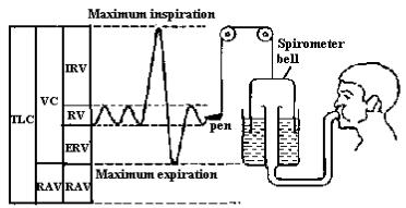
Figure 6 shows how the total lung capacity is broken down into its various volumes.
Fig. 6. The subdivision of the total lung capacity (TLC) with spirometric recording.
RV – respiratory volume, ERV – expiratory reserve volume, IRV – inspiratory reserve volume, RAV – residual air volume, VC – vital capacity.
The respiratory volume (RV) or tidal volume is the total air volume of each normal resting breath (inspiration and expiration). RV varies from 300 to 900 ml; 500 ml on the average. It consists of two parts:
13
1.Alveolar volume: the volume of gas, which reaches the alveoli – the volume of alveolar ventilation;
2.Dead space volume (about 150 ml): the volume of gas, which passes the lips and is present in the larynx, trachea, and bronchi, but does not take part in gas exchange. However, the air of the dead space is mixed with the inspired air to warm and moisten it, which makes it physiologically important.
The expiratory reserve volume (ERV) is the volume of air that can be expired after normal expiration – 1500-
2000 ml.
The inspiratory reserve volume (IRV) is the volume of air that can be inspired after normal inspiration – 1500-
2000 ml.
The vital capacity (VC) is the largest volume that can be expired after full inspiration – 3700 ml on average. The residual air volume (RAV) is the volume of air that remains in the lungs after maximum expiration –
1000-1500 ml.
The total lung capacity (TLC) can be derived by adding RV, ERV, IRV, and RAV. It is about 5000-6000 ml. Studies of the respiratory volumes allow assessing ability of the respiratory failure compensation at the
expense of reserve inspiratory and expiratory volumes. All these volumes, apart from RV, can be measured by spirometer. Spirography gives more reliable information on respiratory volumes. It can be used to measure additional ventilation characteristics such as minute volume, maximum lung ventilation, respiratory reserve, and volume of lung ventilation.
The minute volume (MV) is the volume of gas, which passes the lips in one minute. It can be calculated by multiplying RV by the respiratory rate (frequency, f): MV = f · RV. It is about 5000 ml on the average.
The maximum lung ventilation (MLV) is the amount of air that can be handed by the lungs by maximum efforts of the respiratory system. MLV is determined during deepest breathing at the rate of 50 per minute by spirometer; normally – 80-200 l/ml.
The respiratory reserve (RR) may be calculated by the formula: RR = MLV – MV. Normally RR exceeds the MV by at least 15-20 times; RR is 85% of MLV (in respiratory failure 60% and lower). This value reflects ability of healthy person in considerable load, or of patients with pathology of the respiratory system to compensate significant insufficiency by increasing of minute respiratory volume.
The study of mechanics of the respiratory act allows to evaluate changes in the inspiration and expiration correlation, breath efforts at various respiratory phases, etc.
The forced expiratory vital capacity (FEVC) is determined according to Votchal-Tiffeneau during maximum fast, forced expiration. FEVC is 8-11% (100-300 ml) lower than VC in healthy persons.
The forced inspiratory vital capacity is assesses during maximum fast forced inspiration.
Pneumotachymetry, pneumotachygraphy – methods of speed and pressure measuring at various phases of the breathing by pneumotachygraph. Pneumotachygraphy allows to determined volumetric rate of the airflow during inspiration and expiration (normally in rest breathing it is about 300-500 ml/s; in forced – 5000-8000 ml/s), duration of the respiratory cycle phases, MV, alveolar pressure, airways resistance, elasticity or distensibility or stiffness of the lungs and chest, and some other indices.
Tests for respiratory failure.
Determination of oxygen consumption and oxygen deficit is carried out by spirography with a closed CO2 absorption system. Obtained spirogram compared then with spirogram that records with apparatus filled with O2.
Ergospirography is the method, which allow assessing reserves of the respiratory system. Oxygen consumption and deficit is detected by spirography in the patient at rest and during exercise on ergometer.
Measurement of blood gases
Gas composition of blood samples obtained from warmed up finger is measured on a Van-Slike apparatus. The following is determined:
1.O2 content in units of volume;
2.oxygen capacity of the blood (the amount of O2 that can bound by a blood unit);
3.percentage of O2 saturation of the blood (95% in norm);
4.partial pressure of O2 in the blood (90-100 mm Hg in norm);
5.CO2 content in arterial blood (about 48% v/v);
6.partial pressure of CO2 (about 40 mm Hg in norm).
14
Tests
1.The patient’s position is forced, he is sitting resting his hands against the edge of the chair. There are numerous whistling rales against vesiculotympanic resonance and weak vesicular respiration all over the lungs. What diagnosis can be supposed?
A.Lung cancer
B.Bronchitis
C.Pulmonary emphysema
D.Bronchial asthma
E.Lung abscess
2.In the right subscapular area from the 7th to the 10th ribs there is dull percussion sound, bronchial respiration. What diagnosis can be supposed?
A.Height of lobar pneumonia
B.Lung cancer
C.Lung abscess
D.Pneumosclerosis
E.Exudation pleurisy
3.Solitary coarse moist rales are heard over the left apex of the lung against a background of tympanic sound and amphoric respiration. What diagnosis can be supposed?
A.Bronchial asthma
B.Lung cancer
C.Pneumonia
D.Bronchitis
E.Cavity in the lung
4.The patient complains of pain in the left hemithorax, which becomes worse on breathing in. Lung sound is heard on percussion of the chest. Auscultation demonstrates weak vesicular respiration, pleura friction rub in the left axiliary area. What diagnosis can be supposed?
A.Pneumothorax
B.Exudation pleurisy
C.Pleuropneumonia
D.Dry pleurisy
E.Lung emphysema
5.Dull tympanic sound, weak vesicular respiration and crepitation are heard over the left hemithorax at the level of 4th-10th interspace. What diagnosis can be supposed?
A.Lung abscess
B.Focal pneumonia
C.Initial stage of lobar pneumonia
D.Lung edema
E.Pneumothorax
6.The patient complains of dyspnea on moderate exercise. Acrocyanosis. The ratio of anteroposterior to transverse size of the chest is 0.92; the voice resonance is weak; the chest is rigid. The resonance is vesiculotympanic, the respiration is weak vesicular. ERF investigation demonstrates a “shark’s tooth” curve
and abrupt reduction of the ERF parameters What diagnosis can be supposed?
A.Emphysema
B.Chronic obstructive lung disease.
C.Bronchial asthma
D.Lung cancer
E.Pneumonia
7.The patient complaints of attacks of difficult breathing especially on breathing out, morning cough with some mucous sputum. Microscopy of the sputum demonstrates bronchial epithelium, eosinophils, and Charcot-Leiden crystals. What diagnosis can be supposed?
A.Emphysema
B.Chronic obstructive lung disease.
C.Bronchial asthma
D.Lung cancer
E.Pneumonia
8.A smoker complains of cough with moderate sputum discharge. The sound over the lungs is clear, rigid, vesicular. The rales are disseminated buzzing. Investigation of the sputum demonstrates bronchial epithelium separately and in aggregates, leukocytes in moderate amounts, Churchman’s spirals. X-ray demonstrates increased lung picture. Fibrobronchoscopy shows hyperemia and edema of the bronchial mucosa. ERF has not reveal any ventilation abnormality. What diagnosis can be supposed?
A.Emphysema
B.Chronic obstructive lung disease.
C.Bronchial asthma
D.Lung cancer
E.Pneumonia
9.The patient has tympanic sound on the left of the 2nd and 3rd interspace. X-ray demonstrate a cavity with horizontal fluid level. Laboratory study demonstrates elastic fibers in the sputum. What diagnosis can be suggested?
A.Lung cancer
B.Bronchial asthma
C.Pneumonia
D.Chronic bronchitis
E.Lung abscess
10.The patient with chronic obstructive lung disease has dyspnea at rest, acrocyanosis. RR at rest is 28/min.
Computer spirography demonstrates considerably pronounced disorders of a mixed type (vital lung capacity 55%, forced expiration volume1 50%, Tiffno’s index 60%). What diagnosis can be supposed?
A.Stage 1 respiratory failure.
B.Stage 2 respiratory failure.
C.Stage 3 respiratory failure.
D.Pulmonary emphysema
E.Pneumosclerosis
Keys: Keys: 1D, 2A, 3E, 4D, 5C, 6А , 7С, 8 D, 9 Е, 10В
Methodical instructions
15

RESPIRATORY SYSTEM EXAMINATION. LUNGS PERCUSSION. TECHNIQUE OF COMPARATIVE AND
TOPOGRAPHIC PERCUSSION.
Methodical instructions for students
Authors: Т.V. Ashcheulova
O. M. Kovalyova
G.V. Demydenko
Chief Editor Ashcheulova Т.V.
Редактор____________
Корректор____________
Компьютерная верстка_____________
Пр. Ленина, г. Харьков, 4, ХНМУ, 61022 Редакционно-издательский отдел
16