

книги студ / Color Atlas of Pharmacology
.pdf
|
|
|
Antianemics |
141 |
|
|
Fe III-Salts |
|
|
|
|
Oral |
|
|
|
|
|
intake |
Fe II-Salts |
|
|
|
|
|
Heme-Fe |
|
|
|
|
|
|
|
III |
|
|
|
|
|
Fe |
|
|
Absorption |
|
Fe III |
|
|
|
Duodenum |
|
|
|
|
|
|
|
|
|
|
|
upper jejunum |
|
|
|
|
|
|
|
Ferritin |
|
|
|
|
|
|
Parenteral |
|
|
Transport |
Fe III |
Fe III |
administration |
|
|
|
|
|
|||
plasma |
|
|
|
||
|
|
|
|
|
|
|
Transferrin |
|
|
|
|
|
|
|
i.v. |
i.m. |
|
Uptake into |
Hemoglobin |
Fe III-complexes |
|
||
erythroblast |
|
|
|
||
|
|
|
|
|
|
bone marrow |
|
|
|
|
|
|
|
|
Fe III |
|
|
Erythrocyte |
|
|
Ferritin |
|
|
|
|
|
|
|
|
blood |
|
|
|
|
|
|
|
|
Hemosiderin |
|
|
|
|
|
= aggregated |
|
|
|
|
|
ferritin |
|
|
Loss through |
|
|
Uptake into macrophages |
|
|
bleeding |
|
|
|
||
|
|
spleen, liver, bone marrow |
|
||
|
|
|
|
||
A. Iron: possible routes of administration and fate in the organism
Lüllmann, Color Atlas of Pharmacology © 2000 Thieme
All rights reserved. Usage subject to terms and conditions of license.

142 Antithrombotics
Prophylaxis and Therapy of Thromboses
Upon vascular injury, the coagulation system is activated: thrombocytes and fibrin molecules coalesce into a “plug” (p. 148) that seals the defect and halts bleeding (hemostasis). Unnecessary formation of an intravascular clot – a thrombosis – can be life-threatening. If the clot forms on an atheromatous plaque in a coronary artery, myocardial infarction is imminent; a thrombus in a deep leg vein can be dislodged, carried into a lung artery, and cause complete or partial interruption of pulmonary blood flow (pulmonary embolism).
Drugs that decrease the coagulability of blood, such as coumarins and hep- arin (A), are employed for the prophy- laxis of thromboses. In addition, attempts are directed at inhibiting the aggregation of blood platelets, which are prominently involved in intra-arterial thrombogenesis (p. 148). For the thera- py of thrombosis, drugs are used that dissolve the fibrin meshwork!fibrinolytics (p. 146).
An overview of the coagulation cascade and sites of action for coumarins and heparin is shown in A. There are two ways to initiate the cascade (B): 1) conversion of factor XII into its active form (XIIa, intrinsic system) at intravascular sites denuded of endothelium; 2) conversion of factor VII into VIIa (extrinsic system) under the influence of a tis- sue-derived lipoprotein (tissue thromboplastin). Both mechanisms converge via factor X into a common final pathway.
The clotting factors are protein molecules. “Activation” mostly means proteolysis (cleavage of protein fragments) and, with the exception of fibrin, conversion into protein-hydrolyzing enzymes (proteases). Some activated factors require the presence of phospholipids (PL) and Ca2+ for their proteolytic activity. Conceivably, Ca2+ ions cause the adhesion of factor to a phospholipid surface, as depicted in C. Phos- pholipids are contained in platelet factor 3 (PF3), which is released from ag-
gregated platelets, and in tissue thromboplastin (B). The sequential activation of several enzymes allows the aforementioned reactions to “snowball”, culminating in massive production of fibrin (p. 148).
Progression of the coagulation cascade can be inhibited as follows:
1) coumarin derivatives decrease the blood concentrations of inactive factors II, VII, IX, and X, by inhibiting their synthesis; 2) the complex consisting of heparin and antithrombin III neutralizes the protease activity of activated factors; 3) Ca2+ chelators prevent the enzymatic activity of Ca2+-dependent factors; they contain COO-groups that bind Ca2+ ions (C): citrate and EDTA (ethylenediaminetetraacetic acid) form soluble complexes with Ca2+; oxalate pre- cipitates Ca2+ as insoluble calcium oxalate. Chelation of Ca2+ cannot be used for therapeutic purposes because Ca2+ concentrations would have to be lowered to a level incompatible with life (hypocalcemic tetany). These compounds (sodium salts) are, therefore, used only for rendering blood incoagulable outside the body.
Lüllmann, Color Atlas of Pharmacology © 2000 Thieme
All rights reserved. Usage subject to terms and conditions of license.

|
|
|
Antithrombotics |
143 |
|
XII |
XIIa |
|
|
|
|
|
|
|
Synthesis susceptible to |
||
|
|
|
inhibition by coumarins |
||
|
XI |
XIa |
Reaction susceptible to |
||
|
|
|
|||
|
|
|
inhibition by heparin- |
|
|
|
|
|
antithrombin complex |
|
|
|
IX |
|
IXa |
|
|
|
|
|
VIIa |
VII |
|
|
VIII + Ca2+ + Pl |
|
Ca2+ + Pl (Phospholipids) |
|
|
|
X |
|
Xa |
|
|
|
V + Ca2+ + Pl |
|
|
|
|
|
Prothrombin II |
|
IIa Thrombin |
|
|
Fibrinogen I |
Ia Fibrin |
A. Inhibition of clotting cascade in vivo
Platelets |
Endothelial |
Clotting factor |
|
|
|
|
|
|
|
defect |
|
|
COO– |
|
|
|
|
|
|
|
COO- |
|
|
|
||
|
|
|
|
|
|
|
|
|
XII |
|
|
|
COO– |
|
|
|
|
|
|
|
|
|
|
|
||
|
|
Tissue |
|
|
+ |
|
|
|
|
|
|
Ca+ |
|
|
|||
|
|
thrombo- |
|
|
– |
|||
|
|
kinase |
|
– |
||||
XIIa |
|
|
|
|
|
– |
|
|
|
|
|
|
– |
|
|
||
|
|
|
|
– |
|
|
|
|
PF3 |
VIIa |
VII |
– |
|
|
|
|
|
|
|
Phospholipids |
||||||
|
|
|
|
|
|
|||
|
|
Vessel |
|
|
|
e.g., PF3 |
|
|
|
|
rupture |
Ca2+-chelation |
|
|
|
|
|
|
|
|
|
|
|
|
||
Fibrin |
|
|
Citrate |
|
|
|
|
|
|
|
EDTA |
|
|
|
|
|
|
|
|
|
Oxalate |
|
|
|
|
|
B. Activation of clotting |
|
|
C. Inhibition of clotting by removal of Ca2+ |
|||||
Lüllmann, Color Atlas of Pharmacology © 2000 Thieme
All rights reserved. Usage subject to terms and conditions of license.

144 Antithrombotics
Coumarin Derivatives (A)
Vitamin K promotes the hepatic !-car- boxylation of glutamate residues on the precursors of factors II, VII, IX, and X, as well as that of other proteins, e.g., protein C, protein S, or osteocalcin. Carboxyl groups are required for Ca2+-mediat- ed binding to phospholipid surfaces (p. 142). There are several vitamin K derivatives of different origins: K1 (phytomenadione) from chlorophyllous plants; K2 from gut bacteria; and K3 (menadione) synthesized chemically. All are hydrophobic and require bile acids for absorption.
Oral anticoagulants. Structurally related to vitamin K, 4-hydroxycouma- rins act as “false” vitamin K and prevent regeneration of reduced (active) vitamin K from vitamin K epoxide, hence the synthesis of vitamin K-dependent clotting factors.
Coumarins are well absorbed after oral administration. Their duration of action varies considerably. Synthesis of clotting factors depends on the intrahepatocytic concentration ratio of coumarins to vitamin K. The dose required for an adequate anticoagulant effect must be determined individually for each patient (one-stage prothrombin time). Subsequently, the patient must avoid changing dietary consumption of green vegetables (alteration in vitamin K levels), refrain from taking additional drugs likely to affect absorption or elimination of coumarins (alteration in coumarin levels), and not risk inhibiting platelet function by ingesting acetylsalicylic acid.
The most important adverse effect is bleeding. With coumarins, this can be counteracted by giving vitamin K1. Coagulability of blood returns to normal only after hours or days, when the liver has resumed synthesis and restored sufficient blood levels of clotting factors. In urgent cases, deficient factors must be replenished directly (e.g., by transfusion of whole blood or of prothrombin concentrate).
Heparin (B)
A clotting factor is activated when the factor that precedes it in the clotting cascade splits off a protein fragment and thereby exposes an enzymatic center. The latter can again be inactivated physiologically by complexing with anti- thrombin III (AT III), a circulating glycoprotein. Heparin acts to inhibit clotting by accelerating formation of this complex more than 1000-fold. Heparin is present (together with histamine) in the vesicles of mast cells; its physiological role is unclear. Therapeutically used heparin is obtained from porcine gut or bovine lung. Heparin molecules are chains of amino sugars bearing -COO– and -SO4 groups; they contain approx. 10 to 20 of the units depicted in (B); mean molecular weight, 20,000. Anticoagulant efficacy varies with chain length. The potency of a preparation is standardized in international units of activity (IU) by bioassay and comparison with a reference preparation.
The numerous negative charges are significant in several respects: (1) they contribute to the poor membrane pe- netrability—heparin is ineffective when applied by the oral route or topically onto the skin and must be injected; (2) attraction to positively charged lysine residues is involved in complex formation with ATIII; (3) they permit binding of heparin to its antidote, protamine (polycationic protein from salmon sperm).
If protamine is given in heparin-in- duced bleeding, the effect of heparin is immediately reversed.
For effective thromboprophylaxis, a low dose of 5000 IU is injected s.c. two to three times daily. With low dosage of heparin, the risk of bleeding is sufficiently small to allow the first injection to be given as early as 2 h prior to surgery. Higher daily i.v. doses are required to prevent growth of clots. Besides bleeding, other potential adverse effects are: allergic reactions (e.g., thrombocytopenia) and with chronic administration, reversible hair loss and osteoporosis.
Lüllmann, Color Atlas of Pharmacology © 2000 Thieme
All rights reserved. Usage subject to terms and conditions of license.

|
|
Antithrombotics |
145 |
|
|
Duration of action/days |
|
Vit. K1 |
Phytomenadione |
Phenprocoumon |
|
Vit. K2 |
|
Warfarin |
|
Vit. K3 |
Menadione |
Acenocoumarol |
|
|
Carboxylation of glutamine residues |
|
|
|
VII, IX, X |
|
|
|
|
4-Hydroxy- |
|
|
derivatives |
Coumarin |
|
A. Vitamin K-antagonists of the coumarin type and vitamin K
|
|
|
|
|
|
|
|
|
Activated |
|
|
|
||||
|
|
|
|
|
|
|
|
|
clotting factor |
|
||||||
|
|
|
|
|
|
|
|
|
|
|
|
|
, XIa, |
XII |
|
|
|
|
|
|
|
|
|
|
|
|
|
|
, Xa |
|
|
||
|
|
|
|
|
|
|
|
|
|
|
a |
|
|
a, |
XI |
|
|
|
|
|
|
|
|
|
|
|
|
IX |
|
|
|
|
|
|
|
|
|
|
|
|
|
|
|
, |
|
|
|
|
|
|
|
|
|
|
|
|
|
|
|
a |
|
|
|
|
|
II |
|
|
|
|
|
|
|
|
|
|
II |
|
|
|
|
|
|
a |
|
|
|
|
|
|
|
|
Inacti- |
|
|
|
|
Inacti- |
|||
|
|
|
|
|
|
|
|
vation |
|
|
|
|
vation |
|||
|
|
|
|
AT III |
|
|
|
|
|
AT III |
|
|
||||
|
|
+ + + + |
|
|
+ + + + |
|
||||||||||
|
|
|
|
|
|
|
|
|
|
|
- |
- |
- |
- |
|
|
+ |
- |
- |
|
|
|
|
|
|
|
|
- |
- |
- |
- |
||
++ |
|
- |
|
|
|
|
|
|
||||||||
+ |
|
- |
|
|
|
|
|
|
|
|||||||
|
+ |
|
|
|
|
|
|
|
|
|
||||||
|
+ |
|
|
|
|
|
|
|
|
|
||||||
|
+ |
|
|
|
|
|
|
|
|
|
||||||
|
|
+ |
+ |
|
|
|
|
|
|
|
|
|||||
|
|
+ |
|
|
|
|
|
|
|
|
||||||
|
|
|
|
+ |
|
Protamine |
|
|
|
|||||||
|
|
|
|
|
|
|
|
|
|
|||||||
Mast cell |
Heparin 3 x 5000 IU s.c. |
30 000 IU i.v. |
B. Heparin: origin, structure, and mechanism of action
Lüllmann, Color Atlas of Pharmacology © 2000 Thieme
All rights reserved. Usage subject to terms and conditions of license.

146 Antithrombotics
Low-molecular-weight heparin (average MW ~5000) has a longer duration of action and needs to be given only once daily (e.g., certoparin, dalteparin, enoxaparin, reviparin, tinzaparin).
Frequent control of coagulability is not necessary with low molecular weight heparin and incidence of side effects (bleeding, heparin-induced thrombocytopenia) is less frequent than with unfractionated heparin.
Fibrinolytic Therapy (A)
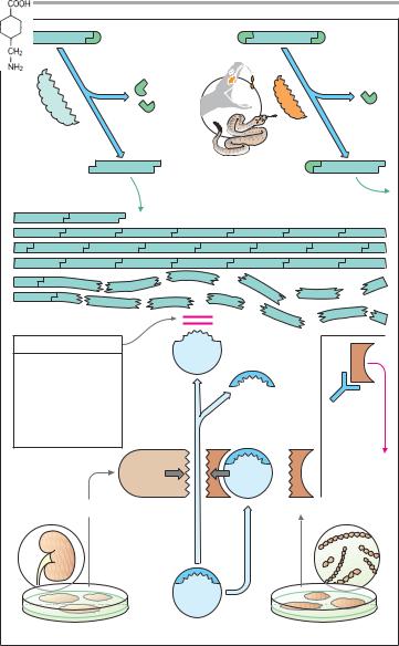
Fibrin is formed from fibrinogen through thrombin (factor IIa)-catalyzed proteolytic removal of two oligopeptide fragments. Individual fibrin molecules polymerize into a fibrin mesh that can be split into fragments and dissolved by plasmin. Plasmin derives by proteolysis from an inactive precursor, plasminogen. Plasminogen activators can be infused for the purpose of dissolving clots (e.g., in myocardial infarction). Thrombolysis is not likely to be successful unless the activators can be given very soon after thrombus formation. Urokinase is an endogenous plasminogen activator obtained from cultured human kidney cells. Urokinase is better tolerated than is streptokinase. By itself, the latter is enzymatically inactive; only after binding to a plasminogen molecule does the complex become effective in converting plasminogen to plasmin. Streptokinase is produced by streptococcal bacteria, which probably accounts for the frequent adverse reactions. Streptokinase antibodies may be present as a result of prior streptococcal infections. Binding to such antibodies would neutralize streptokinase molecules.
With alteplase, another endogenous plasminogen activator (tissue plasminogen activator, tPA) is available. With physiological concentrations this activator preferentially acts on plasminogen bound to fibrin. In concentrations needed for therapeutic fibrinolysis this preference is lost and the risk of bleeding does not differ with alteplase and streptokinase. Alteplase is rather short-
lived (inactivation by complexing with plasminogen activator inhibitor, PAI) and has to be applied by infusion. Reteplase, however, containing only the proteolytic active part of the alteplase molecule, allows more stabile plasma levels and can be applied in form of two injections at an interval of 30 min.
Inactivation of the fibrinolytic system can be achieved by “plasmin inhibitors,” such as !-aminocaproic acid, p-aminomethylbenzoic acid (PAMBA), tranexamic acid, and aprotinin, which also inhibits other proteases.
Lowering of blood fibrinogen concentration. Ancrod is a constituent of the venom from a Malaysian pit viper. It enzymatically cleaves a fragment from fibrinogen, resulting in the formation of a degradation product that cannot undergo polymerization. Reduction in blood fibrinogen level decreases the coagulability of the blood. Since fibrinogen (MW ~340 000) contributes to the viscosity of blood, an improved “fluidity” of the blood would be expected. Both effects are felt to be of benefit in the treatment of certain disorders of blood flow.
Lüllmann, Color Atlas of Pharmacology © 2000 Thieme
All rights reserved. Usage subject to terms and conditions of license.
|
|
Antithrombotics |
147 |
|
Fibrinogen |
|
|
Thrombin |
|
Ancrod |
|
Fibrin |
|
|
|
Plasmin-inhibitors |
|
Plasmin |
|
|
|
|
|
|
|
Antibody from |
|
|
|
prior infection |
|
e.g., Tranexamic acid |
|
|
|
|
|
Fever, |
|
|
|
chills, |
|
|
|
and inacti- |
|
|
|
vation |
|
Urokinase |
|
Streptokinase |
|
Human kidney cell culture |
Plasminogen |
Streptococci |
|
A. Activators and inhibitors of fibrinolysis; ancrod
Lüllmann, Color Atlas of Pharmacology © 2000 Thieme
All rights reserved. Usage subject to terms and conditions of license.

148 Antithrombotics
Intra-arterial Thrombus Formation (A)
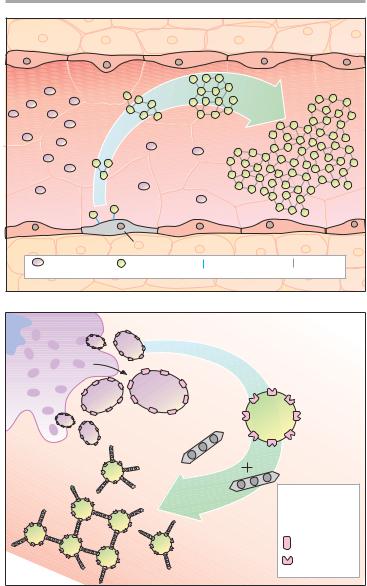
Activation of platelets, e.g., upon contact with collagen of the extracellular matrix after injury to the vascular wall, constitutes the immediate and decisive step in initiating the process of primary hemostasis, i.e., cessation of bleeding. However, in the absence of vascular injury, platelets can be activated as a result of damage to the endothelial cell lining of blood vessels. Among the multiple functions of the endothelium, the production of NO˙ and prostacyclin plays an important role. Both substances inhibit the tendency of platelets to adhere to the endothelial surface (platelet adhesiveness). Impairment of endothelial function, e.g., due to chronic hypertension, cigarette smoking, chronic elevation of plasma LDL levels or of blood glucose, increases the probability of contact between platelets and endothelium. The adhesion process involves GPIB/IX, a glycoprotein present in the platelet cell membrane and von Willebrandt’s factor, an endothelial membrane protein. Upon endothelial contact, the platelet is activated with a resultant change in shape and affinity to fibrinogen. Platelets are linked to each other via fibrinogen bridges: they undergo aggregation.
Platelet aggregation increases like an avalanche because, once activated, platelets can activate other platelets. On the injured endothelial cell, a platelet thrombus is formed, which obstructs blood flow. Ultimately, the vascular lumen is occluded by the thrombus as the latter is solidified by a vasoconstriction produced by the release of serotonin and thromboxane A2 from the aggregated platelets. When these events occur in a larger coronary artery, the consequence is a myocardial infarction; involvement of a cerebral artery leads to stroke.
The von Willebrandt’s factor plays a key role in thrombogenesis. Lack of this factor causes thrombasthenia, a pathologically decreased platelet aggregation. Relative deficiency of the von Wille-
brandt’s factor can be temporarily overcome by the vasopressin anlogue desmopressin (p. 164), which increases the release of available factor from storage sites.
Formation, Activation, and Aggregation of Platelets (B)
Platelets originate by budding off from multinucleate precursor cells, the megakaryocytes. As the smallest formed element of blood (dia. 1–4 µm), they can be activated by various stimuli. Activation entails an alteration in shape and secretion of a series of highly active substances, including serotonin, platelet activating factor (PAF), ADP, and thromboxane A2. In turn, all of these can activate other platelets, which explains the explosive nature of the process.
The primary consequence of activation is a conformational change of an integrin present in the platelet membrane, namely, GPIIB/IIIA. In its active conformation, GPIIB/IIIA shows high affinity for fibrinogen; each platelet contains up to 50,000 copies. The high plasma concentration of fibrinogen and the high density of integrins in the platelet membrane permit rapid cross-linking of platelets and formation of a platelet plug.
Lüllmann, Color Atlas of Pharmacology © 2000 Thieme
All rights reserved. Usage subject to terms and conditions of license.
|
|
Antithrombotics |
149 |
|
|
Aggregation |
|
|
|
Adhesion |
|
|
|
|
|
dysfunctional endothelial cell |
|
|
|
Platelet |
Activated |
von Willebrandt’s |
Fibrinogen |
|
|
platelet |
factor |
|
|
A. Thrombogenesis
Megakaryocyte |
Contact with |
|
|
Activation |
collagen |
ADP |
|
|
Thrombin |
|
Thromboxane A2 |
|
Serotonin |
Activated |
|
platelet |
|
|
Glycoprotein |
Fibrinogen |
IIB/IIIA |
|
Fibrinogen |
|
binding: |
|
impossible |
|
possible |
B. Aggregation of platelets by the integrin GPIIB/IIIA
Lüllmann, Color Atlas of Pharmacology © 2000 Thieme
All rights reserved. Usage subject to terms and conditions of license.

150 Antithrombotics
Inhibitors of Platelet Aggregation (A)
Platelets can be activated by mechanical and diverse chemical stimuli, some of which, e.g., thromboxane A2, thrombin, serotonin, and PAF, act via receptors on the platelet membrane. These receptors are coupled to Gq proteins that mediate activation of phospholipase C and hence a rise in cytosolic Ca2+ concentration. Among other responses, this rise in Ca2+ triggers a conformational change in GPIIB/IIIA, which is thereby converted to its fbrinogen-binding form. In contrast, ADP activates platelets by inhibiting adenylyl cyclase, thus causing internal cAMP levels to decrease. High cAMP levels would stabilize the platelet in its inactive state. Formally, the two messenger substances, Ca2+ and cAMP, can be considered functional antagonists.
Platelet aggregation can be inhibited by acetylsalicylic acid (ASA), which blocks thromboxane synthase, or by recombinant hirudin (originally harvested from leech salivary gland), which binds and inactivates thrombin. As yet, no drugs are available for blocking aggregation induced by serotonin or PAF. ADP-induced aggregation can be prevented by ticlopidine and clopidogrel; these agents are not classic receptor antagonists. ADP-induced aggregation is inhibited only in vivo but not in vitro in stored blood; moreover, once induced, inhibition is irreversible. A possible explanation is that both agents already interfere with elements of ADP receptor signal transduction at the megakaryocytic stage. The ensuing functional defect would then be transmitted to newly formed platelets, which would be incapable of reversing it.
The intra-platelet levels of cAMP can be stabilized by prostacyclin or its analogues (e.g., iloprost) or by dipyrida- mole. The former activates adenyl cyclase via a G-protein-coupled receptor; the latter inhibits a phosphodiesterase that breaks down cAMP.
The integrin (GPIIB/IIIA)-antago- nists prevent cross-linking of platelets. Their action is independent of the ag-
gregation-inducing stimulus. Abciximab is a chimeric human-murine monoclonal antibody directed against GPIIb/IIIa that blocks the fibrinogen-binding site and thus prevents attachment of fibrinogen. The peptide derivatives, eptifibatide and tirofiban block GPIIB/IIIA competitively, more selectively and have a shorter effect than does abciximab.
Presystemic Effect of Acetylsalicylic Acid
(B)
Inhibition of platelet aggregation by ASA is due to a selective blockade of platelet cyclooxygenase (B). Selectivity of this action results from acetylation of this enzyme during the initial passage of the platelets through splanchnic blood vessels. Acetylation of the enzyme is irreversible. ASA present in the systemic circulation does not play a role in platelet inhibition. Since ASA undergoes extensive presystemic elimination, cyclooxygenases outside platelets, e.g., in endothelial cells, remain largely unaffected. With regular intake, selectivity is enhanced further because the anuclear platelets are unable to resynthesize new enzyme and the inhibitory effects of consecutive doses are added to each other. However, in the endothelial cells, de novo synthesis of the enzyme permits restoration of prostacyclin production.
Adverse Effects of Antiplatelet Drugs
All antiplatelet drugs increase the risk of bleeding. Even at the low ASA doses used to inhibit platelet function (100 mg/d), ulcerogenic and bronchoconstrictor (aspirin asthma) effects may occur. Ticlopidine frequently causes diarrhea and, more rarely, leukopenia, necessitating cessation of treatment. Clopidogrel reportedly does not cause hematological problems.
As peptides, hirudin and abciximab need to be injected; therefore their use is restricted to intensive-care settings.
Lüllmann, Color Atlas of Pharmacology © 2000 Thieme
All rights reserved. Usage subject to terms and conditions of license.