

книги студ / Color Atlas of Pharmacology
.pdfCardiac Drugs 131
Helleborus niger
Christmas rose
|
Convallaria |
Digitalis purpurea |
majalis |
Red foxglove |
Lily of the valley |
A. Plants containing cardiac glycosides
Contraction |
|
|
Arrhythmia |
Contracture |
|
|
|
|
|
|
Time |
´therapeutic´ |
´toxic´ |
Dose of cardiac glycoside (CG) |
Na+ |
Na/K-ATPase |
|
CG |
CG |
Na+ |
Na+ |
Na+ |
Ca2+ |
Ca2+ |
Ca2+ |
Coupling |
|
|
K+ K+ |
K+ |
K+ |
Heart muscle cell |
CG |
CG |
CG
B. Therapeutic and toxic effects of cardiac glycosides (CG)
Disturbance of |
color vision |
Excitation of |
N. vagus: |
Heart rate |
Area postrema: |
nausea, vomiting |
C. Cardiac glycoside effects on the CNS
"Re-entrant" |
excitation in |
atrial |
fibrillation |
Cardiac |
glycoside |
Decrease in |
ventricular |
rate |
D.Cardiac glycoside effects in atrial fibrillation
Lüllmann, Color Atlas of Pharmacology © 2000 Thieme
All rights reserved. Usage subject to terms and conditions of license.

132 Cardiac Drugs
Substance |
Fraction |
Plasma concentr. |
Digitalizing |
Elimination |
Maintenance |
||
|
absorbed |
free |
|
total |
dose |
|
dose |
|
% |
|
(ng/mL) |
(mg) |
%/d |
(mg) |
|
|
|
|
|
|
|
|
|
Digitoxin |
100 |
!1 |
|
!20 |
!1 |
10 |
!0.1 |
Digoxin |
50–90 |
!1 |
|
!1.5 |
!1 |
30 |
!0.3 |
Ouabain |
<1 |
!1 |
|
!1 |
0.5 |
no long-term use |
|
|
|
|
|
|
|
|
|
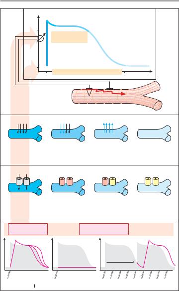
The pharmacokinetics of cardiac glycosides (A) are dictated by their polarity, i.e., the number of hydroxyl groups. Membrane penetrability is virtually nil in ouabain, high in digoxin, and very high in digitoxin. Ouabain (g- strophanthin) does not penetrate into cells, be they intestinal epithelium, renal tubular, or hepatic cells. At best, it is suitable for acute intravenous induction of glycoside therapy.
The absorption of digoxin depends on the kind of galenical preparation used and on absorptive conditions in the intestine. Preparations are now of such quality that the derivatives methyl- digoxin and acetyldigoxin no longer offer any advantage. Renal reabsorption is incomplete; approx. 30% of the total amount present in the body (s.c. full “digitalizing” dose) is eliminated per day. When renal function is impaired, there is a risk of accumulation. Digi- toxin undergoes virtually complete reabsorption in gut and kidneys. There is active hepatic biotransformation: cleavage of sugar moieties, hydroxylation at C12 (yielding digoxin), and conjugation to glucuronic acid. Conjugates secreted with bile are subject to enterohepatic cycling (p. 38); conjugates reaching the blood are renally eliminated. In renal insufficiency, there is no appreciable accumulation. When digitoxin is withdrawn following overdosage, its effect decays more slowly than does that of digoxin.
Other positive inotropic drugs. The phosphodiesterase inhibitor amrinone (cAMP elevation, p. 66) can be administered only parenterally for a maximum of 14 d because it is poorly
tolerated. A closely related compound is milrinone. In terms of their positive inotropic effect, !-sympathomimetics, unlike dopamine (p. 114), are of little therapeutic use; they are also arrhythmogenic and the sensitivity of the !-re- ceptor system declines during continuous stimulation.
Treatment Principles in Chronic Heart
Failure
Myocardial insufficiency leads to a decrease in stroke volume and venous congestion with formation of edema. Administration of (thiazide) diuretics (p. 62) offers a therapeutic approach of proven efficacy that is brought about by a decrease in circulating blood volume (decreased venous return) and peripheral resistance, i.e., afterload. A similar approach is intended with ACE-inhibi- tors, which act by preventing the synthesis of angiotensin II (! vasoconstriction) and reducing the secretion of aldosterone (! fluid retention). In severe cases of myocardial insufficiency, cardiac glycosides may be added to augment cardiac force and to relieve the symptoms of insufficiency.
In more recent times !-blocker on a long term were found to improve cardiac performance — particularly in idiopathic dilating cardiomyopathy — probably by preventing sympathetic overdrive.
Lüllmann, Color Atlas of Pharmacology © 2000 Thieme
All rights reserved. Usage subject to terms and conditions of license.

|
|
|
Cardiac Drugs |
133 |
||
|
|
|
|
|
Plasma |
|
Digoxin |
|
|
|
|
|
|
|
|
|
95% |
|
|
|
|
0% |
|
35% |
|
|
|
|
|
|
|
|
|
|
|
|
|
|
|
cell |
|
|
|
|
Digitoxin |
Digoxin |
||
|
|
|
Cleavage |
|
||
|
|
|
of sugar |
|
|
|
|
|
|
|
Conjugation |
|
|
Intestinal epithelium |
Renal tubular epithelium |
|
Deconjugation |
|
|
|
|
Plasma t1 2 |
9 h |
2 – 3 days |
5 – 7 days |
||
A. Pharmacokinetics of cardiac glycosides
Lüllmann, Color Atlas of Pharmacology © 2000 Thieme
All rights reserved. Usage subject to terms and conditions of license.

134 Cardiac Drugs
Antiarrhythmic Drugs
The electrical impulse for contraction (propagated action potential; p. 136) originates in pacemaker cells of the sinoatrial node and spreads through the atria, atrioventricular (AV) node, and adjoining parts of the His-Purkinje fiber system to the ventricles (A). Irregular- ities of heart rhythm can interfere dangerously with cardiac pumping function.
I. Drugs for selective control of sinoatrial and AV nodes. In some forms of arrhythmia, certain drugs can be used that are capable of selectively facilitating and inhibiting (green and red arrows, respectively) the pacemaker function of sinoatrial or atrioventricular cells.
Sinus bradycardia. An abnormally low sinoatrial impulse rate (<60/min) can be raised by parasympatholytics.
The quaternary ipratropium is preferable to atropine, because it lacks CNS penetrability (p. 107). Sympathomimetics also exert a positive chronotropic action; they have the disadvantage of increasing myocardial excitability (and automaticity) and, thus, promoting ectopic impulse generation (tendency to extrasystolic beats). In cardiac arrest epinephrine can be used to reinitiate heart beat.
Sinus tachycardia (resting rate >100 beats/min). !-Blockers eliminate sympathoexcitation and decrease cardiac rate.
Atrial flutter or fibrillation. An excessive ventricular rate can be decreased by verapamil (p. 122) or cardiac glycosides (p. 130). These drugs inhibit impulse propagation through the AV node, so that fewer impulses reach the ventricles.
II. Nonspecific drug actions on impulse generation and propagation.
Impulses originating at loci outside the sinus node are seen in supraventricular or ventricular extrasystoles, tachycardia, atrial or ventricular flutter, and fibrilla- tion. In these forms of rhythm disorders, antiarrhythmics of the local anesthet-
ic, Na+-channel blocking type (B) are used for both prophylaxis and therapy. Local anesthetics inhibit electrical excitation of nociceptive nerve fibers (p. 204); concomitant cardiac inhibition
(cardiodepression) is an unwanted effect. However, in certain types of arrhythmias (see above), this effect is useful. Local anesthetics are readily cleaved (arrows) and unsuitable for oral administration (procaine, lidocaine). Given judiciously, intravenous lidocaine is an effective antiarrhythmic. Procainamide and mexiletine, congeners endowed with greater metabolic stability, are examples of orally effective antiarrhythmics. The desired and undesired effects are inseparable. Thus, these antiarrhythmics not only depress electrical excitability of cardiomyocytes (negative bathmotropism, membrane stabilization), but also lower sinoatrial rate (neg. chronotropism), AV conduction (neg. dromotropism), and force of contraction (neg. inotropism). Interference with normal electrical activity can, not too paradoxically, also induce cardiac arrhyth- mias–arrhythmogenic action.
Inhibition of CNS neurons is the underlying cause of neurological effects such as vertigo, confusion, sensory disturbances, and motor disturbances (tremor, giddiness, ataxia, convulsions).
Lüllmann, Color Atlas of Pharmacology © 2000 Thieme
All rights reserved. Usage subject to terms and conditions of license.

|
Cardiac Drugs |
135 |
Sinus node |
|
|
|
Para- |
|
Atrium |
sympatholytics |
|
|
|
|
|
!-Sympatho- |
|
AV-node |
mimetics |
|
|
|
|
Bundle of His |
|
|
Tawara´s |
!-Blocker |
|
Verapamil |
|
|
node |
|
|
Purkinje |
Cardiac |
|
fibers |
glycoside |
|
Ventricle |
(Vagal |
|
stimulation) |
|
|
|
|
|
A. Cardiac impulse generation and conduction |
|
|
Main effect
Antiarrhythmic effect
Adverse effects
CNS-disturbances
Arrhythmia
Cardiodepression
Antiarrhythmics of the local anesthetic (Na+-channel blocking) type:
Inhibition of impulse generation and conduction
|
|
|
|
|
|
|
|
|
|
|
|
|
|
|
|
|
|
|
Esterases |
|
|
Procaine |
|
|
|
|
|
|
|
|
|
|
|
|
|
|
|
|
|
|
|
|
|
|
|
|
|
|
|
|
|
|
|
|
|
|
|
|
|
|
|
|
|
|
|
|
|
|
|
|
|
|
|
|
|
|
|
|
|
|
|
|
|
|
|
|
|
|
|
|
|
|
|
|
Procainamide |
|
|
|
|
|
|
|
|
|
|
|
|
|
|
|
|
|
|
|
|
|
|
|
|
|
|
|
|
|
|
|
|
|
|
|
|
|
|
|
|
|
|
|
|
|
|
|
Lidocaine |
|
|
|
|
|
|
|
|
|
|
|
|
|
|
|
|
|
|
|
|
|
|
|
|
|
|
|
|
|
|
|
|
|
|
|
|
|
|
|
|
|
|
|
|
|
|
|
Mexiletine |
|
|
|
|
|
|
|
|
|
|
|
|
|
|
|
|
|
|
|
|
|
|
|
|
B. Antiarrhythmics of the Na+-channel blocking type
Lüllmann, Color Atlas of Pharmacology © 2000 Thieme
All rights reserved. Usage subject to terms and conditions of license.

136 Cardiac Drugs
Electrophysiological Actions of
Antiarrhythmics of the Na+-Channel
Blocking Type
Action potential and ionic currents.
The transmembrane electrical potential of cardiomyocytes can be recorded through an intracellular microelectrode. Upon electrical excitation, a characteristic change occurs in membrane poten- tial—the action potential (AP). Its underlying cause is a sequence of transient ionic currents. During rapid depolarization (Phase 0), there is a short-lived in- flux of Na+ through the membrane. A subsequent transient influx of Ca2+ (as well as of Na+) maintains the depolarization (Phase 2, plateau of AP). A delayed efflux of K+ returns the membrane potential (Phase 3, repolarization) to its resting value (Phase 4). The velocity of depolarization determines the speed at which the AP propagates through the myocardial syncytium.
Transmembrane ionic currents involve proteinaceous membrane pores: Na+, Ca2+, and K+ channels. In A, the phasic change in the functional state of Na+ channels during an action potential is illustrated.
Effects of antiarrhythmics. Antiar- rhythmics of the Na+-channel blocking type reduce the probability that Na+ channels will open upon membrane depolarization (“membrane stabilization”). The potential consequences are (A, bottom): 1) a reduction in the velocity of depolarization and a decrease in the speed of impulse propagation; aberrant impulse propagation is impeded. 2)
Depolarization is entirely absent; patho- logical impulse generation, e.g., in the marginal zone of an infarction, is suppressed. 3) The time required until a new depolarization can be elicited, i.e., the refractory period, is increased; pro- longation of the AP (see below) contributes to the increase in refractory period. Consequently, premature excitation with risk of fibrillation is prevented.
Mechanism of action. Na+-channel blocking antiarrhythmics resemble most local anesthetics in being cationic
amphiphilic molecules (p. 208, exception: phenytoin, p. 190). Possible molecular mechanisms of their inhibitory effects are outlined on p. 204 in more detail. Their low structural specificity is reflected by a low selectivity towards different cation channels. Besides the Na+ channel, Ca2+ and K+ channels are also likely to be blocked. Accordingly, cationic amphiphilic antiarrhythmics affect both the depolarization and repolarization phases. Depending on the substance, AP duration can be increased (Class IA), decreased (Class IB), or remain the same (Class IC).
Antiarrhythmics representative of these categories include: Class IA— quinidine, procainamide, ajmaline, disopyramide, propafenone; Class IB—lido- caine, mexiletine, tocainide, as well as phenytoin; Class IC—flecainide.
Note: With respect to classification, !-blockers have been assigned to Class II, and the Ca2+-channel blockers verapamil and diltiazem to Class IV.
Commonly listed under a separate rubric (Class III) are amiodarone and the !-blocking agent sotalol, which both inhibit K+-channels and which both cause marked prolongation of the AP with a lesser effect on Phase 0 rate of rise.
Therapeutic uses. Because of their narrow therapeutic margin, these antiarrhythmics are only employed when rhythm disturbances are of such severity as to impair the pumping action of the heart, or when there is a threat of other complications. The choice of drug is empirical. If the desired effect is not achieved, another drug is tried. Combinations of antiarrhythmics are not customary. Amiodarone is reserved for special cases.
Lüllmann, Color Atlas of Pharmacology © 2000 Thieme
All rights reserved. Usage subject to terms and conditions of license.
|
|
|
Cardiac Drugs |
137 |
[mV] |
1 |
Membrane potential |
|
|
|
2 |
|
|
|
|
|
|
|
|
0 |
Rate of |
Action |
|
|
|
|
|||
0 |
potential |
|
||
depolarization |
|
|||
|
|
Speed of AP |
(AP) |
|
|
|
3 |
|
|
|
|
propagation |
|
|
-80 |
|
|
4 |
|
|
Refractory period |
|
|
|
0 |
|
250 Time [ms] |
|
|
|
|
|
||
|
|
|
Heart muscle cell |
|
Na+ |
|
Ca2+(+Na+) |
|
|
|
|
|
K+ |
|
Phase 0 |
|
Phases 1,2 |
Phase 3 |
Phase 4 |
Fast |
Slow Ca2+-entry |
|
|
|
Na+-entry” |
|
|
|
|
Ionic currents during action potential |
|
|
||
Na+ |
|
Na+-channels |
|
|
Open (active) |
|
Closed |
|
Closed |
|
|
Opening impossible |
|
Opening possible |
|
|
(inactivated) |
|
(resting, can be |
States of Na+-channels during an action potential |
activated) |
|||
|
||||
Inhibition of |
|
Antiarrhythmics of the |
|
|
Na+-channel opening |
Na+-channel blocking type |
|
||
|
|
|
Inexcitability |
|
Stimulus |
|
|
|
|
Rate of |
|
Suppression |
Prolongation of refractory period = |
|
depolarization |
|
of AP generation |
duration of inexcitability |
|
A. Effects of antiarrhythmics of the Na+-channel blocking type
Lüllmann, Color Atlas of Pharmacology © 2000 Thieme
All rights reserved. Usage subject to terms and conditions of license.

138 Antianemics
Drugs for the Treatment of Anemias
Anemia denotes a reduction in red blood cell count, hemoglobin content, or both. Oxygen (O2) transport capacity is decreased.
Erythropoiesis (A). Blood corpuscles develop from stem cells through several cell divisions. Hemoglobin is then synthesized and the cell nucleus is extruded. Erythropoiesis is stimulated by the hormone erythropoietin (a glycoprotein), which is released from the kidneys when renal O2 tension declines.
Given an adequate production of erythropoietin, a disturbance of erythropoiesis is due to two principal causes: 1. Cell multiplication is inhibited be- cause DNA synthesis is insufficient. This occurs in deficiencies of vitamin B12 or folic acid (macrocytic hyperchromic anemia). 2. Hemoglobin synthesis is impaired. This situation arises in iron deficiency, since Fe2+ is a constituent of hemoglobin (microcytic hypochromic anemia).
Vitamin B12 (B)
Vitamin B12 (cyanocobalamin) is produced by bacteria; B12 generated in the colon, however, is unavailable for absorption (see below). Liver, meat, fish, and milk products are rich sources of the vitamin. The minimal requirement is about 1 µg/d. Enteral absorption of vitamin B12 requires so-called “intrinsic factor” from parietal cells of the stomach. The complex formed with this glycoprotein undergoes endocytosis in the ileum. Bound to its transport protein, transcobalamin, vitamin B12 is destined for storage in the liver or uptake into tissues.
A frequent cause of vitamin B12 deficiency is atrophic gastritis leading to a lack of intrinsic factor. Besides megaloblastic anemia, damage to mucosal linings and degeneration of myelin sheaths with neurological sequelae will occur (pernicious anemia).
Optimal therapy consists in paren- teral administration of cyanocobal- amin or hydroxycobalamin (Vitamin
B12a; exchange of -CN for -OH group). Adverse effects, in the form of hyper-
sensitivity reactions, are very rare. Folic Acid (B). Leafy vegetables and
liver are rich in folic acid (FA). The min- imal requirement is approx. 50 µg/d. Polyglutamine-FA in food is hydrolyzed to monoglutamine-FA prior to being absorbed. FA is heat labile. Causes of deficiency include: insufficient intake, malabsorption in gastrointestinal diseases, increased requirements during pregnancy. Antiepileptic drugs (phenytoin, primidone, phenobarbital) may decrease FA absorption, presumably by inhibiting the formation of monogluta- mine-FA. Inhibition of dihydro-FA reductase (e.g., by methotrexate, p. 298) depresses the formation of the active species, tetrahydro-FA. Symptoms of deficiency are megaloblastic anemia and mucosal damage. Therapy consists in oral administration of FA or in folinic acid (p. 298) when deficiency is caused by inhibitors of dihydro—FA—reductase.
Administration of FA can mask a vitamin B12 deficiency. Vitamin B12 is required for the conversion of methyltet- rahydro-FA to tetrahydro-FA, which is important for DNA synthesis (B). Inhibi- tion of this reaction due to B12 deficiency can be compensated by increased FA intake. The anemia is readily corrected; however, nerve degeneration progresses unchecked and its cause is made more difficult to diagnose by the absence of hematological changes. Indiscriminate use of FA-containing multivitamin preparations can, therefore, be harmful.
Lüllmann, Color Atlas of Pharmacology © 2000 Thieme
All rights reserved. Usage subject to terms and conditions of license.

|
Antianemics |
139 |
Inhibition of DNA |
Inhibition of |
|
synthesis |
hemoglobin synthesis |
|
(cell multiplication) |
|
|
Vit. B12 deficiency |
Iron deficiency |
|
Folate deficiency |
|
|
A very few large |
A few small |
|
hemoglobin-rich |
hemoglobin-poor |
|
erythrocytes |
erythrocytes |
|
A. Erythropoiesis in bone marrow
|
Folic acid H4 |
|
|
DNA |
|
|
|
synthesis |
H3C- Folic acid H4 |
Vit. B12 |
Folic acid |
|
|
|
|
H3C- Vit. B12 |
|
|
|
H3C- |
Vit. B12 |
|
|
|
|
|
|
|
Trans- |
|
HCl |
|
|
|
|
Storage |
cobalamin II |
|
|
|
|
|
|
supply for |
Vit. |
|
Intrinsic |
3 years |
|
||
|
|
factor |
|
|
|
|
|
|
|
|
Parietal cell |
Streptomyces |
|
|
|
griseus |
|
|
|
B. Vitamin B12 and folate metabolism
Lüllmann, Color Atlas of Pharmacology © 2000 Thieme
All rights reserved. Usage subject to terms and conditions of license.

140 Antianemics
Iron Compounds
Not all iron ingested in food is equally absorbable. Trivalent Fe3+ is virtually not taken up from the neutral milieu of the small bowel, where the divalent Fe2+ is markedly better absorbed. Uptake is particularly efficient in the form of heme (present in hemoand myoglobin). Within the mucosal cells of the gut, iron is oxidized and either deposited as ferritin (see below) or passed on to the transport protein, transferrin, a !1-gly- coprotein. The amount absorbed does not exceed that needed to balance losses due to epithelial shedding from skin and mucosae or hemorrhage (so-called “mucosal block”). In men, this amount is approx. 1 mg/d; in women, it is approx. 2 mg/d (menstrual blood loss), corresponding to about 10% of the dietary intake. The transferrin-iron complex undergoes endocytotic uptake mainly into erythroblasts to be utilized for hemoglobin synthesis.
About 70% of the total body store of iron (~5 g) is contained within erythrocytes. When these are degraded by macrophages of the reticuloendothelial (mononuclear phagocyte) system, iron is liberated from hemoglobin. Fe3+ can be stored as ferritin (= protein apoferritin + Fe3+) or returned to erythropoiesis sites via transferrin.
A frequent cause of iron deficiency is chronic blood loss due to gastric/intestinal ulcers or tumors. One liter of blood contains 500 mg of iron. Despite a significant increase in absorption rate (up to 50%), absorption is unable to keep up with losses and the body store of iron falls. Iron deficiency results in impaired synthesis of hemoglobin and anemia (p. 138).
The treatment of choice (after the cause of bleeding has been found and eliminated) consists of the oral administration of Fe2+ compounds, e.g., ferrous sulfate (daily dose 100 mg of iron equivalent to 300 mg of FeSO4, divided into multiple doses). Replenishing of iron stores may take several months. Oral administration, however, is advan-
tageous in that it is impossible to overload the body with iron through an intact mucosa because of its demand-reg- ulated absorption (mucosal block).
Adverse effects. The frequent gastrointestinal complaints (epigastric pain, diarrhea, constipation) necessitate intake of iron preparations with or after meals, although absorption is higher from the empty stomach.
Interactions. Antacids inhibit iron absorption. Combination with ascorbic acid (Vitamin C), for protecting Fe2+ from oxidation to Fe3+, is theoretically sound, but practically is not needed.
Parenteral administration of Fe3+ salts is indicated only when adequate oral replacement is not possible. There is a risk of overdosage with iron deposition in tissues (hemosiderosis). The binding capacity of transferrin is limited and free Fe3+ is toxic. Therefore, Fe3+ complexes are employed that can donate Fe3+ directly to transferrin or can be phagocytosed by macrophages, enabling iron to be incorporated into ferritin stores. Possible adverse effects are, with i.m. injection: persistent pain at the injection site and skin discoloration; with i.v. injection: flushing, hypotension, anaphylactic shock.
Lüllmann, Color Atlas of Pharmacology © 2000 Thieme
All rights reserved. Usage subject to terms and conditions of license.