

Essentials of Orthopedic Surgery, third edition / 05-Children’s Orthopedics
.pdf5
Children’s Orthopedics
JOHN N. DELAHAY and WILLIAM C. LAUERMAN
Children are different! This statement has been presented in many different ways; but it is critically important that this central fact be recognized if one is to successfully diagnose and treat disease in this age group. Even within this rather broad range of ages there are dramatic differences among specific subsets: neonate, child, and adolescent.
These differences are not only biologic, but psychologic, social, and emotional. It is likewise inappropriate to focus only on one aspect of these differences. For example, it would be unwise to ignore a young child’s activity level when treating a fracture: inadequate immobilization or cast removal too early will have disastrous end results.
Recognition of this special group actually gave orthopedics its name. The word means “straight child” and alludes to the interest and time spent correcting deformities in children. These deformities can result not only from injury but also from systemic and local disease states, both congenital and acquired. Because the child is growing, these diseases produce anatomic and physiologic effects not expected in the adult. Before discussing specific entities, it would, therefore, be appropriate to review some of the biologic differences of the child’s musculoskeletal system and the influences that act on the immature skeleton.
Biologic Differences
Growth
As mentioned, the fact that the child’s skeleton is growing, both longitudinally and latitudinally, positions it uniquely for damage resulting from the adverse effects of trauma and disease. The extent of this damage is a reflection of the rate of growth and the immaturity of the skeleton. Hence, an insult will have a greater impact if applied at the time of more rapid growth (a growth spurt) or when the skeleton is very young (neonate).
169

170 J.N. Delahay and W.C. Lauerman
Remodeling
The immature skeleton can remodel to a much greater degree than that of the adult. Because of the presence and activity of multiple cell populations, damage to the skeleton can be repaired more extensively than one should anticipate in the adult. The challenge for the physician is to be able to recognize the limitations of this remodeling process and work within the boundaries of this potential.
Specific Anatomic Structures
Bone
Although a child’s bone is historically lamellar in pattern, there remains enough flexibility in the skeleton to permit what has been called “biological plasticity,” a phenomena not nearly as extensive in adult bone. Essentially, this allows a bone to “bend without breaking”; in point of fact, it is responsible for some of the unique types of fractures seen in the pediatric age groups, specifically, torus and greenstick fractures.
In addition, the mechanical properties of a child’s bone vary from those of the adult. Such characteristics as modulus of elasticity, ultimate tensile strength, and yield point all reflect the elasticity and plasticity unique in this age group. However, the overall “strength” tends to be less than that of the adult in certain modes of loading, such as tension and shear.
Ligament
As a tissue, ligament is one of the most age-resistant tissues in the human body. The tensile strength of the ligaments in the child and the adult is virtually the same. Therefore, these structures remain as a constant in the musculoskeletal system. Although the strength of bone, cartilage, and muscle tends to change, the ligamentous structures remain unchanged with growth and development.
Periosteum
The outer covering of the bone is a dense fibrous layer, which in the child is significantly thicker than that of the adult. The periosteum of the child actually has an outer fibrous layer and an inner cambial or osteogenic layer. Hence, the child’s periosteum confers both mechanical strength as well as biologic activity. The effect of these biologic differences are far reaching when one discusses fractures in children. Because of this thickened periosteum, fractures do not tend to displace to the degree seen in adults, and the intact periosteum can be used as an aid in fracture reduction and maintenance. In addition, fractures will heal significantly faster than

5. Children’s Orthopedics |
171 |
similar injuries in adults because all the cellular precursors are already present. The osteogenic layer supplies active osteoblasts, ready to make bone for the fracture callus. The generation of these precursor elements in adults takes a period of time not required in the child.
Cartilage
As one will recall, the skeleton is developed embryologically within a cartilage model. At birth, large portions of any given bone remain largely cartilaginous. Unfortunately, cartilage is not seen on standard X-rays. The cartilage anlage is very labile and is dramatically affected by external influences such as mechanical loading. It is important to realize, when examining an X-ray, that one should not be lulled into a false sense of security if all appears well; what you do not see (i.e., the cartilage) is more important than what you do! Aberrant cartilaginous growth will drastically affect the ultimate shape of bones and, more importantly, joints. The best example is the proximal femur, where most of the upper end is cartilaginous. Adverse influences caused by eccentric loading seen in developmental dysplasia of the hip can have far-reaching effects when applied to the immature cartilage of the neonatal hip.
The Growth Plate
By far and away the most exceptional characteristic of the immature skel- eton—indeed, the defining component of the immature skeleton—is the growth plate, or the “physis.” The physis is a cartilage plate interposed between the epiphysis (the secondary ossification center) and the metaphysis (Fig. 5-1). It is essential for long bone growth to occur. The downside is that this anatomic structure creates a “normal flaw” in the overall skeletal structure and thus a point of mechanical weakness. The physis historically has four zones (Fig. 5-2), each with its own physiologic role:
a.Resting zone: The top layer of flattened cells are germinal and metabolically store materials for later use, because they will ultimately “move their way” down the plate toward the metaphysis. The chondrocytes in this zone also are synthetic, as they fabricate the matrix within which they lie.
b.Proliferating zone: The cells in this region are actively replicating and extending the plate. They have been described as looking like a “stack of plates.” In this region, the cells are using the materials that they have previously stored for their “trip to the metaphysis.”
c.Hypertrophic zone: Having extended the plate in the former zone, the cells now tend to swell and switch over to a more-catabolic state. They prepare the matrix for calcification and ultimately for conversion to bone. Because of its large swollen cells and the disorganized matrix,

172 J.N. Delahay and W.C. Lauerman
this zone has been cited as being the weakest mechanically; hence, it is here that failure tends to occur. Most, however, would agree that crack propagation can be seen throughout all zones in the case of trauma.
d.Calcified zone: Metabolically, the matrix has been readied for the deposition of calcium salts, and the task of forming the osteoid is left for this lowest region of the plate. In the adjacent metaphysis, small vascular twigs can be seen arborizing toward the basal layers of the plate.
Peripheral Structures of the Plate
Two defined histologic regions have been identified with specific functional roles to play in skeletal development.
FIGURE 5-1. Early secondary ossification center of mature fetus. The formation of the secondary ossification centers in the lower tibia and upper femur coincide with fetal maturity. The secondary center begins not in the center of the epiphysis, but nearer the growth plate. Expansion, therefore, is eccentric. (From Bogumill GP. Orthopaedic Pathology: A Synopsis with Clinical Radiographic Correlation. Philadelphia: Saunders, 1984. Reprinted by permission.)

5. Children’s Orthopedics |
173 |
FIGURE 5-2. Growth plate. Low-power view showing entire plate. (A) Resting zone has isolated cartilage cells in the upper portion together with empty lacunae.
(B) Cell reproduction produces cloning of cells that “stack up” longitudinally. Successive generations occupy more space than each single progenitor cell and thus increase the length of the cartilage model. Secretion of new matrix (C) is followed by dehydration with accompanying fibrillation of the territorial cartilage matrix between the “dinner plates” at (D). Hypertrophy of cartilage cells at
(E) is caused by imbibition of water. Calcification of matrix is followed by vascular invasion at (F). (From Bogumill GP. Orthopaedic Pathology: A Synopsis with Clinical Radiographic Correlation. Philadelphia: Saunders, 1984. Reprinted by permission.)
a.Zone of Ranvier: Around the circumference of the plate is an identifiable clustering of cells that are responsible for latitudinal growth of the plate.
b.Perichondral ring of Lacroix: As the periosteum is continuous around the margins of the plate, this fibrous structure is apparent. Its function arguably has been to serve as a “girdle” for the plate and give mechanical support against translational movement.
Factors Affecting Skeletal Growth
Numerous factors, both intrinsic and extrinsic, affect the way in which the skeleton develops. Some examples are noteworthy, as indicated next.

174 J.N. Delahay and W.C. Lauerman
Genetic Impact
Inborn errors of metabolism (renal rickets) as well as chromosomal alterations (Down syndrome) can cause phenotypic variations in the development of the skeleton. Abnormal histology, aberrational growth, and variational development all affect the ultimate shape and behavior of the skeleton.
Nutrition
Vitamins and proteins are required for normal skeletal development, and without appropriate levels of these, abnormalities are seen. Rickets, for example, will alter the shape of the metaphysis in addition to disrupting normal physeal development.
Endocrine
Hormonal influences play a significant trophic or permissive role in the development of the skeleton. Shortages or excesses, therefore, disrupt the way in which the skeleton matures. Thyroid hormone is a good example. Disrupted epiphyseal development is a hallmark of cretinism.
Environmental Factors
Mechanical effects as well as environmental toxins and drugs can adversely affect the development of the skeleton. Fetal alcohol syndrome and the use of illicit narcotics by the mother are just two examples of the growing compendium of skeletal aberrations caused by externally applied toxins.
Coexistent Disease
Neuromuscular diseases of children, such as cerebral palsy, polio, and muscular dystrophy, provide good examples of the secondary effects seen in the skeleton as a result of extrinsic disease. In these examples, the final common pathway in the pathophysiology of the deformities is muscle imbalance; hence, eccentric and aberrational mechanical loading of the immature skeleton produces changes such as joint dislocations and deformities (e.g., scoliosis).
Developmental Variations in Skeletal Growth
It seems axiomatic to say that children grow and develop at different rates and in different ways. Yet, one of the most common reasons that children are brought to a physician is to evaluate the position of their lower

5. Children’s Orthopedics |
175 |
extremities and the way in which they stand and walk. Toeing in and toeing out, as well as knock-knees and bowlegs, are a major preoccupation of parents and especially grandparents—and a major source of orthopedic referrals. The simple fact is that most of these children, well over 90%, are normal children who are simply reflecting variational growth and development. Dr. Mercer Rang, a preeminent pediatric orthopedist, has tried to emphasize this important fact by referring to these conditions as “non-disease.”
Rang further goes on to suggest that the appropriate management for “non-disease” is “non-treatment.” It is important to recognize the difference between doing nothing and “non-treatment.” As the physician seeing the child, one must be able to recognize the variational patterns and differentiate them from pathologic states. Once that has been accomplished, the physician may embark on a program of aggressive “non-treatment,” which might include such steps as these:
1.Careful examination of the normal child
2.Reassurance of parents and grandparents
3.Supply educational information to strengthen one’s diagnosis and approach
4.Consider use of benign shoe adjustment (scaphoid pad) for the “terminally skeptical”
5.Offer the option of yearly follow-up “to be sure that the non-disease is getting better”
Torsional Variations
The newborn typically reflect the intrauterine position and environment. Therefore, a certain amount of “molding” is to be anticipated. This usually, but not always, results in an internally rotated position of the lower extremities and the ultimate manifestation of this rotation is toeing-in when the child begins to walk. The two most typical variations leading to intoeing follow:
1.Internal tibial torsion: Axial rotation of the tibias can best be identified by examining the child supine with hips and knees flexed and evaluating the transmalleolar axis at the ankle for its relation to the knee axis. Normally, it should lie 10 to 30 degrees externally rotated from that of the knee. Neonates typically have an internally rotated axis that causes intoeing with the initiation of walking and spontaneously corrects after about 1 year of walking. Tibial external rotation can occasionally be seen but is far less common. Neither requires any specific treatment other than those recommended for “non-treatment” (Fig. 5-3).
2.Internal femoral torsion (femoral anteversion): The plane of the femoral head and neck in the normal adult lies 15 degrees externally
176 J.N. Delahay and W.C. Lauerman
FIGURE 5-3. Practical clinical method of measuring tibial torsion (see text for explanation). (From Tachdjian MO. Pediatric Orthopedics, 2nd ed, vol 1. Philadelphia: Saunders, 1990. Reprinted by permission.)
rotated from that of the transcondylar plane of the distal femur. In the newborn, this relationship is more extreme: the head–neck plane is about 45 degrees external to that of the transcondylar plate, and it corrects spontaneously at a rate of about 2 degrees per year (Fig. 5-4). Persistence of this infantile pattern beyond the age of walking will cause intoeing as the leg internally rotates at the hip so that the femoral head sits properly in the acetabulum. The rate of correction varies widely, and non-treatment is usually all that is required. Additionally, two other recommendations might be made: first, the child should be discouraged from sitting in the so-called W position or TV position, because it seems to delay spontaneous correction; and second, lightweight footwear should be encouraged, because the child will toe-in less because of the weight of shoes.
External femoral torsion is described, but most believe this actually represents the persistence of an infantile external rotational contracture of the soft tissues posterior to the hip; despite its etiology, spontaneous correction of this variation can similarly be anticipated.
Examining the child for femoral rotational patterns is best accomplished with the child prone, hips extended, and knees flexed 90 degrees (Fig 5-5). Internal and external rotation of the hips can then be easily estimated using the leg as an angle guide.

5. Children’s Orthopedics |
177 |
Angular Variations
Knock-knees (genu valgum) and bowlegs (genu varum) are another common source of physician referrals. Recognition of the normal allows relatively easy determination of pathologic states.
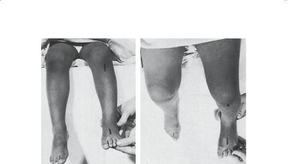
Salenius examined thousands of “normal” children and has provided us with standard expectations for this group (Fig. 5-6). Newborns demonstrate 4 to 10 degrees of genu varus, which tends to spontaneously correct by 18 months of age. Thus, a child who presents with bowlegs would be diagnosed as “physiologic genu varum.” After 18 months of age, a child develops knock-knees, which increases until about age 4 or 5 and then begins to improve. By age 7 or 8, most children have assumed more of an adult pattern: 5 to 7 degrees of valgus in males and 7 to 9 degrees of valgus in females.
FIGURE 5-4. The degree of normal femoral torsion in relation to age. The solid lines represent the mean; the vertical lines represent standard deviation. (From Tachdjian MO. Pediatric Orthopedics, 2nd ed, vol 1. Philadelphia: Saunders, 1990. Reprinted by permission.)
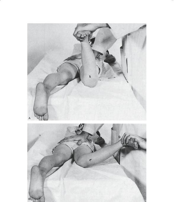
178 J.N. Delahay and W.C. Lauerman
FIGURE 5-5. Range of rotation of the hip in excessive femoral antetorsion. (A) Lateral rotation of the hip in extension is exaggerated. (B) Medial rotation of the hip in extension is limited to neutral. (From Tachdjian MO. Pediatric Orthopedics, 2nd ed, vol 1. Philadelphia: Saunders, 1990. Reprinted by permission.)