

Essentials of Orthopedic Surgery, third edition / 07-The Spine
.pdf7
The Spine
SAM W. WIESEL, WILLIAM C. LAUERMAN, and
STEVEN C. SCHERPING, JR.
The majority of adults are, at some point in their life, affected by disorders of the spine. Every physician should have a basic knowledge of the potential pathology and be able to distinguish a serious problem from a minor condition. Disastrous sequelae such as paralysis can occur if this differentiation is not appreciated. This chapter first addresses the cervical spine and then presents the lumbar spine. In each area, the history, physical, and appropriate diagnostic studies are reviewed. Next, a standardized protocol or algorithm for the diagnosis and management of these patients is described. Finally, several of the most common conservative treatment modalities is presented with special attention given to their efficacy.
Cervical Spine
Disorders of the neck are ubiquitous. Significant problems can arise from various types of arthritis as well as from trauma. In each instance, recovery or improvement is the usual outcome, but poor results can occur with paraplegia or death as the most disastrous. Every physician should be familiar with the signs and symptoms of the various diagnostic entities that occur in the cervical spine and be able to identify the serious problems that require immediate attention.
History
The location of the pain is the major point to obtain from the patient history. The majority of patients complain of localized symptoms in the neck, with and without referral of pain between the scapulae or shoulders. The pain is described as vague, diffuse, axial, nondermatomal, and poorly localized. The pathogenesis of this type of complaint is attributed to structures innervated by the sinuvertebral nerve or the nerves innervating the paravertebral soft tissues and is generally a localized injury.
276

7. The Spine |
277 |
Another group of patients complains of neck pain with the addition of arm involvement. This arm pain is secondary to nerve root irritation and is termed radicular pain. The degree of nerve root involvement can vary from a monoradiculopathy to multiple levels of involvement. It is described as a deep aching, burning, or shooting arm pain, often with associated paresthesias. The pathogenesis of radicular pain can derive from soft tissue (herniated disk), bone (spondylosis), or a combination of these two.
Finally, a third group of patients complains of symptoms secondary to cervical myelopathy, which is compression of the spinal cord and usually secondary to degenerative changes. The clinical complaints vary considerably. The onset of symptoms usually begins after 50 years of age, and males are more often affected. Onset is usually insidious, although there is occasionally a history of trauma. The natural history is that of initial neurologic deterioration followed by a plateau period lasting several months. The resulting clinical picture is often one of an incomplete spinal lesion with a patchy distribution of deficits. Disability varies with the number of vertebrae involved and with the degree of changes at each level.
Common presenting symptoms of cervical myelopathy include numbness and paresthesias in the hands, clumsiness of the fingers, weakness (greatest in the lower extremities), and gait disturbances. Abnormalities of micturition are seen in about one-third of cases and indicate more severe cord involvement. Symptoms of radiculopathy can coexist with myelopathy and confuse the clinical picture. Sensory disturbances may show a patchy distribution. Spinothalamic tract (pain and temperature) deficits may be seen in the upper extremities, the thorax, or the lumbar region and may be in a stocking or glove distribution. Posterior column deficits (vibration and proprioception) are more commonly seen in the feet than in the hands. Usually there is no gross sensory impairment, but a diminished sense of appreciation of light touch and pinprick. A characteristic broad-based, shuffling gait may be seen, signaling the onset of functionally significant deterioration.
Physical Examination
The physical examination should begin with observation of the cervical spine and upper torso unencumbered by clothing. The physical findings are of two different types. One set can be categorized as nonspecific and found in most patients with neck pain but will not help to localize the type or level of the pathologic process. A decreased range of motion is the most frequent nonspecific finding. It can be secondary to pain or, structurally, to distorted bony or soft tissue elements in the cervical spine. Hyperextension and excessive lateral rotation, however, usually cause pain, even in a normal individual.
Tenderness is another nonspecific finding that can be quite helpful. There are two types of tenderness that must be considered. One is diffuse,

278 S.W. Wiesel et al.
elicited by compression of the paravertebral muscles, and is found over a wide area of the posterolateral muscle masses. The second type of tenderness is more specific and may help localize the level of the pathology. It can be localized by palpation over each intervertebral foramen and spinous process.
The next goal of the physical examination is to isolate the level or levels in the cervical spine responsible for the symptomatology. The exam is also important to rule out other sources of pain, which include compression neuropathies, thoracic outlet syndrome, and chest or shoulder pathology.
The major focus of the exam is directed at finding a neurologic deficit (Table 7-1). A motor deficit (most commonly weak triceps, biceps, or deltoid) or diminished deep tendon reflex is the most likely objective
TABLE 7-1. Cervical radiculopathy symptoms and findings.
Disk |
Nerve |
|
level |
root |
Symptoms and findings |
C2–C3 |
C3 |
Pain: Back of neck, mastoid process, pinna of ear |
|
|
Sensory change: Back of neck, mastoid process, pinna of ear |
|
|
Motor deficit: None readily detectable except by EMG |
|
|
Reflex change: None |
C3–C4 |
C4 |
Pain: Back of neck, levator scapula, anterior chest |
|
|
Sensory change: Back of neck, levator scapula, anterior chest |
|
|
Motor deficit: None readily detectable except by EMG |
|
|
Reflex change: None |
C4–C5 |
C5 |
Pain: Neck, tip of shoulder, anterior arm |
|
|
Sensory change: Deltoid area |
|
|
Motor deficit: Deltoid, biceps |
|
|
Reflex change: Biceps |
C5–C6 |
C6 |
Pain: Neck, shoulder, medial border of scapula, lateral arm, dorsal |
|
|
forearm |
|
|
Sensory change: Thumb and index finger |
|
|
Motor deficit: Biceps |
|
|
Reflex change: Biceps |
C6–C7 |
C7 |
Pain: Neck, shoulder, medial border of scapula, lateral arm, dorsal |
|
|
forearm |
|
|
Sensory change: Index and middle fingers |
|
|
Motor deficit: Triceps |
|
|
Reflex change: Triceps |
C7–T1 |
C8 |
Pain: Neck, medial border of scapula, medial aspect of arm and |
|
|
forearm |
|
|
Sensory change: Ring and little fingers |
|
|
Motor deficit: Intrinsic muscles of hand |
|
|
Reflex cange: None |
|
|
|
Source: From Boden S, Wiesel SW, Laws E, et al. The Aging Spine. Philadelphia: Saunders, 1991:46. Reprinted by permission.

7. The Spine |
279 |
finding in a patient with a radiculopathy. Although less reproducible, manual tests and maneuvers that increase or decrease radicular symptoms may be helpful. In the neck compression test, the patient’s head is flexed laterally, slightly rotated toward the symptomatic side, and then compressed to elicit reproduction or aggravation of the radicular symptoms. The axial manual traction test is performed in the presence of radicular symptoms in the supine position. With 20 to 25 lb axial traction, a positive test is the decrease or disappearance of radicular symptoms. All these tests are highly specific (low false-positive rate) for the diagnosis of root compression, but the sensitivity (false-negative rate) is less than 50%.
Myelopathic physical findings should also be specifically checked. These patients can have a gait disturbance, so they should be observed walking. The extent of motor disability can vary from mild to severe. Pyramidal tract weakness and atrophy are more commonly seen in the lower extremities and are the most common abnormal signs. The usual clinical findings in the lower extremities are spasticity and weakness.
Weakness and wasting of the upper extremities and hands may also be due to combined spondylotic myelopathy and radiculopathy. In this situation, the patient usually complains of hand clumsiness. A diminished or absent upper-extremity deep tendon reflex can indicate compressive radiculopathy superimposed on spondylotic myelopathy.
Sensory deficits in spinothalamic (pain and temperature) and posterior column (vibration and proprioception) function should be documented. Usually there is no gross impairment of sensation; rather, a patchy decrease in light touch and pinprick is seen. Hyperreflexia, clonus, and positive Babinski’s signs are seen in the lower extremities. Hoffman’s sign and hyperreflexia may be observed in the upper extremities.
Diagnostic Studies
In evaluating any pathologic process, one usually has a choice of several diagnostic tests. The cervical spine is no exception. This section presents the most common ones that are routinely used. In general, all these tests play a confirmatory role. In other words, the core of the information derived from a thorough history and physical examination should be the basis for a diagnosis; the additional tests are obtained to confirm this clinical impression. Trouble develops when these tests are used for screening purposes because most of them are overly sensitive and relatively nonselective. Thus, the studies discussed should never be interpreted in isolation from the overall clinical picture.
Plain Radiographs
Radiographic evaluation of the cervical spine is helpful in assessing patients with neck pain, and the routine study should include anteroposterior,

280 S.W. Wiesel et al.
lateral, oblique, and odontoid views. Flexion–extension X-rays are necessary in defining stability. The generally accepted radiographic signs of cervical disk disease are loss of height of the intervertebral disk space, osteophyte formation, secondary encroachment of the intervertebral foramina, and osteoarthritic changes in the apophyseal joints.
It should be stressed that the identification of “some pathology” on plain cervical X-rays does not, per se, indicate the cause of the patient’s symptoms. In several series, large numbers of asymptomatic patients have shown radiographic evidence of advanced degenerative disk disease. At approximately age 40, some degeneration (narrowing) can be expected, particularly at the C5–C6 and C6–C7 levels, and this is considered to represent a normal aging process. The difficult problem with regard to radiographic interpretation is not in the identification of these changes but rather in determining how much significance should be attributed to them.
Radiographic abnormalities of alignment in the cervical spine may also be of clinical significance, but they need to be correlated with the whole clinical picture; listhesis or slipping forward or backward (retrolisthesis) of one vertebra upon the vertebra below it is such a finding.
If “instability” is suspected, functional X-rays may be taken. These view the spine from the side, with the head flexed (bent forward) or extended (arched back); the spine normally flexes equally at each spinal level. If one vertebral level is “unstable,” that particular vertebra moves more or less and disrupts the symmetry of motion. Again, this finding must be correlated with the whole clinical picture, as its mere presence may be asymptomatic.
Magnetic Resonance Imaging
Magnetic resonance imaging (MRI) provides an image on film that is obtained by measuring the differences in proton density between the various tissues evaluated. With the use of the computer, multiplanar images are obtainable. It is a safe test because it uses neither ionizing radiation nor invasive contrast agents.
The technical advances for MRI have rapidly changed. The distinction between soft tissues and bone and the relationship of both to the neural foramen are excellent (Fig. 7-1). MRI can also accurately detect rare conditions such as infection, tumor, or intrinsic abnormalities of the spinal cord. An excellent test, MRI can be combined with plain films to permit an accurate noninvasive evaluation of a cervical radiculopathy or myelopathy. It is currently the diagnostic study of choice in the cervical spine.
MRI should be used as a confirmatory test to substantiate a clinical impression; it should not be used as a screening test because there are many false-positive as well as false-negative results. Thus, some normal people have abnormal MRI findings whereas some abnormal people are found to have normal MRIs.
7. The Spine |
281 |
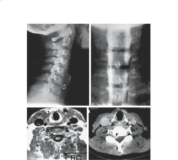
A
FIGURE 7-1. This 33-year-old man presented with right triceps weakness, C7 radicular pain, and absent triceps reflex. (A) Lateral radiograph of C6–C7 shows loss of disk height (arrow). (B) Anteroposterior myelogram confirms right C7 root sleeve cutoff. (C) Axial magnetic resonance imaging (left) and computerized tomography (right) show occlusion of the right C6–C7 foramen (arrows). (From Boden S, Wiesel SW, Laws E, et al. The Aging Spine. Philadelphia: Saunders, 1991. Reprinted by permission.)
Myelography
A myelogram is performed by injecting a water-soluble dye into the spinal sac so that the outline of the sac itself, as well as each nerve root sleeve, can be evaluated. If there is pressure upon the nerve root or dural sac from either a bony spur or disk herniation, it will be seen as a constriction on

282 S.W. Wiesel et al.
the X-ray picture. Complications from myelography are rare, and it can be performed on an outpatient basis. The major disadvantages are its invasive nature, radiation exposure, and the lack of diagnostic specificity. Watersoluble myelography does provide excellent contrast for subsequent examination by computerized tomography (CT) (see Fig. 7-1).
Computerized Tomography
Computerized tomography (CT) permits one to create cross-sectional imaging of the cervical spine at any desired level. It is currently used after the instillation of water-soluble dye, which is termed a “CTmyelogram.” The advantages of CT-myelography include excellent differentiation of bone and soft tissue (disk or ligament) lesions, direct demonstration of spinal cord and spinal cord dimensions, assessment of foraminal encroachment, and visualization of regions distal to a myelographic blockade.
Unfortunately, when combined with myelography, CT becomes an invasive procedure and involves radiation exposure. It does, however, provide very good information and is especially useful for patients who, for a variety of reasons, cannot undergo an MRI investigation.
Electromyography
Electromyography (EMG) is an electric test that confirms the interaction of nerve to muscle. The test is performed by placing needles into muscles to determine if there is an intact nerve supply to that muscle. The EMG is particularly useful in localizing a specific abnormal nerve root. It should be appreciated that it takes at least 21 days for an EMG to show up as abnormal. After 21 days of pressure on a nerve root, signs of denervation with fibrillation can be observed. Before 21 days, the EMG will be negative in spite of nerve root damage. It should be noted that there is no quantitative interpretation of this test. Thus, it cannot be said that the EMG is 25% or 75% normal.
The EMG is an electronic extension of the physical examination. Although it is 80% to 90% accurate in establishing cervical radiculopathy as the cause of pain, false-negative results do occur. If cervical radiculopathy affects only the sensory root, the EMG will be unable to demonstrate an abnormality. A false-negative examination can occur if the patient with acute symptoms is examined early (4–28 days from onset of symptoms). A negative study should be repeated in 2 to 3 weeks if symptoms persist. The accuracy of the EMG increases if both the paraspinal and extremity muscles innervated by the suspected root demonstrate abnormalities.
The EMG is not part of the routine evaluation of the cervical spine. It is indicated to confirm a clinical impression or to rule out other sources of pathology such as peripheral neuropathies or compressive neuropathies in the upper extremities.

7. The Spine |
283 |
Clinical Conditions
There are many conditions that may present as neck pain, with or without arm pain, in any particular individual. However, several that are quite common are presented here in detail.
Neck Sprain–Neckache
Neck sprain, although a misnomer, describes a clinical condition involving a nonradiating discomfort or pain about the neck area associated with a concomitant loss of neck motion (stiffness). Although the clinical syndrome may present as a headache, most often the pain is located in the middle to lower part of the back of the neck. A history of injury is rarely obtained, but the pain may start after a night’s rest or on simply turning the head. The source of the pain is most commonly believed to be the ligaments about the cervical spine and/or the surrounding muscles. The axial pain may also be produced by small annular tears without disk herniation or from the facet joints.
The pain associated with a neck sprain is most often a dull aching pain that is exacerbated by neck motion. The pain is usually abated by rest or immobilization. The pain may be referred to other mesenchymal structures derived from a similar sclerotome during embryogenesis. Common referred pain patterns include the scapular area, the posterior shoulder, the occipital area, or the anterior chest wall (cervical angina pectoris). Those referred pain patterns do not connote a true radicular pain pattern and are not usually mechanical in origin.
Physical examination of patients with neckache usually reveals nothing more than a locally tender area or areas, usually just lateral to the spine. The intensity of the pain is variable and the loss of cervical motion correlates directly with the pain intensity. The presence of true spasm, defined as a continuous muscle contraction, is rare except in severe cases where the head may be tilted to one side (torticollis).
Because the radiograph in cervical sprain is usually normal, a plain X-ray is usually not warranted on the first visit. If the pain continues for more than 2 weeks or the patient develops other physical findings, then an X-ray should be taken to rule out other more serious causes of the neck pain such as neoplasia or instability. The prognosis for these individuals is excellent because the natural history is one of complete resolution of the symptoms over several weeks. The mainstay of therapy includes rest and immobilization, usually in a soft cervical orthosis. Although medications such as antiinflammatory agents or muscle relaxants may aid in the acute management of pain, they do not seem to alter the natural history of the disorder.
Acute Herniated Disk
A herniated disk is defined as the protrusion of the nucleus pulposus through the fibers of the annulus fibrosus (Fig. 7-2). Most acute disk her-

284 S.W. Wiesel et al.
FIGURE 7-2. Types of soft disk protrusion. (A) Intraforaminal, most common. (B) Posterolateral, produces mostly motor signs. (C) Midline, may manifest as myelopathy. (Modified from DePalma AF, Rothman RH. The Intervertebral Disc. Philadelphia: Saunders, 1970. From Wiesel S, Delahay J (eds) Essentials of Orthopaedic Surgery, 2nd ed. Philadelphia: Saunders, 1997. Reprinted by permission.)

7. The Spine |
285 |
niations occur posterolaterally and in patients around the fourth decade of life when the nucleus is still gelatinous. The most common areas of disk herniation are C5–C6 and C6–C7, whereas C7–T1 and C3–C4 are infrequent. Disk herniation of C2–C3 is very, very rare. In contrast to the lumbar herniated disk, the cervical herniated disk may cause myelopathy in addition to radicular pain because of the presence of the spinal cord in the cervical region.
The disk herniation usually affects the root numbered lowest for the given disk level; for example, a C3–C4 disk affects the C4 root, C4–C5 the fifth cervical root, C5–C6 the sixth cervical root, C6–C7 the seventh nerve root, and C7–T1 the eighth cervical root. In contrast to the lumbar region, the disk herniation does not involve other roots, but more commonly presents some evidence of upper motor neuron findings secondary to spinal cord local pressure.
Not every herniated disk is symptomatic. The presence of symptoms depends on the spinal reserve capacity, the presence of inflammation, and the size of the herniation as well as the presence of concomitant disease such as osteophyte formation.
Clinically, the patient’s major complaint is arm pain, not neck pain. The pain is often perceived as starting in the neck area, but then radiates from this point down the shoulder, arm, forearm, and usually into the hand, commonly in a dermatomal distribution. The onset of the radicular pain is often gradual, although there can be a sudden onset associated with a tearing or snapping sensation. As time passes, the magnitude of the arm pain clearly exceeds that of the neck or shoulder pain. The arm pain may vary in intensity from severe enough to preclude any use of the arm without severe pain to a dull cramping ache in the arm muscles with use of the arm. The pain is usually severe enough to awaken the patient at night.
Physical examination of the neck usually shows some limitation of motion, and on occasion the patient may tilt his head in a “cocked-robin” position (torticollis) toward the side of the herniated cervical disk. Extension of the spine will often exacerbate the pain because it further narrows the intervertebral foramina. Axial compression, Valsalva maneuver, and coughing may also exacerbate or recreate the pain pattern.
The presence of a positive neurologic finding is the most helpful aspect of the diagnostic workup, although the neurologic exam may remain normal despite a chronic radicular pattern. Even when a deficit exists, it may not be temporally related to the present symptoms but rather to a prior attack at a different level. To be significant, the neurologic exam must show objective signs of reflex diminution, motor weakness, or atrophy. Subjective sensory changes are often difficult to interpret and require a coherent and cooperative patient to be of clinical value. The presence of sensory changes alone is usually not sufficient to make a firm diagnosis.
Nerve root sensitivity can be elicited by any method that increases the tension of the nerve root. Radicular arm pain is often increased by the