

Essentials of Orthopedic Surgery, third edition / 04-Tumors of the Musculoskeletal System
.pdf116 M. Malawer and K. Kellar-Graney
FIGURE 4-4. Biopsy technique. This clinical photograph illustrates a trochar needle utilized for biopsy and frozen-section diagnosis of soft tissue sarcomas. Soft tissue extension of bone tumors may also be sampled using this device. Multiple ‘cores’ of tissue may be obtained through one puncture site by varying the angle at which the trochar is inserted. Ideally, the orthopedic oncologist should be present during this biopsy to be certain that the biopsy tract is within the plane of dissection for any planned resection in an attempt to prevent contamination of surrounding tissues.
that adequate tissue is obtained to provide a diagnosis. Radiographs should be obtained to document the position of the trocar. Core biopsy is preferable if a limb-sparing option exists because it entails less local contamination than does open biopsy.
Open Incisional Biopsy
Proper techniques for open biopsies are necessary to minimize contamination. A tourniquet is used if feasible to facilitate visualization of the tumor. Transverse incisions are to be avoided at all cost, and consideration of subsequent surgery for limb salvage should guide positioning of the biopsy incision. Because sarcomas are characteristically surrounded by the most immature cells, biopsy of the lesion peripheral tissue is recommended. If a soft tissue component is present, there is no need to biopsy the underlying bone. If it is necessary to biopsy the underlying bone, a small, rounded cortical window should be used, especially for a tumor that requires primary radiotherapy. Large segments do not reossify, leading to fracture and subsequent amputation.

4. Tumors of the Musculoskeletal System |
117 |
Classification of Surgical Procedures of Bone and Soft
Tissue Tumors
Surgical removal—including curettage, resection, and amputation—is the traditional method of managing skeletal neoplasms. The advent of advanced imaging techniques, improved understanding of the biologic behavior of sarcomas, and adoption of effective adjuvant therapy have led to widespread acceptance of limb-sparing techniques. Retrospective analyses of disease-free survival and overall survival have shown no difference between limb salvage and amputation for osteosarcoma (the most common bone sarcoma) of the distal femur.
A classification scheme of surgical procedures based on the surgical plane of dissection (Fig. 4-5) in relation to the tumor and the method of accomplishing the removal has recently been developed. This system, summarized next, permits meaningful comparisons of various operative procedures and gives surgeons a common language.
1.Intralesional. An intralesional procedure passes through the pseudocapsule and directly into the lesion. Macroscopic tumor is left, and the entire operative field is potentially contaminated. Biopsies are by definition intralesional.
2.Marginal. A marginal procedure is one in which the entire lesion is removed in one piece. The plane of dissection passes through the
Distal femoral osteosarcoma: soft tissue resection
Wide excision
Marginal excision
FIGURE 4-5. Schematic diagram of the planes of surgical resection in terms of the biology of the tumor (see text). The distal femur is the most common site of most primary bone sarcomas.

118 M. Malawer and K. Kellar-Graney
pseudocapsule, or reactive zone, around the lesion. When performed for a sarcoma, it leaves macroscopic disease because of tumor involvement of the pseudocapsule.
3.Wide (intracompartmental). A wide excision is commonly termed en bloc resection. A wide excision includes the entire tumor, the reactive zone, and a marginal cuff of normal tissue. The entire structure of origin of the tumor is not removed. In patients with high-grade sarcomas, this procedure may leave skip nodules.
4.Radical (extracompartmental). The entire tumor and the structure of origin of the lesion are removed. The plane of dissection is beyond the limiting fascial or bony borders.
It is important to note that any of these procedures may be accomplished either by local (i.e., limb-sparing) surgery or by amputation. An amputation may entail a marginal, wide, or radical excision, depending upon the plane through which it passes in relationship to the tumor. Therefore, an amputation is not automatically an adequate cancer operation; careful consideration to the desired final margin is required before selection of the amputation level. The local anatomy dictates how a specific margin can be obtained surgically, and proper preoperative staging (as already discussed) is necessary to assess both local tumor extent and relevant local anatomy. In general, benign bone tumors can be adequately treated with either an intralesional procedure (curettage) or a marginal excision. Figure 4-6 demon-
Autograft
(on subchondral bone)
PMMA Reinforced with
IM rods
FIGURE 4-6. Surgical reconstruction. Schematic demonstrates reconstruction of a tumor cavity, utilizing subchondral bone graft, intramedullary hardware, and polymethyl acrylate (PMMA). This type of reconstruction is frequently utilized following curettage and cryosurgery to permit early mobilization, and it can be used in all anatomic locations. (Source: Bickels J, Meller I, Shmookler BM, Malawer MM. The role and biology of cryosurgery in the treatment of bone tumors. A review. Acta Orthop Scand. 1999 Jun;70(3):308–15, reprinted by permission of Taylor & Francis AS, http://www.tandf.no/ortho.)

4. Tumors of the Musculoskeletal System |
119 |
strates reconstruction of a tumor cavity following curettage and cryosurgery. Malignant tumors require a minimum of wide (intracompartmental) excision or radical (extracompartmental) resection, which can be accomplished by amputation or by an en bloc procedure (limb salvage). Similarly, benign soft tissue tumors are treated by marginal excision, aggressive tumors by wide excision, and malignant tumors by wide or radical resection.
Malignant Bone Tumors
Primary malignancies of bone arise from mesenchymal cells (sarcoma) and bone marrow cells (myeloma and lymphoma). Bone is also a common site of metastasis from a variety of carcinomas. Osteosarcoma and Ewing’s sarcoma, the most common malignant mesenchymal bone tumors, usually occur during childhood and adolescence. Other mesenchymal tumors [malignant fibrous histiocytoma (MFH), fibrosarcoma, chondrosarcoma], while occasionally seen in childhood, are more common in adults. Multiple myeloma and metastatic carcinoma typically increase in frequency with increasing patient age and are usually seen in patients over 40 years of age. This section describes the clinical, radiographic, and pathologic characteristics and treatment of the primary bone sarcomas.
Osteosarcoma provides the model on which treatment of all other sarcomas is based. The effectiveness of multiagent chemotherapy regimens has been proved by increasing overall survival rates from the bleak picture of 15% to 20% with surgery alone in the 1970s to 55% to 80% by the 1980s. In parallel with improved survival, dramatic advances in reconstructive surgery have made it possible for limb salvage to supplant amputation as the standard method of treatment.7
Classic Osteosarcoma
Osteosarcoma (OS) is a high-grade malignant spindle cell tumor arising within a bone. Its distinguishing characteristic is the production of “tumor” osteoid, or immature bone, directly from a malignant spindle cell stroma.
Clinical Characteristics and Physical Examination
OS typically occurs during childhood and adolescence. In patients over the age of 40, it is usually associated with a preexistent disease such as Paget’s disease, irradiated bones, multiple hereditary exostosis, or polyostotic
fibrous dysplasia. The most common sites are bones of the knee joint (50%) and the proximal humerus (25%). Between 80% and 90% of OS occur in the long tubular bones; the axial skeleton is rarely affected.
With the exception of the level of serum alkaline phosphatase, which is elevated in 45% to 50% of patients, laboratory findings are usually not helpful. Furthermore, an elevated alkaline phosphatase level per se is not

120 M. Malawer and K. Kellar-Graney
diagnostic because it is also found in association with other skeletal diseases such as hyperparathyroidism (brown tumor), fibrous dysplasia, and Paget’s disease. Pain is the most common complaint on presentation, with a firm, soft tissue mass fixed to the underlying bone found on physical examination. Systemic symptoms are rare. Incidence of pathologic fracture is less than 1%.
Radiographic Characteristics
Typical radiographic findings in OS include increased intramedullary sclerosis (caused by tumor bone or calcified cartilage), an area of radiolucency (caused by nonossified tumor), a pattern of permeative destruction with poorly defined borders, cortical destruction, periosteal elevation, and extraosseous extension with soft tissue ossification. This combination of characteristics is not seen with any other lesion. There are three broad categories: sclerotic (Fig. 4-7A) OS (32%), osteolytic OS (22%), and mixed (Fig. 4-7B) (46%). Although there is no statistically significant difference among overall survival rates of these types, it is important to recognize the patterns. The sclerotic and mixed types offer few diagnostic problems. Errors of diagnosis most often occur with pure osteolytic tumors. The differential diagnosis of osteolytic OS includes giant cell tumor, aneurysmal bone cyst, fibrosarcoma, and MFH.
Microscopic Characteristic
The diagnosis of OS is based on the identification of a malignant stroma that produces unequivocal osteoid matrix. The stroma consists of a haphazard arrangement of highly atypical cells. The pleomorphic cells contain hyperchromatic, irregular nuclei. Mitotic figures, often atypical, are usually easy to identify. Between these cells is a delicate, lacelike eosinophilic matrix, assumed to be malignant osteoid. The term osteoblastic osteosarcoma is used for those tumors in which the production of malignant osteoid prevails. Calcification of the matrix is variable. Some tumors reveal a predominance of malignant cartilage production; hence, the term chondroblastic osteosarcoma. Even though the malignant cartilaginous elements may be overwhelming, the presence of a malignant osteoid matrix warrants the diagnosis of OS. Yet another variant is characterized by large areas of proliferating fibroblasts, arranged in intersecting fascicles. Such areas are indistinguishable from fibrosarcoma, and thorough sampling may be necessary to identify the malignant osteoid component.
Natural History, Prognosis, and Chemotherapy
Before the development of adjuvant chemotherapy, effective treatment was limited to radical margin amputation. Metastasis to the lungs and other

4. Tumors of the Musculoskeletal System |
121 |
bones generally occurred within 24 months. Overall survival rates 2 years after surgery ranged from 5% to 20%. No significant correlation between overall survival and histologic subtypes, tumor size, patient age, or degree of malignancy was seen. The most significant clinical variable was anatomic site: pelvic and axial lesions had a lower survival rate than extremity tumors, whereas tibial lesions had a better survival rate than femoral lesions.
The dismal outcome associated with osteosarcoma has been dramatically altered by adjuvant chemotherapy as well as by aggressive thoracotomy for pulmonary disease. A recent update of 227 patients showed that 48% remained alive at an average 11 years after surgery. Of critical importance was that no difference in local recurrence or overall survival was seen between patients undergoing amputation versus limb-sparing surgery.
Chemotherapy protocols have typically included various combinations and dosage schedules of high-dose methotrexate (HDMTX), doxorubicin hydrochloride (adriamycin), and cisplatin. Ifosfamide, which is as effective as adriamycin in single-agent studies, recently has supplanted methotrexate in many ongoing protocols. Multiagent chemotherapy, using various dosing schedules, is now considered standard treatment for osteosarcoma. Success with adjuvant chemotherapy led to investigation of treatment in the neoadjuvant (preoperative) setting. When used in that setting, tumor response results in shrinkage of the soft tissue components, facilitating surgical excision and subsequent limb salvage (Fig. 4-8A,B).
Limb-Sparing Resection
Limb salvage surgery is a safe operation for approximately 85% to 90% of individuals. This technique may be used for all spindle cell sarcomas, regardless of histogenesis. The majority of OS can be treated safely by a limb-sparing resection combined with effective adjuvant treatments.7 The successful management of localized OS and other sarcomas requires careful coordination and timing of staging studies, biopsy, surgery, and preoperative and postoperative chemotherapy and/or radiation therapy. The site of the lesion is evaluated as previously described. Preoperative studies allow the surgeon to conceptualize the local anatomy and the volume of tissue to be resected and reconstructed.
Successful limb-sparing surgery consists of three phases:
1.Resection of tumor. Resection strictly follows the principles of oncologic surgery. Avoiding local recurrence is the criterion of success and the main determinant of the amount of bone and soft tissue to be removed.
2.Skeletal reconstruction. The average skeletal defect following adequate bone tumor resection measures 15 to 20 cm. Techniques of

122 M. Malawer and K. Kellar-Graney
FIGURE 4-7. Osteosarcoma. (A) Anteroposterior (AP) radiograph of the right distal femur shows a densely sclerotic (new bone formation) lesion of the lateral condyle. This is a typical radiograph of a bone producing sarcoma (osteosarcoma). (B) AP radiograph of the knee shows an aggressive mixed blastic (new bone formation) and lytic (bonedestroying) lesion of the proximal tibia with soft tissue extension. Osteoid matrix formation is visible in the proximal portion of this tumor. The proximal tibia is the second most common site for osteosarcoma. It is the most difficult region to reconstruct because of the lack of soft tissues to be utilized for endoprosthetic coverage.
A
B

4. Tumors of the Musculoskeletal System |
123 |
C
D
FIGURE 4-7. (Continuted)

124 M. Malawer and K. Kellar-Graney
A 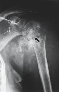
 B
B
FIGURE 4-8. Osteosarcoma. (A) AP radiograph of the left proximal humerus shows a large expansile, lytic osteosarcoma with marked bony destruction. The arrow indicates a pathologic fracture, and there is clear evidence of a soft tissue component in the medial tissues; 95% of bone sarcomas have a soft tissue component. (B) AP radiograph shows a large osteosarcoma of the proximal humerus following induction chemotherapy. Healing of pathologic fractures and ossification of the tumor following use of chemotherapy are good prognostic signs. There is substantial ossification of this lesion.
reconstruction [prosthetic replacement (Fig. 4-9), arthrodesis, allograft, or combination] vary and are independent of the resection, although the degree of resection may favor one technique over the other.
3. Soft tissue and muscle transfers. Muscle transfers are performed to cover and close the resection site and to restore lost motor power. Adequate skin and muscle coverage is mandatory to decrease postoperative morbidity.
Guidelines for Surgical Resection
The surgical guidelines and technique of limb-sparing surgery used by the authors and by surgeons at most cancer centers in the United States are summarized as follows:

4. Tumors of the Musculoskeletal System |
125 |
A
B
FIGURE 4-9. Segmental prostheses. (A) AP postoperative radiograph of a patient who underwent limb-sparing resection for osteosarcoma of the proximal humerus. A segmental prosthesis was inserted for reconstruction of the bony defect. Reconstruction with a segmental system provides better mobilization and improved function when compared with patients who have no reconstruction and are left with a flail extremity. The shoulder is the third most common site for bone sarcomas. (B) Segmental prosthesis of the distal femur. Lateral radiograph shows flexion of the knee following distal femoral resection and reconstruction with a modular segmental prosthesis. The distal femur is the most common site for bone sarcomas (osteosarcoma) to arise. The technique of reconstruction with a prosthesis is referred to as limb-sparing surgery, in lieu of an amputation.