

PS-2020a / part17
.pdfDICOM PS3.17 2020a - Explanatory Information |
Page 691 |
XA 2D Projection SOP Instance “C”
Non-Contrast Frames
1... |
|
|
...N |
|||
|
Acquisition Context #1 |
|||||
|
Reconstruction Z1 |
|
Reconstruction Z2 |
|||
|
Full field of view |
|
Object of Interest |
|||
|
|
|
|
|
|
|
|
X-Ray 3D SOP Instance “Z1” |
|
X-Ray 3D SOP Instance “Z2” |
|||
|
|
|
|
|
|
|
|
Full field of view |
|
|
|
Object of interest |
|
1... ...M 1... ...P
Reconstruction Z1 |
Reconstruction Z2 |
- From Acquisition Context #1 |
- From Acquisition Context #1 |
Figure TTT.2.3-1. Encoding of two 3D reconstructions of different regions of the anatomy
TTT.2.3.3 Encoding Details
TTT.2.3.3.1 X-Ray 3D Angiographic Image IOD
TTT.2.3.3.1.1 Frame of Reference Module Recommendations
Sincethetwovolumesarereconstructedfromthesameprojections,thereconstructionapplicationwillusethesamepatientcoordinate system on both volumes so that the spatial location of the object of interest in both volumes will be the same. Therefore the two X- Ray 3D Instances will have the same Frame of Reference (FoR) UID. If the originating 2D Instances do not deliver a value of FoR UID, a new FoR UID has to be created for the reconstructed volumes.
Table TTT.2.3-1. Frame of Reference Module Recommendations
Attribute Name |
Tag |
Recommendation |
Frame of Reference UID |
(0020,0052) |
Use the same value for the full field of view volume and the |
|
|
sub-region volume. |
TTT.2.3.3.1.2 Pixel Measures Macro Recommendations
The detailed size of the volume element (Pixel Spacing for x/y dimension and Slice Thickness for z dimension) may be different between the full field of view reconstruction and the sub-region reconstruction.
Table TTT.2.3-2. Pixel Measures Macro Recommendations
Attribute Name |
Tag |
Recommendation |
Pixel Measures Sequence |
(0028,9110) |
Thepixelsizesand/orslicethicknessarenotnecessarilyequalinthetwo |
|
|
reconstructed volumes. Within each individual volume this sequence is |
|
|
encoded as "shared". |
TTT.2.3.3.1.3 Plane Position (Patient) Macro Recommendations
The plane position of the first slice in the first volume may have a different value than in the second volume, as the sub-region volume can be smaller and shifted with respect to the full field of view volume.
- Standard -

Page 692 |
DICOM PS3.17 2020a - Explanatory Information |
TTT.2.3.3.1.4 Plane Orientation (Patient) Macro Recommendations
The plane orientation could be different in the second volume depending on the application needs, e.g., to align the slices with the object of interest.
TTT.2.3.3.1.5 Frame Content Macro Recommendations
This module encodes the timing information of the frames, as well as dimension and stack index values.
Table TTT.2.3-3. Frame Content Macro Recommendations
Attribute Name |
Tag |
Recommendation |
Frame Content Sequence |
(0020,9111) |
Provides details for each frame. |
>Frame Acquisition Duration |
(0018,9220) |
Use the duration of the rotational acquisition in the two |
|
|
reconstructed volumes. |
TTT.2.3.3.1.6 Frame Anatomy Macro Recommendations
The volume directly reconstructed from a sub-region of each of the original projection X-Ray frames does not necessarily reflect the same anatomy or laterality as the full field of view volume. Therefore the Frame Anatomy macro may point to a different anatomic context than the one documented for the originating frames.
TTT.2.3.4 Example
In this example, the slices of the two volumes are reconstructed in the axial plane of the patient; the row direction is aligned in the positive x-direction of the patient (right-left) and the column direction is aligned in the positive y-direction of the patient (anterior-pos- terior).
The full field of view reconstruction in encoded with the Instance UID "Z1" and consists of a 512 cube volume of 0.2 mm of voxel size. The sub-region reconstruction in encoded with the Instance UID "Z2" and consists of a 256 cube volume of the voxel size of 0.1 mm.
Both volumes share the same Frame of Reference UID.
SOP Instance UID |
(0008,0018) |
|
= UID “Z1” |
||||
|
|
|
|||||
Rows |
(0028,0010) |
= 512 |
|||||
|
|
|
|
||||
Columns |
(0028,0011) |
= 512 |
|||||
|
|
|
|
|
|
|
|
... |
|
|
|
|
|
|
|
|
|
|
|
||||
Frame of Reference UID |
(0020,0052) |
|
= UID “D” |
||||
|
|
|
|
|
|
|
|
... |
|
|
|
|
|
|
|
|
|
|
|
|
|
|
|
Shared Functional Group Sequence |
(5200,9229) |
|
|
||||
|
|
|
|
|
|
|
|
|
Item 1 |
|
|
Common to all frames of the X-Ray 3D |
|||
|
|
... |
|
|
|
|
|
|
|
|
|
|
|
|
|
|
|
>Pixel Measures Sequence |
(0028,9110) |
|
|
||
|
|
|
|
|
|
|
|
|
|
|
Item 1 |
|
|
|
|
|
|
|
|
>>Slice Thickness |
(0018,0050) |
= 0.2 |
|
|
|
|
|
>>Pixel Spacing |
(0028,0030) |
= 0.2 |
|
|
|
>Plane Orientation Sequence |
(0028,9116) |
|
|
||
|
|
|
Item 1 |
|
|
|
|
|
|
|
|
>>Image Orientation (Patient) |
(0020,0037) |
= 1\0\0\0\1\0 |
|
|
|
>Frame Anatomy Sequence |
(0020,9071) |
|
|
||
|
|
|
|
|
|
|
|
|
|
|
Item 1 |
|
|
|
|
|
|
|
|
>>Frame Laterality |
(0020,9072) |
|
provide value |
|
|
|
|
>>Anatomic Region Sequence |
(0008,2218) |
|
provide anatomic region |
|
|
... |
|
|
|
|
|
|
|
|
|
|
|
|
|
... |
|
|
|
|
|
|
|
|
|
|
|
||||
Per-Frame Functional Groups Sequence |
(5200,9230) |
|
“M” frames |
||||
|
|
|
|
|
|
||
|
|
... |
|
|
|
|
|
|
|
|
|
|
Frame “i” of the X-Ray 3D |
||
|
Item i |
|
|
||||
|
|
>Plane Position Sequence |
(0020,9113) |
|
|
||
|
|
|
|
|
|
|
|
|
|
|
Item 1 |
|
|
|
|
|
|
|
|
>>Image Position (Patient) |
(0020,0032) |
|
|
|
|
... |
|
|
|
|
|
|
|
|
|
|
|
|
|
Figure TTT.2.3-2. Attributes of 3D Reconstruction of the full field of view of the projection frames
- Standard -

DICOM PS3.17 2020a - Explanatory Information |
Page 693 |
SOP Instance UID |
(0008,0018) |
|
= UID “Z2” |
||||
|
|
|
|||||
Rows |
(0028,0010) |
= 256 |
|||||
|
|
|
|
||||
Columns |
(0028,0011) |
= 256 |
|||||
|
|
|
|
|
|
|
|
... |
|
|
|
|
|
|
|
|
|
|
|
||||
Frame of Reference UID |
(0020,0052) |
|
= UID “D” |
||||
|
|
|
|
|
|
|
|
... |
|
|
|
|
|
|
|
|
|
|
|
|
|
|
|
Shared Functional Group Sequence |
(5200,9229) |
|
|
||||
|
|
|
|
|
|
|
|
|
Item 1 |
|
|
Common to all frames of the X-Ray 3D |
|||
|
|
... |
|
|
|
|
|
|
|
|
|
|
|
|
|
|
|
>Pixel Measures Sequence |
(0028,9110) |
|
|
||
|
|
|
|
|
|
|
|
|
|
|
Item 1 |
|
|
|
|
|
|
|
|
>>Slice Thickness |
(0018,0050) |
= 0.1 |
|
|
|
|
|
>>Pixel Spacing |
(0028,0030) |
= 0.1 |
|
|
|
>Plane Orientation Sequence |
(0028,9116) |
|
|
||
|
|
|
Item 1 |
|
|
|
|
|
|
|
|
>>Image Orientation (Patient) |
(0020,0037) |
= 1\0\0\0\1\0 |
|
|
|
>Frame Anatomy Sequence |
(0020,9071) |
|
|
||
|
|
|
|
|
|
|
|
|
|
|
Item 1 |
|
|
|
|
|
|
|
|
>>Frame Laterality |
(0020,9072) |
|
May be different value vs. full reconstruction |
|
|
|
|
>>Anatomic Region Sequence |
(0008,2218) |
|
May be different value vs. full reconstruction |
|
|
... |
|
|
|
|
|
|
|
|
|
|
|
|
|
... |
|
|
|
|
|
|
|
|
|
|
|
||||
Per-Frame Functional Groups Sequence |
(5200,9230) |
|
“P” frames |
||||
|
|
|
|
|
|
||
|
|
... |
|
|
|
|
|
|
|
|
|
|
Frame “i” of the X-Ray 3D |
||
|
Item i |
|
|
||||
|
|
>Plane Position Sequence |
(0020,9113) |
|
|
||
|
|
|
|
|
|
|
|
|
|
|
Item 1 |
|
|
|
|
|
|
|
|
>>Image Position (Patient) |
(0020,0032) |
|
May be different value vs. full reconstruction |
|
|
... |
|
|
|
|
|
|
|
|
|
|
|
|
|
Figure TTT.2.3-3. Attributes of 3D Reconstruction using a sub-region of all frames
TTT.2.4 Case #4: Multiple Rotations, One Or More 2D Instances, One Reconstruction, One X- Ray 3D Instance
This application case is related to a high resolution reconstruction from several rotations around the same anatomy.
TTT.2.4.1 User Scenario
Theimageacquisitionsystemperformsmultiple2Drotationalacquisitionsaroundthepatientwithmovementsinthesameoropposite directions in the patient's transverse plane. A single volume is reconstructed from the acquired data (e.g., through "back-projection" algorithm). The reconstruction can either occur on the same system (e.g., Acquisition Modality) or a secondary processing system (e.g., Co-Workstation).
The reconstructed Volume needs to be encoded and saved for further use.
TTT.2.4.2 Encoding Outline
The rotational acquisitions can be encoded either as a single instance (e.g., "C") containing several rotations or as several instances (e.g., "C1", "C2", etc.) containing one rotation per instance. The rotational acquisitions can either be encoded as XA Image(s) with limited frame-specific Attributes or as Enhanced XA Image(s), with frame-specific Attributes encoded that inform the algorithms to reconstruct a volume.
The reconstructed volume data is encoded as a single X-Ray 3D Angiographic instance. The reconstructed region covers typically the full field of view of the projected matrix size.
All frames of the original XA Images or Enhanced XA Images are used to reconstruct the volume.
TheX-Ray3DinstancereferencestheoriginalacquisitioninstancesandrecordsAttributesoftheprojectionsdescribingtheacquisition context.
- Standard -

Page 694 |
DICOM PS3.17 2020a - Explanatory Information |
XA 2D Projection SOP Instance “C”
|
|
|
|
Rotation #1 |
|
|
|
|
|
|
|
Rotation #2 |
|
|
|
|
|
|
|
Rotation #3 |
|||||||||||||||
|
|
|
|
|
|
|
|
|
|
|
|
|
|
|
|
|
|
|
|
|
|
|
|
|
|
|
|
|
|
|
|
|
|
|
|
|
|
|
|
|
|
|
|
|
|
|
|
|
|
|
|
|
|
|
|
|
|
|
|
|
|
|
|
|
|
|
|
|
|
|
|
N1... |
|
...N2 N3... |
|
...N4 N5... |
|
|
|
|
|
|
...N6 |
||||||||||||||||||||||||
Acquisition Context #1 Acquisition Context #2 Acquisition Context #3
Reconstruction #1
X-Ray 3D SOP Instance “Z”
1... |
...M |
Reconstruction #1
- From Acquisition Context #1, #2, #3
Figure TTT.2.4-1. Encoding of one 3D reconstruction from three rotational acquisitions in one instance
Study “A”
Series “B1”
XA 2D Projection
SOP Instance “C1”
Rotation #1
Series “B2”
XA 2D Projection
SOP Instance “C2”
Rotation #2
1... |
...N1 |
1... |
...N2 |
Acquisition Context #1 |
Acquisition Context #2 |
||
Reconstruction #1
X-Ray 3D SOP Instance “Z”
1... |
...M |
Reconstruction #1
- From Acquisition Context #1, #2
Figure TTT.2.4-2. Encoding of one 3D reconstruction from two rotational acquisitions in two instances
TTT.2.4.3 Encoding Details
TTT.2.4.3.1 2D X-Ray Angiographic Image IOD
This scenario is based on the encoding of the different rotations in one or more 2D instance(s), which can be encoded either as X- Ray Angiography or Enhanced XA Images.
- Standard -

DICOM PS3.17 2020a - Explanatory Information |
Page 695 |
TTT.2.4.3.1.1 Frame of Reference Module Recommendations
In the case of multiple source 2D Instances, the acquisition equipment assumes that the patient has not moved between the different rotations. This module encodes the same FoR UID in all the rotations, identifying a common spatial relationship between them, thus allowing the 3D reconstruction to use the projections of all the rotations to perform a single volume reconstruction.
If the source 2D Instances do not provide a value of FoR UID, it has to be created for the reconstructed volume.
Table TTT.2.4-1. Frame of Reference Module Recommendations
Attribute Name |
Tag |
Recommendation |
Frame of Reference UID |
(0020,0052) |
All XA Images or Enhanced XA Images created from the rotational |
|
|
acquisitions share the same spatial relationship. |
Position Reference Indicator |
(0020,1040) |
No recommendation to set a value, unless a system is capable to derive |
|
|
such information from the anatomy or has a mandatory user interface to |
|
|
enter such information. |
Note
The case where all the source 2D Instances have the same FoR UID is the "lucky" case. If no FoR UID value is provided in the 2D Instances, or if the FoR UIDs are different, there should be an additional 2D registration step before performing the 3D reconstruction.
TTT.2.4.3.2 X-Ray 3D Angiographic Image IOD
TTT.2.4.3.2.1 X-Ray 3D Angiographic Image Contributing Sources Module Recommendations
This module encodes the source SOP instance(s) used to create the X-Ray 3D Angiographic instance.
Table TTT.2.4-2. X-Ray 3D Angiographic Image Contributing Sources Module Recommendations
Attribute Name |
Tag |
Recommendation |
Contributing Sources Sequence |
(0018,9506) |
One item for each of the originating instances that was used for the |
|
|
reconstruction of the X-Ray 3D Angiographic image. |
TTT.2.4.3.2.2 X-Ray 3D Angiographic Acquisition Module Recommendations
There are multiple acquisition contexts, one per rotation of the equipment. This module encodes the frame numbers of the source SOP instance that belong to each acquisition context, as well as the important technical and physical parameters of the source SOP instances used to create the X-Ray 3D Angiographic instance.
Table TTT.2.4-3. X-Ray 3D Angiographic Acquisition Module Recommendations
Attribute Name |
Tag |
Recommendation |
X-Ray 3D Acquisition Sequence |
(0018,9507) |
One item for each acquisition context (i.e., each rotation) that |
|
|
contributed to the reconstruction of the X-Ray 3D Angiographic |
|
|
image pixel data contents. |
… |
|
|
>Source Image Sequence |
(0008,2112) |
One item for each acquisition context. |
>>Referenced SOP Class UID |
(0008,1150) |
|
>>Referenced SOP Instance UID |
(0008,1155) |
The source SOP instance where this rotation belongs. |
>>Referenced Frame Number |
(0008,1160) |
The frame numbers of the projections corresponding to this |
|
|
rotation. |
…
- Standard -

Page 696 |
DICOM PS3.17 2020a - Explanatory Information |
||
Attribute Name |
|
Tag |
Recommendation |
>Per Projection Acquisition Sequence |
(0018,9538) |
The content of this sequence needs to be carefully aligned with |
|
|
|
|
the list of frame numbers in the Referenced Frame Numbers |
|
|
|
(0008,1160) Attribute in the Source Image Sequence |
|
|
|
(0008,2112). |
TTT.2.4.3.2.3 Frame Content Macro Recommendations
This module encodes the timing information of the frames, as well as dimension and stack index values.
Table TTT.2.4-4. Frame Content Macro Recommendations
Attribute Name |
Tag |
Recommendation |
Frame Content Sequence |
(0020,9111) |
Provides details for each frame. |
>Frame Acquisition Duration |
(0018,9220) |
Use the elapsed time from the first projection frame time of the |
|
|
first rotation to the last projection frame time of the last rotation |
|
|
used for this reconstruction. |
TTT.2.4.4 Example
Thisexampleisthereconstructionofavolumebyaback-projectionfromallframesofarotationalacquisitionwithtworotationsencoded as two XA Images.
- Standard -

DICOM PS3.17 2020a - Explanatory Information |
Page 697 |
SOP Instance UID |
(0008,0018) |
||||||||
|
|
|
|
|
|
|
|
|
|
... |
|
|
|
|
|
|
|
|
|
|
|
|
|
|
|
|
|
|
|
Contributing Sources Sequence |
(0018,9506) |
||||||||
|
|
|
|
|
|
|
|
|
|
|
Item 1 |
|
|||||||
|
|
>Contributing SOP Instances Reference Sequence |
(0020,9529) |
||||||
|
|
|
|
|
|
|
|
|
|
|
|
|
Item 1 |
|
|||||
|
|
|
|
>>Reference Series Sequence |
(0008,1115) |
||||
|
|
|
|
|
Item 1 |
|
|||
|
|
|
|
|
|
|
|
|
|
|
|
|
|
|
|
>>>Referenced Instance Sequence |
(0008,114a) |
||
|
|
|
|
|
|
|
Item 1 |
|
|
|
|
|
|
|
|
|
|
>>>>Referenced SOP Class UID |
(0008,1150) |
|
|
|
|
|
|
|
|
|
|
|
|
|
|
|
|
|
|
>>>>Referenced SOP Instance UID |
(0008,1155) |
|
|
|
|
|
|
|
|
|
|
|
|
|
|
|
|
>>>Series Instance UID |
(0020,000e) |
||
|
|
|
|
|
|
|
|
|
|
|
|
|
|
>>Study Instance UID |
(0020,000d) |
||||
|
|
|
|
|
|
|
|
|
|
|
|
>Acquisition Datetime |
(0008,002A) |
||||||
|
|
|
|
||||||
|
|
>Acquisition Protocol Name |
(0018,9423) |
||||||
|
|
|
|
||||||
|
|
>... (other attributes from the contributing image sources) |
|
||||||
|
|
|
|
|
|
|
|
|
|
|
Item 2 |
|
|||||||
|
|
>Contributing SOP Instances Reference Sequence |
(0020,9529) |
||||||
|
|
|
|
|
|||||
|
|
|
Item 1 |
|
|||||
|
|
|
|
>>Reference Series Sequence |
(0008,1115) |
||||
|
|
|
|
|
Item 1 |
|
|||
|
|
|
|
|
|
>>>Referenced Instance Sequence |
(0008,114a) |
||
|
|
|
|
|
|
|
Item 1 |
|
|
|
|
|
|
|
|
|
|
>>>>Referenced SOP Class UID |
(0008,1150) |
|
|
|
|
|
|
|
|
|
|
|
|
|
|
|
|
|
|
>>>>Referenced SOP Instance UID |
(0008,1155) |
|
|
|
|
|
|
|
|
|
|
|
|
|
|
|
|
>>>Series Instance UID |
(0020,000e) |
||
|
|
|
|
|
|
|
|
||
|
|
|
|
>>Study Instance UID |
(0020,000d) |
||||
|
|
|
|
|
|
||||
|
|
>Acquisition Datetime |
(0008,002A) |
||||||
|
|
|
|
||||||
|
|
>Acquisition Protocol Name |
(0018,9423) |
||||||
|
|
|
|
||||||
|
|
>... (other attributes from the contributing image sources) |
|
||||||
|
|
|
|
|
|
|
|
|
|
... |
|
|
|
|
|
|
|
|
|
|
|
||||||||
X-Ray 3D Acquisition Sequence |
(0018,9507) |
||||||||
|
|
|
|||||||
|
Item 1 |
|
|||||||
|
|
>Source Image Sequence |
(0008,2112) |
||||||
|
|
|
Item 1 |
|
|||||
|
|
|
|
>>Referenced SOP Class UID |
(0008,1150) |
||||
|
|
|
|
>>Referenced SOP Instance UID |
(0008,1155) |
||||
|
|
>... (other attributes of this source: kVp, mA, FOV, filter...) |
|
||||||
|
|
|
|
||||||
|
Item 2 |
|
|||||||
|
|
>Source Image Sequence |
(0008,2112) |
||||||
|
|
|
Item 1 |
|
|||||
|
|
|
|
>>Referenced SOP Class UID |
(0008,1150) |
||||
|
|
|
|
>>Referenced SOP Instance UID |
(0008,1155) |
||||
|
|
>... (other attributes of this source: kVp, mA, FOV, filter...) |
|
||||||
|
|
|
|
|
|
|
|
|
|
... |
|
|
|
|
|
|
|
|
|
|
|
||||||||
X-Ray 3D Reconstruction Sequence |
(0018,9530) |
||||||||
|
|
|
|||||||
|
Item 1 |
|
|||||||
|
|
>Acquisition Index |
(0020,9518) |
||||||
|
|
|
|
||||||
|
|
>... (other attributes related to this reconstruction) |
|
||||||
... |
|
|
|
|
|
|
|
|
|
|
|
||||||||
Shared Functional Group Sequence |
(5200,9229) |
||||||||
|
|
|
|||||||
|
Item 1 |
|
|||||||
|
|
... |
|
|
|
|
|
|
|
|
|
|
|
||||||
|
|
>X-Ray 3D Frame Type Sequence |
(0018,9504) |
||||||
|
|
|
|
|
|||||
|
|
|
Item 1 |
|
|||||
|
|
|
|
>>Reconstruction Index |
(0020,9536) |
||||
|
|
|
|
>>... |
|
|
|
|
|
|
|
... |
|
|
|
|
|
|
|
|
|
|
|
|
|
|
|
|
|
= UID “Z”
Two contributing sources in this example
=XA Image or Enhanced XA Image
=UID “C1”
=UID “B1”
=UID “A”
=Date and time of the Instance “C1”
=Protocol 1
=XA Image or Enhanced XA Image
=UID “C2”
=UID “B2”
=UID “A”
=Date and time of the Instance “C2”
=Protocol 2
Acquisition context #1
one source image for this acquisition context
=XA Image or Enhanced XA Image
=UID “C1”
Acquisition context #2
one source image for this acquisition context
=XA Image or Enhanced XA Image
=UID “C2”
Reconstruction #1 =1\2
Common to all frames of the X-Ray 3D
= 1 (all frames share the same reconstruction data)
Figure TTT.2.4-3. Attributes of 3D Reconstruction using multiple rotation images
TTT.2.5 Case #5: One Rotation, One 2D Instance, Multiple Reconstructions, One X-Ray 3D Instance
This application case is related to a rotational acquisition of several cardiac cycles with related ECG signal information.
- Standard -
Page 698 |
DICOM PS3.17 2020a - Explanatory Information |
TTT.2.5.1 User Scenario
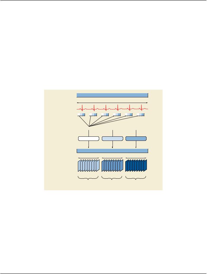
The image acquisition system performs one 2D rotational acquisition of the heart in a cardiac procedure. The gantry is continuously rotating at a constant speed. The ECG is recorded during the rotation, and the cardiac trigger delay time is known for each frame of the rotational acquisition allowing it to be assigned to a given cardiac phase.
Several 3D volumes are reconstructed, one for each cardiac phase.
TTT.2.5.2 Encoding Outline
The rotational acquisition can either be encoded as XA Image or as Enhanced XA Image. The XA instance (let's call it "C") is encoded
in the Series "B" of the Study "A".
Eachreconstructionisrelatedtoonecardiacphasecorrespondingtoasub-setofframesoftherotationalacquisition.Therefore,each cardiac phase represents one acquisition context.
Each reconstruction leads to one volume, all volumes are encoded in one single X-Ray 3D Angiographic instance ("Z"). Each volume is for a different cardiac phase. All volumes share the same stack id.
|
|
XA 2D Projection SOP Instance “C” |
|
|||
|
|
|
ECG-triggered acquisition |
|
|
|
|
... |
... |
... |
... |
... |
... |
|
1... |
|
|
|
|
...N |
|
Acquisition Context #1 |
Acquisition Context #2 |
Acquisition Context #3 |
|||
|
Reconstruction #1 |
Reconstruction #2 |
Reconstruction #3 |
|||
|
|
|
X-Ray 3D SOP Instance “Z” |
|
|
|
|
Cardiac phase A |
Cardiac phase B |
Cardiac phase C |
|||
Frame Number: |
1... |
...M |
M+1... |
...2xM |
2xM+1... |
...3xM |
In-Stack Position Number: |
1... |
...M |
1... |
...M |
1... |
...M |
|
Reconstruction #1 |
|
Reconstruction #2 |
|
Reconstruction #3 |
|
|
- From Acquisition Context #1 |
- From Acquisition Context #2 |
- From Acquisition Context #3 |
|||
|
- Stack ID #1 |
|
- Stack ID #1 |
|
- Stack ID #1 |
|
Figure TTT.2.5-1. Encoding of various 3D reconstructions at different cardiac phases
Note
1.Thisfigureshowsonlythefirstthreecardiacphases.Animplementationmaychosehowmanyphasesitwillreconstruct.
2.Projection frames are assigned to a phase based on their cardiac trigger delay time. The rotation speed and acquisition pulseratewillnotnecessarilyalignuniformlywiththecardiaccycle(especiallyiftheheartbeatisirregular).Thusdifferent phases may end up with different number of projections assigned to them. The reconstructed volumes will have the same space.
- Standard -

DICOM PS3.17 2020a - Explanatory Information |
Page 699 |
TTT.2.5.3 Encoding Details
TTT.2.5.3.1 2D X-Ray Angiographic Image IOD
This scenario is based on the encoding of a single rotational acquisition in one 2D instance, together with the information of the ECG and/or the cardiac trigger delay times of each frame of the rotational image.
TTT.2.5.3.2 X-Ray 3D Angiographic Image IOD
TTT.2.5.3.2.1 Image Pixel Module Recommendations
Thismoduleencodesthedescriptionofthepixelsoftheslicesofthevolumes,eachslicebeingoneframeoftheX-Ray3DAngiographic instance. The pixel data encodes all the frames of the first cardiac phase followed by all the frames of the second cardiac phase and so on. Within one cardiac phase, the order of the frames is aligned with the Image Position (Patient) Attribute.
TTT.2.5.3.2.2 Multi-frame Dimension Module Recommendations
This module encodes the dimensions for the presentation order of the image frames.
Table TTT.2.5-1. Multi-frame Dimension Module Recommendations
Attribute Name |
Tag |
Recommendation |
Dimension Organization Sequence |
(0020,9221) |
There will be a single Dimension UID. |
Dimension Organization Type |
(0020,9311) |
The value will be "3D". |
Dimension Index Sequence |
(0020,9222) |
Two items are defined: the first one related to the cardiac phase, the |
|
|
second one related to the spatial position of the slices. All frames of |
|
|
the same reconstructed volume have the same cardiac phase. |
>Dimension Index Pointer |
(0020,9165) |
In the first item, the Attribute Nominal Percentage of Cardiac Phase |
|
|
(0020,9241)isused.Intheseconditem,theAttributeImagePosition |
|
|
(Patient) (0020,0032) is used. |
>Functional Group Pointer |
(0020,9167) |
Contains the tags (0018,9118) Cardiac Synchronization Sequence |
|
|
and (0020,9113) Plane Position Sequence respectively in the first |
|
|
and second item. |
>Dimension Organization UID |
(0020,9164) |
Same value for both items. |
TTT.2.5.3.2.3 X-Ray 3D Angiographic Acquisition Module Recommendations
There are multiple acquisition contexts, one per cardiac phase. This module encodes the frame numbers of the source SOP instance that belong to each acquisition context and have the same cardiac phase.
Table TTT.2.5-2. X-Ray 3D Angiographic Acquisition Module Recommendations
Attribute Name |
Tag |
Recommendation |
X-Ray 3D Acquisition Sequence |
(0018,9507) |
One item for each acquisition context (i.e., each cardiac phase). |
>Source Image Sequence |
(0008,2112) |
|
>>Referenced Frame Number |
(0008,1160) |
The frame numbers of the source SOP instance that belong to this |
acquisition context (i.e., that have the same cardiac phase). Note
The number of projection frames may be different for each acquisition context. See Note 2 of Section TTT.2.5.2.
TTT.2.5.3.2.4 X-Ray 3D Reconstruction Module Recommendations
This module encodes the identification of the reconstructions performed to create the X-Ray 3D Angiographic Instance.
- Standard -

Page 700 DICOM PS3.17 2020a - Explanatory Information
Table TTT.2.5-3. X-Ray 3D Reconstruction Module Recommendations
Attribute Name |
Tag |
Recommendation |
X-Ray 3D Reconstruction Sequence |
(0018,9530) |
One item for each single reconstruction, i.e., for each cardiac phase. |
>Acquisition Index |
(0020,9518) |
Number of the acquisition context for this reconstruction. As there is |
|
|
one reconstruction for each cardiac phase, the acquisition index is |
|
|
equal to the reconstruction index. |
>Reconstruction Description |
(0018,9531) |
Free text description of the purpose of the reconstruction. It's |
|
|
recommended to identify the cardiac phase. |
TTT.2.5.3.2.5 Frame Content Macro Recommendations
This module encodes the timing information of the frames, as well as dimension and stack index values. All frames forming a volume of one cardiac phase have the same time reference, and a single dimension index value for the first dimension. All volumes for all cardiac phases share the same stack id because they span the same space.
Table TTT.2.5-4. Frame Content Macro Recommendations
Attribute Name |
Tag |
Recommendation |
Frame Content Sequence |
(0020,9111) |
|
>Frame Reference DateTime |
(0018,9151) |
Use the date and time of the first 2D frame used for the reconstruction |
|
|
of this 3D frame. In practice it will be the time of the first projection of |
|
|
this cardiac phase. |
>Frame Acquisition DateTime |
(0018,9074) |
Use the same value as the Frame Reference DateTime (0018,9151). |
>Frame Acquisition Duration |
(0018,9220) |
Use the elapsed time from the first to the last projection frame time |
|
|
used for the reconstruction of this 3D frame. |
>Cardiac Cycle Position |
(0018,9236) |
Use the most representative position in the cardiac cycle. |
>Dimension Index Values |
(0020,9157) |
ThefirstvalueofthisAttributecontainsthesameindexforalltheframes |
|
|
of the same volume (i.e., same cardiac phase). The second value |
|
|
indexes the spatial position of each frame in the volume. |
>Stack ID |
(0020,9056) |
Same ID for all the frames of all cardiac phases. |
>In-Stack Position Number |
(0020,9057) |
From 1 to M for each cardiac phase, where M is the number of frames |
|
|
in each reconstructed phase. |
The spatially corresponding frames in different cardiac phases share the same In-Stack Position Number.
TTT.2.5.3.2.6 Cardiac Synchronization Macro Recommendations
This module encodes a value representing the cardiac phase of the 3D frames (i.e., the time of the frame relative to the R-peak).
Table TTT.2.5-5. Cardiac Synchronization Macro Recommendations
Attribute Name |
Tag |
Recommendation |
Cardiac Synchronization Sequence |
(0018,9118) |
|
>Nominal Percentage of Cardiac Phase |
(0020,9241) |
All the frames belonging to the same reconstruction will have |
|
|
the same value. This Attribute is used as a dimension index. |
>Nominal Cardiac Trigger Delay Time |
(0020,9153) |
Use the average time in ms from the time of the previous |
|
|
R-peak to the value of the Frame Reference DateTime |
|
|
(0018,9151). |
TTT.2.5.3.2.7 X-Ray 3D Frame Type Macro Recommendations
This macro encodes the context of the volume slices. In this scenario of multi-volume encoding, it is encoded "per frame", since the slices belong to different volumes depending on the cardiac phase.
- Standard -