

2.4 Cytochrome c oxidase and nitric oxide release
Absorption spectra obtained for cytochrome c oxidase (Cox) in different oxidation states were recorded and found to be very similar to the action spectra for biological responses to light [9]. Therefore it was proposed that Cox is the primary photoacceptor for the red-NIR range in mammalian cells.
 Nitric
oxide produced in the mitochondria can inhibit respiration by binding
to Cox and competitively displacing oxygen, especially in stressed or
hypoxic cells. Increased nitric oxide (NO) concentrations can
sometimes be measured in cell culture or in animals after LLLT due to
its photo release from the mitochondria and Cox. It has been
proposed that LLLT might work by photodissociating NO from Cox,
thereby reversing the mitochondrial inhibition of respiration due to
excessive NO binding [12].
Nitric
oxide produced in the mitochondria can inhibit respiration by binding
to Cox and competitively displacing oxygen, especially in stressed or
hypoxic cells. Increased nitric oxide (NO) concentrations can
sometimes be measured in cell culture or in animals after LLLT due to
its photo release from the mitochondria and Cox. It has been
proposed that LLLT might work by photodissociating NO from Cox,
thereby reversing the mitochondrial inhibition of respiration due to
excessive NO binding [12].
Figure 4 illustrates the photodissociation of NO from its binding sites on the heme iron and copper centers where it cometively inhibits oxygen binding and reduces necessary enzymic activity, thus allowing an immediate influx of oxygen and resumption of respiration and generation of reactive oxygen species.
2.5 No signaling
In addition to NO being photodissociated from Cox as described, it may also be photo-released from other intracellular stores such as nitrosy-lated hemoglobin and nitrosylated myoglobin. Light mediated vasodilation was first described in 1968 by R F Furchgott, in his nitric oxide research that lead to his receipt of a Nobel Prize thirty years later in 1998 [14]. Later studies conducted by other researchers confirmed and extended Furchgott's early work and demonstrated the ability of light to influence the localized production or release of NO and stimulate vasodilation through the effect NO on cyclic guanine monophosphate (cGMP). This finding suggested that properly designed illumination devices may be effective, noninvasive therapeutic agents for patients who would benefit from increased localized NO availability [1].
2.6 Downstream cellular response
Although the underlying mechanism of LLLT are still not completely understood, in vitro studies, animal experiments and clinical studies have all tended to indicate that LLLT delivered at low doses may produce a better result when compared to the same light delivered at high doses. LLLT can prevent cell apoptosis and improve cell proliferation, migration and adhesion at low levels of red/NIR light illumination (see Figure 6).
LLLT at low doses has been shown to enhance cell proliferation in vitro in several types of cells: fibroblasts, keratinocytes, endothelial cells, and lymphocytes. The mechanism of proliferation was proposed to involve photostimulatory effects in mitochondria processes, which enhanced growth factor release, and ultimately led to cell proliferation [15]. The attachment and proliferation of human gingival fibroblasts were enhanced by LLLT in a dose-dependent manner. LLLT modulated matrix metalloproteinase activity and gene expression in porcine aortic smooth muscle cells [1]. LLLT could activate skeletal muscle satellite cells, enhancing their proliferation, inhibiting differentiation and regulating protein synthesis [15].
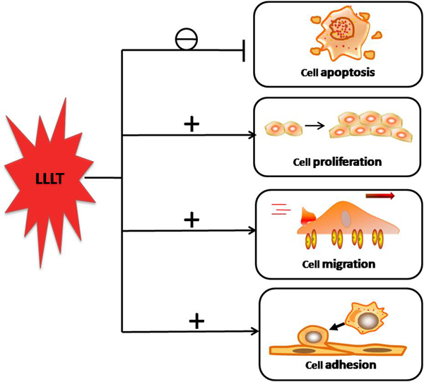
FIGURE 6. The downstream cellular effects of LLLT signaling include increases in cell proliferation, migration and adhesion molecules. Cell survival is increased and cell death reduced by expression of proteins that inhibit apoptosis.