

It carries are removed, by the phagocytic activity of macrophages that span the
sinuses.
2) Antibodies production:
Antigens carried to the lymph nodes, activate its B-lymphocytes to change into plasma cells which produce the specific antibodies.
About 1% of the antigens carried into the lymph node by the lymph pass through the cortical follicles. These antigens stimulate the B-lymphocytes to become active.
Activated B-lymphocytes proliferate in the germinal centres of the cortical follicles. Some of the daughter cells are preserved as memory cells. The other cells change to plasma cells which migrate to the medulla and secrete the specific antibodies. The antibodies pass with the lymph to the circulation. Memory cells leave the node with the lymph to the circulation. If they are stimulated again by the same antigen, they migrate to the surrounding CT. and change to plasma cells and secrete the antibodies.
3) Site of proliferation of lymphocytes:
Activated lymphocytes, arising from the non-capsulated lymphoid tissues, migrate through the blood vessels to the lymph nodes. The cells pass in-between the high endothelial cells of the post-capillary venules to the nodal parenchyma where they proliferate. Daughter cells get out of the node through the efferent lymphatics to be finally poured back, to the circulation.
Lymphatic tissue and the immune response
The lymphatic system is specialized form of connective tissue that consists of cells, tissues, and organs that monitor body surfaces and internal fluid compartments and react to the presence of potentially harmful antigenic substances.
Included in this system are thymus, spleen, lymph nodes, lymphatic nodules, and diffuse lymphatic tissue.
The several forms of lymphatic organs and tissue are often collectively referred to as the immune system.
Lymphatic vessels connect parts of the system to the blood vascular system.
Lymphocytes are the chief cellular constituent of lymphatic tissue.
Functionally, two types of lymphocytes are identified: T lymphocytes and B lymphocytes.
T lymphocytes or T cells, involved in cell-mediated immunity
B lymphocytes or B cells, involved in humoral immunity and the production of antibodies
The bone marrow and thymus have been identified as primary or central lymphatic organs. Lymphocytes undergo their antigen independent proliferation and differentiation into immunocompetent cells in these organs.
Immunocompetent lymphocytes, together with plasma cells derived from B lymphocytes and with macrophages, organize around mesenchymal reticular cells and their reticular fibers to form the adult effector lymphatic tissues and organs, i. e. lymphatic nodules, lymph nodes, tonsils and spleen. It is in these secondary or peripheral lymphatic organs that T and B lymphocytes undergo antigen-dependent proliferation and differentiation into effector lymphocytes and memory cells.
Cells of the lymphatic system
B lymphocytes may differentiate into plasma cells that produce antibodies
or into memory cells.
B lymphocytes that have been activated by contact with antigen transform into immunoblasts (plasmoblasts) that proliferate and then differentiate into:
-plasma cells, which synthesize and secrete a specific antibody
-memory cells, which are able to respond more quickly to the next encounter with the same antigen
The specific antibody produced by the plasma cell binds to the stimulating antigen, forming an antigen-antibody complex.
These complexes are eliminated in a number of ways, including phagocytosis by macrophages and eosinophils.
The complexes may also activate a system of plasma proteins, the complement system, and cause one of the components, to bind to an antigenic bacterium and act as a ligand for its phagocytosis by macrophages.
Memory cells do not participate in the initial or primary response to a specific antigen. However, memory cells are programmed and ready to respond to the same antigen should it appear again. The response to the same antigen on second exposure is called the secondary response. It is more rapid and intense than the primary response, because of the presence of a population of specific memory B lymphocytes already programmed to respond to that antigen.
T lymphocytes
Immunocompetent T lymphocytes that have been activated by interaction with an antigen also transform into lymphoblasts that proliferate and differentiate into several types of effector.
Three types of T lymphocytes have been identified:
-Cytotoxic lymphocytes (CTL) or killer T cells, which serve as primary effector cells in cell-mediated immunity.
-Helper T lymphocytes (TH cells) which assist B cells as well as other T cells in their response to antigens
-Suppressor T lymphocytes (TS), which suppress the activity of B cells
The primary function of cytotoxic lymphocytes is to screen other cells for signs of viral infection or other signs of abnormality, such as development into cancer cells. The antigen receptors on the killer cells enable them to recognize viral or cancer-related peptides on the surface of cells and to kill the cells by causing them to lyse.
Helper T cells assist in the stimulation of B lymphocytes to produce antibodies. TH cells have surface receptors that bind to a surface peptide complex on the membrane of macrophages or B cells that have “processed” an antigen. Thus, stimulates the helper T cells to become active, dividing and producing polypeptide hormones called interleukins that, in turn, stimulate B cells to divide and produce their antibodies.
Lymphokines are soluble substances released by sensitized lymphocytes on contact with a specific antigen. Along with other functions, these substances stimulate the activity of monocytes and macrophages in the cell-mediated immune response.
In persons infected with AIDS-causing human immunodeficiency virus, the number of helper T cells is dramatically reduced.
Antigen presenting cells
Antigen presenting cells interact with helper T cells to facilitate immune responses.
The interaction between most antigens and the antibodies on the surface of B cells is insufficient to stimulate B cell growth, differentiation, and secretion of soluble antibody. For the lymphocytes to function effectively, antigens are processed and presented to them by antigen-presenting cells.
The group of antigen presenting cells includes:
-Langerhans cells of the epidermis
-Lymphatic dendritic cells
-Perisinusoidal macrophages (Kupffer cells) of the liver
-Tissue macrophages
Macrophages and the immune response
Macrophages are closely associated with lymphocytes in both types of immune response. Macrophages can process and present the antigen to the B cells or helper T cells and destroy the antigen after it has been processed by other cells of the immune system
Endocrine organs
Communication between cells is necessary to maintain homeostasis and coordinate growth and development.
The primary function of two major organ systems, the endocrine system and the nervous system is intercellular communication.
The endocrine system communicates through the release of hormones, secretory products of endocrine cells and organs that pass into the circulatory system for transport to target cells that possess receptors for the hormones.
Functionally, the endocrine system and the nervous system are closely interrelated and may overlap in function. The hypothalamus, a part of the brain, coordinates most endocrine functions of the body and serves as one of the major controlling centers of the autonomic nervous system. The hypophysis and the hypothalamus the portion of the brain to which the hypophysis is attached, are morphologically and functionally linked in the endocrine and neuroendocrine control of other endocrine glands. They are often called the “master organs” of the endocrine system.
Endocrine hormones include three classes of compounds.
Steroids are synthesized and secreted by cells of ovaries, testes and adrenal cortex.
Small peptides, proteins and glycoproteins are synthesized and secreted by the cells of the hypothalamus, hypophysis (pituitary), thyroid, parathyroid, pancreas and scattered endocrine cells of the gastrointestinal tract and lungs.
Amino acid analogues and derivatives, including the catecholamines are synthesized and secreted by many neurons as well as cells of the adrenal medulla.
Hypophysis (pituitary gland)
The hypophysis has two functional components:
-adenohypophysis (anterior pituitary) the glandular epithelial tissue
-neurohypophysis (posterior pituitary) the neural secretory tissue
The adenohypophysis consists of:
-pars distalis
-pars intermedia
-pars tuberalis
The neurohypophysis consists of:
-pars nervosa
-pars infundibulum
Adenohypophysis
The adenohypophysis is the master gland of the endocrine system, regulating other endocrine glands.
Pars distalis
Figure 40. Photomicrograph of the human pars distalis, Ac, acidophils; Bas, basophils; Ch, chromophobes
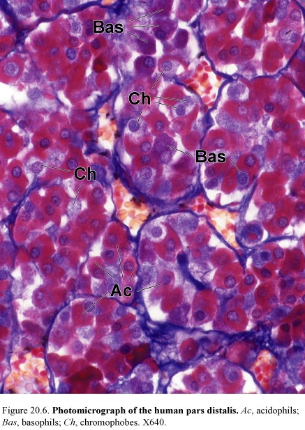
Descriptions of the cells within the pars distalis were based only on the staining properties of secretory granules within the cells.
Histologists identified three types of cells according to their staining reaction, such as, basophiles 10%, acidophils 40%, and chromophobes 50%.
Basophiles is divided into:
-the adrenocorticolipotropes (the most common basophiles) which produce adrenocorticotropic hormone (ACTH) and lipotropic hormone (LPH)
-the gonadotropes (small basophiles) which produce luteinizing hormone (LH) and follicle-stimulating hormone (FSH)
-the thyrotropes (large basophiles) which produce thyroid-stimulating hormone (TSH)
Acidophils is divided into:
-the somatotropes (small acidophils) which produces somatotropin, a growth hormone (GH)
-the lactotropes (mammotropes) are variable and scattered acidophils that produces prolactin (PR) or lactogenic hormone (LTH) and undergo hypertrophy during lactation.
Chromophobes have undifferentiated cells and follicular cells.
The long branching processes of follicular cells form a supporting network for the other cells.
Pars intermedia
The pars intermedium is in humans, a rudimentary region made up of cords and follicles of weakly basophilic cells that contain small secretory granules and chromophobic cells. The function of these cells is not known. In the basophilic cells are found small amount of melanocyte-stimulating hormone.
Pars tuberalis
The endocrine cells are arranged in short clusters or cords in association with the blood vessels. Nests of squamous cells and small follicles lined with cuboidal cells and scattered in the region. Some functional gonadotropes are present in this region.
Neurohypophysis
The neurohypophysis is a nerve tract whose terminals store and release secretory product from the hypothalamus.
The neurohypophysis contains pituicytes associated with the fenestrated capillaries. These cells are irregular in shape, with many branches, and resemble glial cells. Their nuclei are round or oval, and pigment granules are present in the cytoplasm.
The neurohypophysis consists of the pars nervosa and the infundibulum that connects it to the hypothalamus.
The pars nervosa contains nonmyelinated axons and nerve endings of approximately 100.000 neurosecretory neurons whose cell bodies lie in the supraoptic and paraventricular nuclei of the hypothalamus.
Dilatations of the axons are called Herring bodies. The membrane-bounded neurosecretory granules that aggregate to form the Herring bodies contain either oxytocin or vasopressin (antidiuretic hormone) (ADH).
Antidiuretic hormone (vasopressin) increases blood pressure by promoting the contraction of smooth muscle in small arteries and arterioles. ADH decreases urine volume by altering the permeability of kidney collecting tubules, decreases the rate of perspiration.
Oxytocin promotes contraction of smooth muscle of the uterus during copulation and parturition and myoepithelial cells of the breast.
The hypothalamus
The hypothalamus regulates hypophyseal function.
The hypothalamus has supraoptic and paraventricular nuclei. Antidiuretic hormone and vasopressin are secreted by these nucleuses.
The hypothalamus is the site of production of a number of neurosecretory proteins.
In addition to oxytocin and ADH, the hypothalamic neurons secrete proteins that promote and inhibit the secretion and release of adenohypophyseal hormones.
These other hypothalamic peptides also accumulate in nerve endings near the median eminence and infundibular stalk and are released into the first capillary bed of the hypophyseal portal system for transport to the pars distalis.
Hypothalamic regulatory peptides
-growth hormone releasing factor
-growth hormone inhibiting factor
-prolactin releasing factor
-prolactin inhibiting factor
-gonadotropin releasing factor
-corticotropin releasing factor
-thyrotropin releasing factor
Pineal gland
Figure 41. Photomicrograph of the human pineal gland

The pineal gland (pineal body) is now described as an endocrine or neuroendocrine gland but it is functions in humans are not clearly defined.
In humans, pineal activity, as indicated by changes in the plasma level of melatonin, rises during darkness and falls during light. Clinically, tumors that destroy the pineal gland are associated with precocious (early onset) puberty. Pineal gland function then influences seasonal sexual activity and circadian (24 hour) biorhythms. The pineal gland contains two basic types of parenchymal cells:
-pinealocytes and interstitial (glial) cells
Pinealocytes are the most common parenchymal cell in the pineal gland. They secrete 2 kinds of amines, serotonin and melatonin. They are arranged in clumps or cord within the lobules formed by connective tissue septa that extend into the gland from the pia matter that covers its surface. These cells have a large, deeply infolded nucleus with one or more prominent nucleoli and contain lipid droplets within their cytoplasm.
Pinealocytes show typical cytoplasmic organelles along with numerous, dense-cored, membrane-bounded granules in their elongated cytoplasmic processes. The expanded club- like endings of the processes are associated with the blood capillaries. This feature is strongly suggestive of neuroendocrine activity.
The interstitial (glial) cells comprise about 5% of the cells in the gland. They have staining and ultrastructural features that closely resemble those of astrocytes and are reminiscent of the pituicytes of the neurohypophysis.
In addition to the two cell types, the human pineal gland is characterized by the presence of calcified concretions, called brain sand.
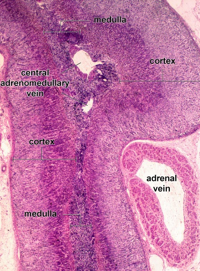
ADRENAL GLANDS
The adrenal (suprarenal) glands secrete both steroid hormones and catecholamines. They have a flattened triangular shape and are embedded in the perirenal fat at the superior poles of the kidneys.
The glands are covered with a thick connective tissue capsule from which trabeculae extend into the parenchyma, carrying blood vessels and nerves. The secretory parenchymal tissue is organized in cortical and medullary regions:
The cortex is the steroid-secreting portion. It lies beneath the capsule and constitutes nearly 90% of the gland by weight.
The medulla is the catecholamine-secreting portion. It lies deep to the cortex and forms the center of the gland.
Parenchymal Cells of the Cortex and Medulla Are of Different Embryologic Origin
Embryologically, the cortical cells originate from mesodermal mesenchyme, whereas the medulla originates from neural crest cells that migrate into the developing gland.
Blood Supply
The adrenal glands are supplied with blood by the superior, middle, and inferior adrenal arteries. In the capsule, the arteries branch to give rise to three principal patterns of blood distribution.
Zonation of the Adrenal Cortex
Figure 42. Diagram illustrating the organization of the cells within the adrenal gland and their relationship to the blood vessels
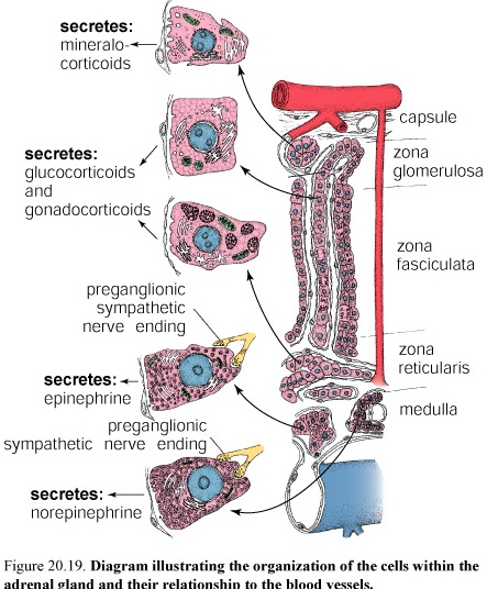
The adrenal cortex is divided into three zones on the basis of the arrangement of its parenchymal cells. The zones are designated
Zona glomerulosa, the narrow outer zone
Zona fasciculata, the thick middle zone
Zona reticularis, the inner zone
Zona Glomerulosa
The cells are relatively small columnar or pyramidal shaped. In the human, some areas of the cortex may lack a recognizable zona glomerulosa.
The Zona Glomerulosa Secretes Aldosterone That Functions in the Control of Blood Pressure
The cells of the zona glomerulosa secrete mineralocorticoids, compounds that function in the controlling electrolyte homeostasis (act in distal tubule cells of kidney to increase sodium resorption and decrease potassium resorption) and also maintaining the osmotic balance in the urine. The principal secretion is aldosterone (95% of mineralocorticoids).
Zona Fasciculata
The cells of the zona fasciculata are large and polyhedral. The cells of the zona fasciculata have a lightly staining spherical nucleus. The generally acidophilic cytoplasm contains numerous lipid droplets. The lipid droplets contain neutral fats, fatty acids, cholesterol, and phospholipids that are precursors for the steroid hormones secreted by these cells. The cells have highly developed sER and mitochondria with tubular cristae. They also have a well-developed Golgi complex and numerous profiles of rER.
The Principal Secretion of the Zona Fasciculata Is Glucocorticoids That Regulate Glucose and Fatty Acid Metabolism
The zona fasciculata secretes glucocorticoids, so called because of their role in regulating gluconeogenesis and glycogenesis. The most important of the glucocorticoids secreted by the zona fasciculata is hydrocortisone (cortisol). Also they secrete cortisone and corticosteron. This compound acts on many different cells and tissues to increase the metabolic availability of glucose and fatty acids.
Glucocorticoids depress the immune response and the inflammatory response and, as a result of the latter, inhibit wound healing. They depress the inflammatory response by inhibiting macrophage recruitment and migration. They also stimulate destruction of lymphocytes in lymph nodes and inhibit mitosis by transformed lymphoblasts. They also provide resistance to stress and suppress some allergic reaction.
Figure 43. Photomicrograph of the adrenal gland
Zona Reticularis
The cells of the zona reticularis are noticeably smaller than those of the zona fasciculata, and their nuclei are more deeply stained. They are arranged in anastomosing cords, separated by fenestrated capillaries. The cells have relatively few lipid droplets. Both light and dark cells are seen. They have a well-developed sER and numerous mitochondria with tubular cristae, but they have little rER.
The Principal Secretions of the Zona Reticularis Are Weak Androgens and Glucocorticoids
The principal secretion of the cells in the zona reticularis consists of weak androgens, mostly dehydroepiandrosterone (DHA). The cells also secrete some glucocorticoids, in much smaller amounts than those of the zona fasciculata. Here, too, the principal glucocorticoid secreted is hydrocortisone. The zona reticularis is also under feedback control of the CRF-ACTH system.
Adrenal Medulla
Adrenal Medullary Cells Are Modified Postganglionic Neuronal Cells That Have a Secretory Function
The central portion of the adrenal gland, the medulla, is composed of a parenchyma of large, epithelioid cells, called chromaffin cells, connective tissue, numerous sinusoidal blood capillaries, and nerves.
The catecholamines epinephrine (80%) and nor-epinephrine (20%) secreted by the medullary cells are produced by different cell types. Two populations of cells are distinguished by the nature of the membrane-bounded granules.
One population of cells contains only large dense core granules. These cells secrete norepinephrine. The other population of cells contains granules that art smaller, more homogeneous, and less dense. These сеlls secrete epinephrine.
Catecholamines have sympathomimetic functions (increases heart rate, blood pressure, sweating, rate of respiration and decreases blood flow to viscera and skin, digestion, urine production and others.
Thyroid gland
Figure 44. Thyroid gland
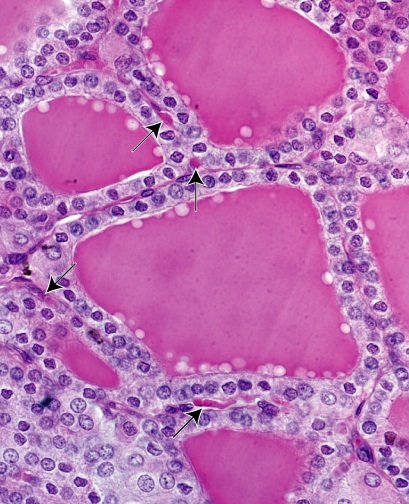
The thyroid is a bilobed endocrine gland located in the neck, anterolateral to the larynx and upper trachea.
The lobes are connected by a thin band of thyroid tissue, the isthmus that crosses anterior to the upper part the trachea. A thin connective tissue capsule surrounds
the gland. It sends trabeculae into the parenchyma to partially outline irregular lobes and lobules. Secretory follicles constitute the functional units of the gland.
Thyroid gland function is essential to normal growth and development.
The thyroid gland produces three hormones, each of which is essential to normal metabolism and homeostasis.
-Thyroxin (tetroidothyronine T4) and triidothyronin T3 regulate cell and tissue metabolism, influences body and tissue growth and development of the nervous system in the fetus and young child.
-Calcitonin regulates blood calcium level.
Follicular cells
The follicle is the structural unit of the thyroid gland. A follicle is a spheroidal cyst-like compartment with a wall formed by a simple squamous or cuboidal epithelium, the follicular epithelium.
The lumina of the follicles are filled with a gel-like mass called colloid. The apical surfaces of the follicular epithelial cells are in contact with the colloid, and the basal surfaces rest on a typical basal lamina.
Two basic cell types are present in the follicles principal or follicular cells and parafollicular or C cells.
Follicular cells secrete T4 and T3. Parafollicular cells secrete calcitonin.
The follicles are surrounded by an extensive network of fenestrated capillaries.
Lymphatic capillaries are present in the interfollicular connective tissue and may also provide a second route for conveying the hormones from the gland.
The nuclei of follicular epithelial cells are spherical. In the basophilic cytoplasm presents Golgi complex and lipid droplets.
Figure 45. Diagram of steps in thyroid hormone synthesis
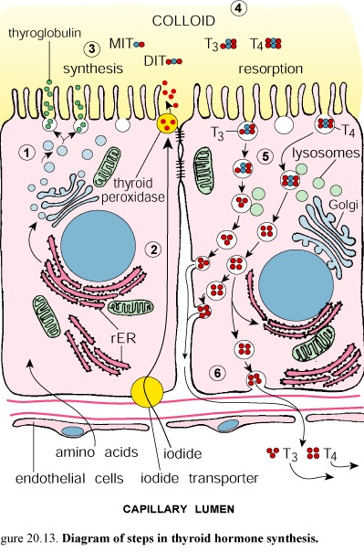
The active cuboidal or columnar cells reveal the presence of organelles commonly associated with both secretory and absorptive cells. The organelles include typical junctional complexes at the apical end of the lateral plasma membrane, short microvilli on the apical of the cells, rough surfaced endoplasmic reticulum in the basal region, supranuclear Golgi complex, numerous small vesicles in the apical cytoplasm, that are morphologically similar to the vesicles associated with the Golgi complex, abundant lysosomes and multivesicular bodies, and membrane limited vesicles, identified as colloidal resorption droplets in the apical region. Colloid contains thyroglobulin, the inactive storage form of the thyroid hormones.
The protein portion of thyroglobulin is synthesized in the rER of the follicular epithelial cells and glycosylated there and in the Golgi complex before it is secretes by exocytosis into the lumen of the follicle.
The follicular epithelial cells actively transport iodide from the blood into their cytoplasm. The iodide is oxidized to iodine by thyroperoxidase in the cytoplasm. It is then released into the follicular lumen where iodination of some tyrosine residues of the thyroglobulin occurs near the microvilli of the apical cell surface.
The synthesis, storage and release of thyroid hormones are controlled by thyroid-stimulating hormone from the adenohypophysis.
The thyroid hormones are formed by the coupling of two iodinated tyrosine residues to produce thyroxine (T4 and T3).
These active thyroid hormones (in T4 : T3 ratio of 20: 1) cross the basal membrane and enter blood and lymphatic capillaries.
Parafollicular cells
Parafollicular cells are present within the follicular epithelium or are scattered in the connective tissue.
The parafollicular cells (C cells, calcitonin cells) are the second type, of endocrine cell found in the thyroid. In the follicles they are located near the basal lamina and do not extend to the follicular lumen to the contact the colloid. They have numerous, small, membrane-bounded secretory granules.
Parafollicular cells produce calcitonin, a hormone that lowers blood calcium level.
Calcitonin (thyrocalcitonin) is the physiologic antagonist to parathyroid hormone.
Calcitonin lowers blood calcium levels by suppressing bone resorption and increasing the rate of osteoid calcification. Secretion of calcitonin is regulated directly by blood calcium levels. High levels of calcium stimulate secretion, low levels inhibit it.
Parathyroid glands
The parathyroid glands are small endocrine glands closely associated with the thyroid. They are ovoid, a few millimeters in diameter, and arranged in two pairs, to comprise the superior and inferior parathyroid glands. They are usually located in the connective tissue on the posterior surface of the thyroid.
Development and Structure
Embryologically, the inferior parathyroid glands (and the thymus) derive from the third branchial pouch; the superior glands, from the fourth branchial pouch.
Structurally, each parathyroid gland is surrounded by a thin connective tissue capsule that separates it from the thyroid. Septa extend from the capsule into the gland to divide it into lobules and to separate the densely packed cords of cells.
Principal (Chief) Cells and Oxyphil Cells Constitute the Epithelial Cells of the Parathyroid Gland
Principal cells, the more numerous of the parenchymal cells of the parathyroid, are responsible for the secretion of parathyroid hormone (PTH). The cytoplasm contains small granules with PTH. They are small, polygonal cell.
Oxyphil cells constitute a minor portion of the parenchymal cells. They are more rounded and larger than the principal cells.
Function
The parathyroids function in the regulation of blood ion levels of calcium and phosphate. Parathyroid hormone is essential for life.
Parathyroid Hormone Regulates Calcium and Phosphate Levels in the Blood
The parathyroids produce PTH, also known as parathormone. Release of this hormone causes the level of calcium in the blood to increase. Simultaneously, it reduces the concentration of phosphate. Bone resorption is stimulated by PTH. During osteolysis, calcium and phosphate are both released from calcified bone matrix into the extracellular fluid.
Parathyroid Hormone and Calcitonin Have Reciprocal Effects in the Regulation of Blood Calcium Levels
Digestive system
The digestive system consists of alimentary canal and its associated organs, namely, the tongue, teeth, salivary glands, pancreas, liver and gallbladder.
The alimentary mucosa is the surface across which most substances enter the body.
The alimentary mucosa has numerous functions in its role as an interface between the body and the environment. These include its
1. Barrier function: The mucosa serves as a barrier to the entry of noxious substances, antigens, and pathogenic organisms.
2. Immunologic function: Lymphatic tissue in the mucosa serves as a first line of defense of the body by the immune system.
3. Secretory function: The lining of the alimentary canal secretes, at specific sites, digestive enzymes, hydrochloric acid, mucin, and antibodies.
4. Absorptive function: The epithelium of the mucosa absorbs metabolic substrates, i.e., the breakdown products of digestion, as well as vitamins, water, electrolytes, and other substances.
ORAL CAVITY
The oral cavity consists of the mouth and its contents, i.e., the tongue, teeth, salivary glands, and tonsils.
Tongue
The tongue is a muscular organ. The striated muscles of the tongue are arranged in bundles that generally run in three planes and usually separated by connective tissue.
The dorsal surface of the tongue is divided into an anterior two-thirds and a posterior one-third by a V shaped depression, the sulcus terminalis.
Papillae cover the dorsal surface of the anterior portion of the tongue. There are four types:
-filiform
-fungiform
-circumvallate
-foliate
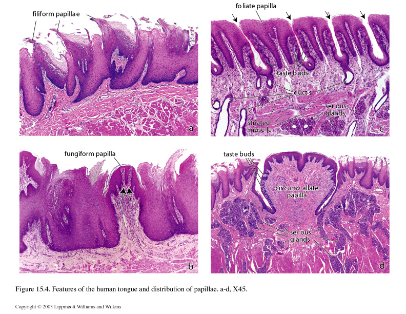
Filiform papillae are the most numerous in humans and the smallest. They are conical, elongated projections of connective tissue that are covered with partly keratinized stratified squamous epithelium.
Fungiform papillae are mushroom shaped projections located on the dorsal surface of the tongue. They are more numerous near the tip of the tongue. Taste buds are present on the dorsal surface of these papillae.
Circumvallate papillae are the large, dome-shaped structures that reside in the mucosa just anterior to the sulcus terminalis. Each papilla is surrounded by a moat-like invagination lined with stratified squamous epithelium that contains numerous taste buds.
Foliate papillae occur on the lateral edge of the tongue in aged humans; the foliate papillae may not be recognized. In younger individuals, they are easily found and contain many taste buds in the epithelium.
Lingual tonsils
Lingual tonsils are located in the lamina propria of the root or base of the tongue. They are found posterior to the sulcus terminalis. The lingual tonsils contain lymphatic nodules. Epithelial crypts usually invaginate into the lingual tonsil.
Gingiva
Figure 46. Schematic diagram of gingiva
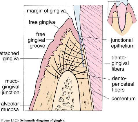
The gingival is the part of the mucous membrane commonly called the gums. The gingival is firmly attached to the teeth and underlying bony tissue. It is composed of stratified squamous epithelium and numerous connective tissue papillae. This epithelium is bound to the tooth enamel by means of a cuticle that resembles a thick basal lamina and forms the epithelial attachment of Gottlieb. Between the enamel and the epithelium is the gingival crevice a small deepening surrounding the crown.
Teeth and supporting tissues
Teeth are a major component of the oral cavity and are essential for the digestive process. Teeth are embedded in and attached to the maxilla and mandible. A child has 10 deciduous teeth in each jaw, consisting, on each side of
A central incisor
A lateral incisor
A canine tooth
Two molar teeth
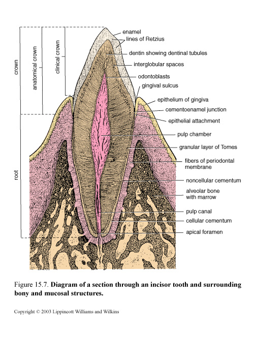
Figure 47. Diagram of a section through an incisor tooth and surrounding bony and mucosal structures
Over a period of years, usually beginning at about age 6, these are gradually replaced by 16 permanent teeth in each jaw, consisting, on each side, of
A central incisor
A lateral incisor
A canine
Two premolar teeth
Three molar teeth
Incisors and canines have one root each, premolars usually have two, and molars may have three or four roots. All teeth have the same basic structure.
The adult tooth consists of four distinct structural components, the enamel and the cementum on the outside, the dentin beneath them, and the pulp in a central pulp cavity.
Enamel
Enamel covers the crown of the tooth. That part of the crown that is exposed and visible above the gum line is called the clinical crown; the anatomical crown describes all of the tooth that is covered by enamel, some of which is below the gum line. Enamel varies in thickness over the crown and may be as thick as 2.5 mm on the cusps (biting and grinding surfaces) of some teeth. The enamel layer ends at the neck or cervix of the tooth at the cementoenamel junction; the root of the tooth is then covered by cementum.
Enamel Is the Hardest Substance in the Body;
F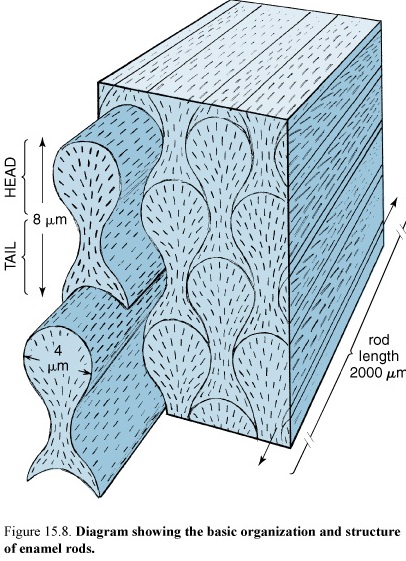 igure
48. Diagram showing the basic organization and structure of enamel
rods
igure
48. Diagram showing the basic organization and structure of enamel
rods
It Consists of 96-98% Hydroxyapatite. The hydroxyapatite of the enamel is arranged in rods or prisms that vary in diameter from 4-8 µm. The structural and functional unit of the tooth is rods or prisms. Each enamel rod spans the full thickness of the enamel layer from the dentinoenamel junction to the enamel surface; the spaces between the rods are also filled with hydroxyapatite. Striations observed on enamel rods (contour lines of Retzius) may represent evidence of rhythmic growth of the enamel in the developing tooth. The enamel in a mature tooth is acellular and nonreplaceable.
Although the enamel of an erupted tooth is devoid of cells and cell processes, it is not a static tissue. It is under the influence of substances in saliva, the secretion of the salivary glands, that are essential to its maintenance. The substances in saliva that affect teeth include
Digestive enzymes
Antibacterial enzymes
Antibodies
Inorganic (mineral) components
Mature enamel contains very little organic material. Despite its hardness, enamel can be damaged by decalcification by acid-producing bacteria acting on food products trapped on the surface of the enamel. This is the basis of the initiation of dental caries.
Enamel Formation (Amelogenesis)
Enamel is produced by ameloblasts with the close cooperation of other enamel organ cells
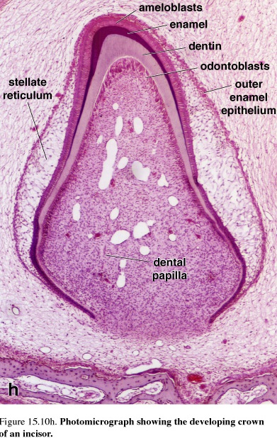
Figure 49. Photomicrograph showing the developing crown of an incisor
 The
major stages of amelogenesis are the period
of matrix production, or the secretory stage, and the period
of maturation. In the formation of mineralized tissues
of the tooth, dentin is produced first. Then, partially mineralized
enamel matrix is deposited directly on the surface of the
previously formed dentin. The cells
producing this partially mineralized organic matrix are referred
to as secretory
ameloblasts.
The major stages of amelogenesis are the period
of matrix production, or the secretory stage, and the period
of maturation. In the formation of mineralized tissues
of the tooth, dentin is produced first. Then, partially mineralized
enamel matrix is deposited directly on the surface of the
previously formed dentin. The cells
producing this partially mineralized organic matrix are referred
to as secretory
ameloblasts. As
do osteoblasts in bone,
these cells produce an organic proteinaceous matrix by
activity of the rough endoplasmic reticulum (rER), Golgi apparatus,
and secretory granules.
The
major stages of amelogenesis are the period
of matrix production, or the secretory stage, and the period
of maturation. In the formation of mineralized tissues
of the tooth, dentin is produced first. Then, partially mineralized
enamel matrix is deposited directly on the surface of the
previously formed dentin. The cells
producing this partially mineralized organic matrix are referred
to as secretory
ameloblasts.
The major stages of amelogenesis are the period
of matrix production, or the secretory stage, and the period
of maturation. In the formation of mineralized tissues
of the tooth, dentin is produced first. Then, partially mineralized
enamel matrix is deposited directly on the surface of the
previously formed dentin. The cells
producing this partially mineralized organic matrix are referred
to as secretory
ameloblasts. As
do osteoblasts in bone,
these cells produce an organic proteinaceous matrix by
activity of the rough endoplasmic reticulum (rER), Golgi apparatus,
and secretory granules.
Figure 50. Schematic diagram of a partially formed tooth showing details of amelogenesis
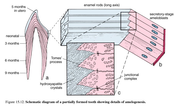
The major stages of amelogenesis are the period of matrix production, or the secretory stage, and the period of maturation. In the formation of mineralized tissues of the tooth, dentin is produced first. Then, partially mineralized enamel matrix is deposited directly on the surface of the previously formed dentin. The cells producing this partially mineralized organic matrix are referred to as secretory ameloblasts. As do osteoblasts in bone, these cells produce an organic proteinaceous matrix by activity of the rough endoplasmic reticulum (rER), Golgi apparatus, and secretory granules. The secretory ameloblasts continue to produce enamel matrix until the full thickness of the future enamel is achieved.
Maturation of the partially mineralized enamel matrix involves the removal of organic material as well as continued influx of calcium and phosphate into the maturing enamel. Cells involved in this second stage of enamel formation are referred to as maturation ameloblasts. Maturation ameloblasts differentiate from secretory ameloblasts and function primarily as a transport epithelium, moving substances into and out of the maturing enamel.
Secretory ameloblasts are narrow, highly polarized, columnar cells. They are directly adjacent to the developing enamel. At the apical pole of the cell is a process, Tomes' process, that is surrounded by the developing enamel. Adjacent to the mitochondria is the nucleus; in the main column of cytoplasm are the rER, Golgi, secretory granules, and other cell elements. Junctional complexes are present at both apical and basal extremities. Contractile filaments joined to these junctional complexes may be involved in moving the secretory ameloblast over the developing enamel. The rod produced by the ameloblast follows in the wake of the cell. Thus, in mature enamel, the direction of the enamel rod is a record of the path taken earlier by the secretory ameloblast.
At their basal poles, the secretory ameloblasts are adjacent to a layer of enamel organ cells called the stratum intermedium. The plasma membrane of these cells and that of the basal pole of the ameloblasts is positive for alkaline phosphatase, an enzyme active in calcification. Stellate enamel organ cells are external to the stratum intermedium and are separated from the adjacent blood vessels by a basal lamina.
The histologic feature that marks the cycles of maturation ameloblasts is the presence of a striated or ruffled border. Maturation ameloblasts with a striated border occupy about 70% of a specific cycle, and those that are smooth ended are about 30% of a specific cycle. There is no stratum intermedium in the enamel organ during enamel maturation; stellate papillary cells are adjacent to the maturation ameloblasts.
The maturation ameloblasts and the adjacent papillary cells are characterized by the presence of numerous mitochondria. This is indicative of cellular activity that requires large amounts of energy and is a reflection of the fact that the maturation ameloblasts and the adjacent papillary cells function as a transporting epithelium.
The matrix of developing enamel contains three major proteins:
Amelogenins
Enamelins
Tuft protein
Mature enamel contains only enamelins and tuft protein.
Amelogenins are removed during enamel maturation. Tuft protein is located near the dentinoenamel junction. It is present in enamel tufts and accounts for the fact that the enamel tufts are hypomineralized; i.e., they have a higher percentage of organic material than the remainder of the mature enamel. The maturation of the developing enamel results in the continued mineralization of enamel, so that it becomes the hardest substance in the body. The ameloblasts degenerate after the enamel is fully formed, at about the time of tooth eruption from the gum.
Cementum
Cementum covers the root of the tooth. The root is that part of the tooth that fits into its socket or alveolus in the maxilla or mandible. Cementum is a thin layer of bone-like material that is secreted by cementocytes, cells that closely resemble osteocytes. Like bone, cementum has a mineral content of 45-50%. The lacunae and canaliculi in the cementum contain the cementocytes and their processes, respectively. They resemble those structures in bone that contain osteocytes and osteocyte processes.
Unlike bone, cementum is avascular. Also, the canaliculi in cementum do not appear to form an interconnecting network. A layer of cementoblasts (cells that resemble the osteoblasts of the surface of growing bone) is seen on the outer surface of the cementum, adjacent to the periodontal ligament.
Figure 51. EM of Sharpey’s fibers
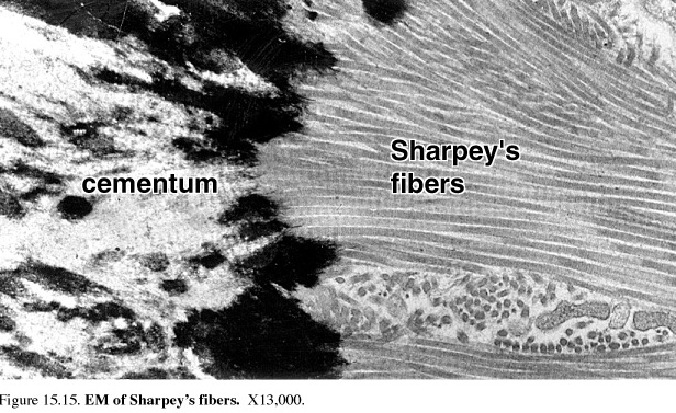
Collagen fibers that project out of the matrix of the cementum and embed in the bony matrix of the socket wall form the bulk of the periodontal ligament. These fibers are another example of Sharpey's fibers. Oxytalan fibers that resemble developing elastic fibers and stain with elastic stains are also a component of the periodontal ligament. This mode of attachment of the tooth in its socket allows slight movement of the tooth to occur naturally and forms the basis of the orthodontic procedures that are used to straighten teeth and reduce malocclusion of the biting and grinding surfaces of the maxillary and mandibular teeth. During such corrective tooth movement, the alveolar bone of the1 socket is resorbed and resynthesized, but the cementum is not.
Dentin
Figure 51.Photomicrograph of a decalcified tooth showing dental pulp and structure of dentin
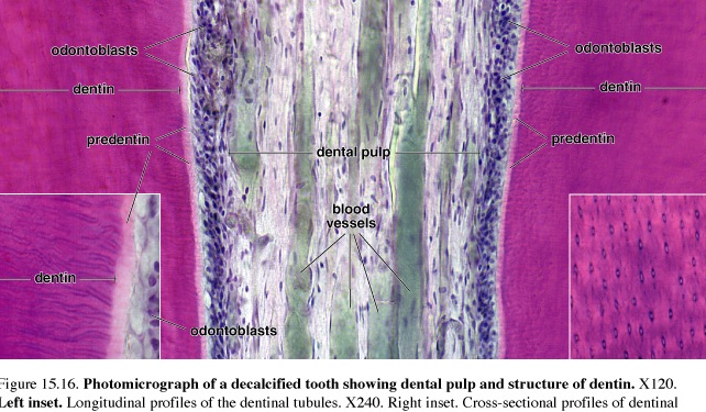
Dentin is calcified material that forms most of the tooth substance Dentin lies deep to the enamel and cementum. It contains less hydroxyapatite than does enamel, about 70%, but more than is found in bone and cementum. Dentin is secreted by odontoblasts that form an epithelial layer over the inner surface of the dentin, i.e., that surface that is in contact with the pulp. Odontoblasts, too, are elongated cylindrical cells that contain a well-developed rER, a large Golgi apparatus, and other organelles associated with the synthesis and secretion of large amounts of protein. The apical surface of the odontoblast is in contact with the forming dentin; junctional complexes between the odontoblasts at the level separate the dentinal compartment from the pulp compartment.
The layer of odontoblasts retreats as the dentin is laid down, leaving odontoblasts processes embedded in the dentin is narrow channels called dentinal tubules. The tubules and processes continue to elongate as the dentin continues to thicken by rhythmic growth. The rhythmic growth of dentin produces certain "growth lines" in the dentin (incremental lines of von Ebner and the thicker lines of Owen) that can identify significant developmental times such as birth (neonatal line) and may identify when unusual substances such as lead were incorporated into the growing tooth. Study of growth lines has proved useful in forensic medicine.
Predentin is newly secreted organic matrix, closest to the cell body of the odontoblasts that has yet to be mineralized. An unusual feature of the secretion of collagen and hydroxyapatite by odontoblasts is the presence, in Golgi vesicles, of arrays of a formed filamentous collagen precursor to which granules believed to contain calcium attach. This gives rise to structures called abacus bodies. The abacus bodies become more condensed as they mature into secretory granules.
Dentinogenesis
Dentin is produced by odontoblasts.
Dentin is the first mineralized component of the tooth to be deposited. During the formation of the very outermost dentin, which is referred to as mantle dentin, it is also formed by subodontoblastic cells that produce small bundles of collagen fibers (von Korff’s fibers). The odontoblasts differentiate from cells at the periphery of the dental papilla. The progenitor cells have the appearance of typical mesenchymal cells; i.e., they contain little cytoplasm. During their differentiation into odontoblasts, cytoplasmic volume and organelles characteristic of collagen-producing cells increase. The cells form a layer at the periphery of the dental papilla, and they secrete the organic matrix of dentin, called predentin, at their apical pole (the end of the cell away from the dental papilla). As the thickness of the predentin increases, the odontoblasts move or are displaced centrally. A wave of mineralization follows the receding odontoblasts; this mineralized product is the dentin. As the cells move centrally, the odontoblastic process becomes increasingly long, with its greatest length being surrounded by the mineralized dentin. In newly formed dentin, the wall of the dentinal tubule is simply the edge of the mineralized dentin. With time, the dentin immediately surrounding the dentinal tubule becomes more highly mineralized; this more mineralized sheath of dentin is referred to as the peritubular dentin. The reminder of the dentin is then referred to as the intertubular dentin.
Dental Pulp and Pulp Cavity
The Dental Pulp Cavity Is a Connective Tissue Compartment Bounded by the Tooth Dentin
The pulp cavity is the space within a tooth that is occupied by pulp, a loose connective tissue that is richly vascularized and supplied by abundant nerves. The pulp cavity has the general shape of the tooth. The blood vessels and nerves enter the pulp cavity at the tip (apex) of the root, at a site called the apical foramen. (The designations apex and apical in this context refer only to the narrowed tip of the root of the tooth rather than to a luminal (apical) surface, as used in describing secretory and absorptive epithelia.)
The blood vessels and nerves extend to the crown of the tooth where they form vascular and neural networks beneath and within the layer of odontoblasts. Some bare nerve fibers also enter the proximal portions of the dentinal tubules and contact odontoblast processes. The odontoblast processes are believed to serve a transducer function in transmitting stimuli from the tooth surface to the nerves in the dental pulp. In teeth with more than one cusp, pulpal horns extend into the cusps and contain large numbers of nerve fibers. More of these fibers extend into the dentinal tubules than at other sites. Because dentin continues to be secreted throughout life, the pulp cavity decreases in volume with age.
Alveolar Process and Alveolar Bone
The Alveolar Processes of the Mandible and Maxilla Contain the Sockets or Alveoli for the Roots of the Teeth
The alveolar bone proper, a thin layer of compact bone, forms the wall of the alveolus and is the bone to which the periodontal ligament is attached. The rest of the alveolar process is supporting bone. The surface of the alveolar bone proper usually shows regions of bone resorption and bone deposition, particularly when a tooth is being moved. Periodontal disease usually leads to loss of alveolar bone, as does the absence of functional occlusion of a tooth with its normal opponent.
Periodontal Ligament
The periodontal ligament is the fibrous connective tissue joining the tooth to its surrounding bone. The ligament is also called the periodontal membrane, but neither term describes its structure and function adequately. The periodontal ligament provides for
Attachment
Support
Bone remodeling (during movement of a tooth)
Nutrition of adjacent structures
Proprioception
Tooth eruption
Attachment and support are the most apparent functions.
A histological section of the periodontal ligament shows it to contain areas of both dense and loose connective tissue. The dense connective tissue contains collagen fibers and fibroblasts that appear elongated parallel to the long axis of collagen fibers. The fibroblasts are believed to move back and forth, leaving behind a trail of collagen fibers. Periodontal fibroblasts have also been shown to contain internalized collagen fibrils that are digested by the hydrolytic enzymes of the cytoplasmic lysosomes. These observations indicate that these fibroblasts not only produce collagen fibrils but also resorb collagen fibrils, thereby adjusting continuously to the demands of tooth movement.
The loose connective tissue in the periodontal ligament contains blood vessels and nerve endings in addition to the cells and thin collagenous fibers. The periodontal ligament also contains longitudinally disposed oxytalan fibers. They are attached to bone or cementum at each end. Some appear to be associated with the adventitia of blood vessels. As in other connective tissue, oxytalan fibers stain with special elastic stains; ultrastructurally they resemble developing elastic fibers, with their chief structural component being the microfibril that is characteristic of developing elastic fibers.
Salivary glands
Exocrine glands in the mouth produce saliva, which has digestive, lubricating and immunologic functions.
The minor salivary glands are located in the submucosa of different parts of the oral cavity. They include the lingual, labial, buccal, molar and palatine glands.
The major salivary glands are paired glands with long ducts. They consist of parotid, submandibular and sublingual glands. These glands consist of two general types of secretory cells-serous and mucous one and a duct system.
Serous cells are usually pyramidal in shape, with a broad base resting on the basal lamina and a narrow apical surface with short, irregular microvilli facing the lumen. They are protein -secreting cells.
Mucous cells are usually cuboidal to columnar in the shape. They are mucus-secreting cells.
Each salivary gland arises from developing ora; cavity epithelium.
Figure 52. Diagram comparing the components of the salivon in the three major salivary glands

Secretory gland acini
The acini of salivary gland contain either serous cells, mucous cells or both.
Thus, three types of acini are described:
-serous acini
-mucous acini
-mixed acini
Myoepithelial cells are instrumental in moving secretory products toward the excretory duct.
Salivary ducts
The lumen of the salivary acinus is continuous with that of a duct system that may have as many as three sequential segments.
These are referred to as:
-intercalated duct
-striated duct
-excretory duct
Intercalated ducts are located between a secretory acinus and a larger duct and are lined by low cuboidal epithelial cells. Several of these ducts join to form an intralobular duct, the striated duct.
Striated duct cells have numerous infoldings of the basal plasma membrane with numerous elongated mitochondria. Striated ducts are lined by a simple cuboidal epithelium that gradually becomes columnar.
The infoldings of the basal plasma membrane are seen in histologic sections as “striations”. The striated ducts of each lobule converge and drain into the connective tissue septae separating the lobules, where they become interlobular or excretory.
Excretory ducts travel in the interlobular and inter lobar connective tissue. Excretory ducts constitute the principal ducts of each of the major glands. They connect with oral cavity. The epithelium of small excretory ducts is simple cuboidal. It gradually changes to stratified cuboidal or pseudostratified columnar.
Parotid gland
The parotid glands are branched acinar and totally serous. The paired parotid glands are the largest of the major salivary glands. The parotid duct travels from the gland, which is located below and in front of the ear, to enter the oral cavity opposite the second upper molar tooth.
The secretory units in the parotid glands are serous and surround numerous, long, narrow intercalated ducts. Striated ducts are large. Large amounts of adipose tissue may be one of its distinguishing features.
Submandibular gland
The submandibular glands are branched tubuloacinar gland; its secretory portion contains both mucous and serous cells. Serous cells are the main component of this gland. The paired, large, mixed submandibular glands are located under either side of the floor of the mouth, close to the mandible. A duct from each of the two glands runs toward and medially to a papilla located on the floor of the mouth just lateral to the frenulum of the tongue.
Intercalated ducts are less extensive than in the parotid gland.
Sublingual gland
The small sublingual glands are branched tubuloacinar gland; its secretory portion contains both mucous and serous cells. Mucous cells are the main component of this gland. The sublingual gland the smallest of the paired major salivary glands, are located in the floor of the mouth anterior to the submandibular glands. Their multiple small sublingual ducts empty into the submandibular duct as well as directly onto the floor of the mouth. Intercalated ducts and striated ducts are difficult to locate or may be absent.
Alimentary canal structure and functions
The wall of the tract is formed by four distinctive layers. From the lumen outward they are:
-mucosa, consisting of a lining epithelium, an underlying connective tissue called lamina propria, and a muscularis mucosae, composed of smooth muscle
-submucosa, consisting of dense irregular connective tissue
-muscularis externa, consisting of two layers of muscle
-serosa or adventitia, a serous membrane consisting of a simple squamous epithelium, the mesothelium, and a small amount of underlying connective tissue, where the wall is directly attached to adjoining structures, the outer layer is the adventitia and is composed of connective tissue
Figure 53.Diagram of general organization of the alimentary canal
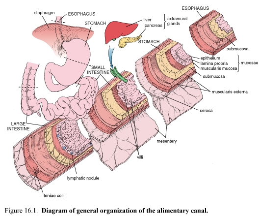
The wall of the tract is formed by four distinctive layers. From the lumen outward they are:
-mucosa, consisting of a lining epithelium, an underlying connective tissue called lamina propria, and a muscularis mucosae, composed of smooth muscle
-submucosa, consisting of dense irregular connective tissue
-muscularis externa, consisting of two layers of muscle
-serosa or adventitia, a serous membrane consisting of a simple squamous epithelium, the mesothelium, and a small amount of underlying connective tissue, where the wall is directly attached to adjoining structures, the outer layer is the adventitia and is composed of connective tissue
The mucosa of the digestive tract has three principal functions, a barrier, a secretory and an absorptive function.
The lamina propria contains glands, vessels that receive absorbed substances; and elements of the immune system. The immunologic barrier consists of diffuse lymphatic tissue, lymphatic nodules and eosinophils.
The muscularis mucosa forms the boundary between mucosa and submucosa. This sublayer consists of smooth muscle cells arranged as an inner circular and an outer longitudinal layer.
The submucosa consists of dense, irregular connective tissue. It contains the larger blood vessels and the nerve network, which constitute the submucosal plexus.
Muscularis externa
In most parts of the digestive tract, the muscularis externa consists of two concentric and relatively thick layers of smooth muscle. The cells in the inner layer are described as a circularly oriented layer, and those in the outer layer is described as a longitudinally oriented layer.
Contractions of the muscularis externa mix and propel the luminal contents of the digestive tract. The circular muscle layer forms sphincters along the digestive tract.
Serosa and adventitia
Large blood vessels and lymphatic vessels and nerve trunks travel through the serosa.
Esophagus
Figure 54. Photomicrograph of the esophagus
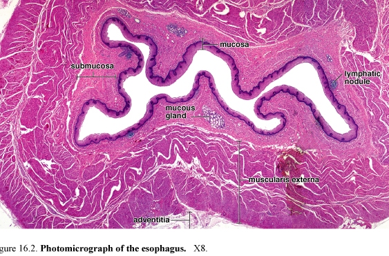
The esophagus is a muscular tube that delivers food and liquid from the oropharynx to the stomach. The esophagus is lined with a nonkeratinized stratified squamous epithelium.
The underlying lamina propria and the muscularis mucosae are not unique.
The submucosa along with the muscularis mucosae forms a number of longitudinal folds and creates a highly irregular luminal profile. The muscularis externa differs from that of the rest of the digestive tract in that upper one-third is striated muscle.
Striated muscle and smooth muscle are interwoven in the muscularis externa of the middle third of the esophagus; the muscularis externa of the distal third consists of smooth muscle, as in the rest of the digestive tract.
The outer layer of esophagus in the thoracic cavity is composed of adventitia. After entering the abdominal cavity it is covered by serosa.
Glands of the esophagus
Both two types of glands are mucous secreting. Esophageal glands proper occur in the submucosa. They are small compound tubuloalveolar glands.
Esophageal cardiac glands (so named because of their similarity to the cardiac glands of the stomach) occur in the lamina propria of the mucosa. They are present in the terminal part of the esophagus and frequently in the beginning portion of the esophagus. They are simple tubular gland.
Stomach
The stomach is an expanded part of the digestive tube that lies under the diaphragm. Mixing and partial digestion of the food in the stomach by its gastric secretions produces a pulpy fluid mix called chime.
Structural organization
The stomach has mucosae, submucosa, muscularis externa and a serosa.
The inner surface of the empty stomach has a number of longitudinal folds or ridges called rugae. In the mucosal surface is present numerous openings. These are the gastric pits or foveolae.
The smaller regions of the mucosa are formed by grooves or shallow trenches that divide the stomach surface into bulging irregular areas called mamillated areas.
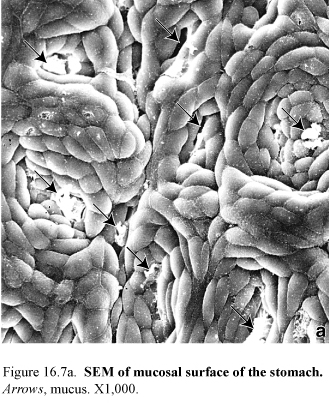
Figure 55.Photograph of hemisected human stomach (left) and SEM of mucosal surface of the stomach (right)
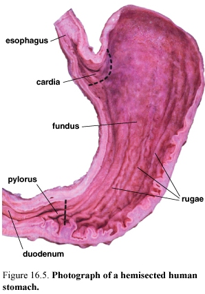
The stomach is divided into three distinct into three distinct parts:
-the cardia (cardiac region) the part near the esophageal orifice, contains the cardiac glands
-the pylorus (pyloric region) the part proximal to the pyloric sphincter, contains the pyloric glands
-the fundus (fundic region) sometimes called the body, the largest part of the stomach, is situated between the cardia and pylorus and contains the fundic or gastric glands.
Gastric secretions
The gastric secretions include pepsinogen, hydrochloric acid and intrinsic factor.
In addition, the hormone gastrin and other hormones and hormone like secretions are produced by enteroendocrine glands in the gastric epithelium.
Gastric mucosa
The epithelium that lines the surface and the gastric pits of the stomach is simple columnar. These columnar cells are designated surface mucous cells. The mucus secretion is described as visible mucus because of its cloudy appearance.
Fundic glands of the gastric mucosa
Figure 56. Diagram of the fundic gland
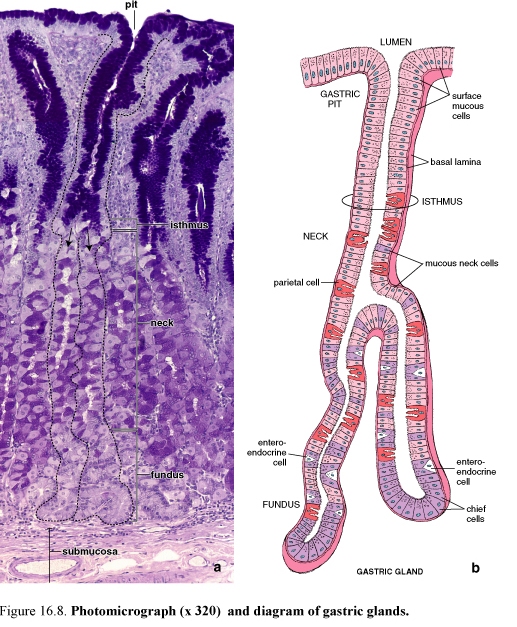
Fundic glands produce the digestive juice of the stomach. The fundic glands, also referred to as gastric glands, are present throughout the entire gastric mucosa except for the relatively small regions occupied by the cardiac pyloric glands. They are simple, branched tubular glands.
Each gland has a narrow, relatively long neck segment and a shorter and wider base or fundic segment. Typically, several glands open into a single gastric pit.
Fundic glands are composed of four functionally different cell types. In addition, undifferentiated cells that give rise to these cells are also present. Thus, the various cells are:
-mucous neck cells
-chief cells
-parietal cells, also called oxyntic cells
-enteroendocrine cells
-undifferentiated cells present in the upper neck region of the gland that give rise to the mature cells listed
1. Mucous neck cells are localized in the neck region, interspersed with parietal cells. The cell secretes soluble mucus compared with insoluble or cloudy mucus produced by the surface mucous cells.


2. Chief cells are located in the deepest part of the fundic glands. Chief cells are typical protein-secreting cells. Chief cells secrete pepsin in an inactive precursor form designated pepsinogen and a weak lipase
3. Parietal cells are found in the neck of the fundic glands, among the mucous neck cells and in the deeper part of the gland. Parietal cells secrete HCL and intrinsic factor. Parietal cells have an extensive intracellular canalicular system that communicates with the lumen of the gland. Intrinsic factor, a glycoprotein that is essential for the absorption of vitamin B12.
4. Enteroendocrine cells secrete their product into the lamina propria. These cells secrete gastrin, one of the gastrointestinal polypeptide hormones, is the principal effective agent for stimulating the secretion of HCL.
II. Cardiac glands of the gastric mucosa
The glands are simple tubular, and branched. They are composed mainly of mucus-secreting cells, with occasional interspersed enteroendocrine cells.
III. Pyloric glands of the gastric mucosa
Pyloric glands are located in the pyloric antrum (the part of the stomach between the fundus and the pylorus) and the pylorus. They are branched, tubular glands that are coiled. Enteroendocrine cells are found interspersed within the gland epithelium along with occasional parietal cells.
Lamina propria and muscularis mucosae are not unique.
Gastric submucosa
Gastric submucosa is composed of a dense connective tissue and submucosal (Meissner’s) plexus.
Gastric muscularis externa
The muscularis externa of the stomach consists of an outer longitudinal layer, a middle circular layer, and an inner oblique layer.
Gastric serosa is not unique.
Small intestine
It is divided into three anatomic segments:
-duodenum
-jejunum
-ileum
Intestinal lining
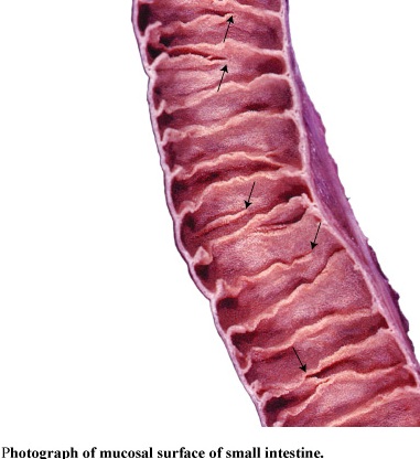
Plicae circulares, villi and microvilli increase the absorptive surface of the small intestine.
Plicae circulares are permanent transverse folds that contain a core of submucosa. Each semilunar fold is circularly arranged and extends about one-half to two-thirds around the circumference of the lumen.
Villi are finger-like and leaf-like projections of the mucosa that extend into the intestinal lumen.
Microvilli of the enterocytes provide the major amplification of the luminal surface. They give the apical region of the cell a striated appearance, the so-called striated border.
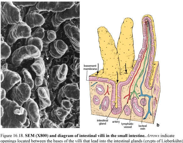
Figure 57. Photograph of mucosal surface of the small intestine (left) and SEM of intestinal villi in the small intestine (right); arrows indicate openings located between the bases of the villi that lead into the lead into the intestinal glands
Mucosa
The villi and intestinal glands along with lamina propria and associated GALT (Gut Associated lymphoid tissue) and muscularis mucosae constitute the essential features of the small intestinal mucosa.
Figure 58.Photomicograph (left) and diagram (right) of intestinal villus
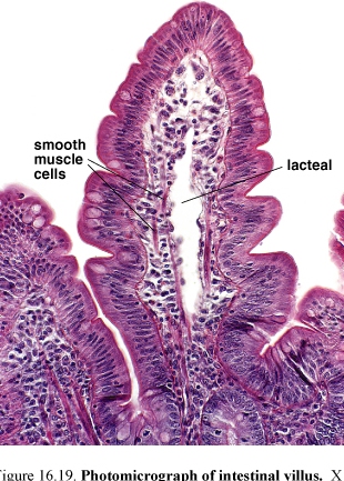
Villi completely cover the surface of the small intestine. The core of the villus consists of an extension of the lamina propria with a network of fenestrated capillaries located just under the epithelial basal lamina.
The lamina propria of the villus also contains a central, blind ending lymphatic capillary, the lacteal. Smooth muscle cells and myofibroblasts present here and help villi to contract and shorten intermittently.
The intestinal glands or crypts of Liberkühn are simple tubular structures. They open on to the luminal surface of the intestine of the base of the villi.
The lamina propria surrounds the glands and contains numerous cells of the immune system, particularly in the villi. The lamina propria also contains numerous nodules of lymphatic tissue that represent a major component of the GALT. They are large and numerous in the ileum, where they are located on the side of the intestine opposite the mesenteric attachment. It is called aggregated nodules or Peyer’s patches.
The muscularis mucosae consist of two thin layers of smooth muscle cells, an inner circular and an outer longitudinal layer. Strands of smooth muscle cells extend from the muscularis mucosae into the lamina propria of the villi.
Cells of the mucosal epithelium
They include:
-enterocytes, whose primary function is absorption
-goblet cells, unicellular mucin-secreting glands
-paneth cells
-enteroendocrine cells
-M cells (microfold cell)
Enterocytes
Figure 59. Diagrams of an enterocyte in different phases of absorption
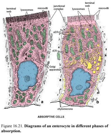
Enterocytes are specialized for the transport of substances from the lumen of the intestine to the circulatory system. They are tall columnar cells with a basally positioned nucleus.
Microvilli of the enterocytes increase the apical surface area as much as 600 times. They are formed striated border on the luminal surface. Each microvilli has a core of vertically oriented actin microfilaments.
Enterocytes are bound to one another and to the other cells of the epithelium by junctional complexes. The junction establishes a barrier between the lumen and the intercellular compartment. The lateral membranes of the enterocytes show elaborate development of flattened processes (plications) that implicate with processes of adjacent cells, thus increasing the amount of plasma membrane containing transport enzymes. During active absorption, especially of electrolytes, water, and lipids the lateral plications separate, allowing the development of an enlarged intercellular compartment.
In addition to the membrane specializations associated with absorption and transport, the cytoplasm of the enterocytes is also specialized for these functions. Elongated mitochondria that provide energy for the transport function of the cells are concentrated in the apical cytoplasm. Tubules and cisternae of the sER, which are involved in the absorption of fatty acids and glycerol and in the resynthesis of neutral fat, are found in the apical cytoplasm under the terminal web.
Enterocytes are also secretory cells producing glycoprotein enzymes needed for terminal digestion and absorption. Small secretory vesicles containing glycoproteins destined for the cell surface are located in the apical cytoplasm along the lateral plasma membrane. Free ribosomes, rER, and Golgi complex provide the secretory function of the enterocytes.
G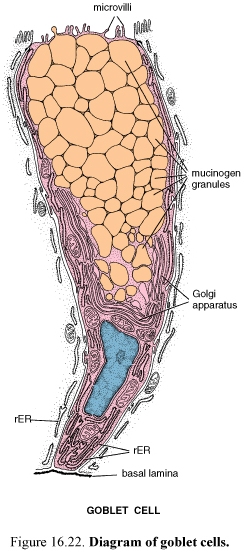 oblet
cells
oblet
cells
Goblet cells increase in number from the proximal to the distal small intestine and are most numerous in the terminal ileum. As in other epithelia, goblet cells produce mucus. There are a large accumulation of mucinogen granules in the apical cytoplasm that distends the apex of the cell and distorts the shape of neighboring cells. An extensive array of flattened Golgi saccules forms a wide cup around the newly formed mucinogen granules near the basal part of the cell.
Goblet cells have microvilli that are restricted to a thin rim of cytoplasm (the theca) that surrounds the apical-lateral portion of the accumulation of mucinogen granules. The large apical accumulation of mucinogen granules leaves the rest of the cell as a narrow stem forming the basal portion of the cell.
Paneth cells
Paneth cells are found in the bases of the mucosal glands. They are occasionally found in the normal colon in small numbers. The acidophilic secretory granules contain the antibacterial enzyme lysozyme, other glycoproteins, an arginine rich protein and zinc. Lysozyme digests the cell walls of certain groups of bacteria. This antibacterial action and the phagocytosis of certain bacteria and protozoa by Paneth cells suggest that Paneth cells have a role in regulating the normal bacterial flora of the small intestine.
Enteroendocrine cells
They are concentrated in the lower portion of the intestinal crypt but migrate slowly and can be found at all levels of the villus unit.
Cholecystokinin, secretin and gastric inhibitory peptide are the most active regulators of gastrointestinal physiology that are released in this portion of the gut.
These three hormones increase pancreatic and gallbladder activity and inhibit gastric secretory function and motility.
M cells (microfold cell)
Figure 60. Diagram (a) and SEM (b) of M cells in a lymphatic nodule of the intestine
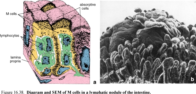
The epithelial cells that overlie Peyer’s patches and other large lymphatic nodules are different from the surrounding intestinal cells.
They are nearly squamous have microfolds rather than microvilli on their apical surface and take up macromolecules from the lumen in endocytic vesicles.
Intermediate cells
Intermediate cells constitute the majority of the cells in the lower half of the intestinal crypt. Intermediate cells have characteristics of both immature absorptive cells and goblet cells. These cells are still capable of cell division. These cells have short, irregular microvilli and small mucin-like secretory droplets which form a column in the center of the supranuclear cytoplasm.
Submucosa
A distinguishing characteristic of the duodenum is the presence of submucosal glands.
The submucosa consists of a dense connective tissue and localized sites that contain aggregates of adipose cells.
A conspicuous feature in the duodenum is the presence of submucosal glands (of Brunner).
The branched tubuloalveolar submucosal glands of the duodenum have secretory cells with characteristics of both zymogen-secreting and mucus-secreting cells.
The secretion of these glands has a pH of 8,1-9,3 and contains neutral and alkaline glycoproteins and bicarbonate ions. This probably serves to protect the proximal small intestine by neutralizing the acid-containing chime that is delivered to it and serves to bring the Ph of the intestinal contents close to the optimal pH for the pancreatic enzymes that are also delivered to the duodenum.
Muscularis externa
The muscularis externa consists of an inner layer of circularly arranged smooth muscle cells and an outer layer of longitudinally arranged smooth muscle cells.
Two kinds of muscular contraction occur in the small intestine. Local contractions displace intestinal contents both proximally and distally are designated as segmentation.
These contractions are primarily of the circular muscle layer.
They serve to circulate the chime locally, mixing it with digestive juices and moving it into contact with the mucosa for absorption.
Peristalsis, the second type of contraction, largely involves the longitudinal muscle layer and moves the intestinal contents distally.
Serosa
Serosa is not unique.
Large intestine
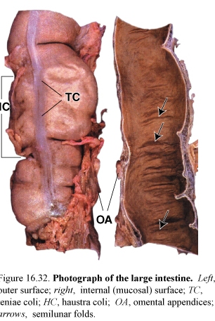
Figure 61. Photograph of the large intestine (left) outer surface; TC, teniae coli; HC, haustra coli, OA, omental appendices; (middle) internal (mucosal) surface; arrows, semilunar folds; (right) photomicrograph of the mucosa and part of the submucosa of the large intestine; arrows, openings of the glands at the intestinal surface

The large intestine is composed of the cecum, ascending colon, sigmoid colon, rectum, and anal canal.
The four layers characteristics of the alimentary canal are present throughout. There are, however several distinctive features at the gross level
-The mucosa has a smooth surface; neither plicae circulares nor villi are present.
-The outer longitudinal layer of the muscularis externa exhibits three equally spaced bands.
Mucosa
The mucosa of the large intestine contains numerous straight tubular glands
that extends through the full thickness of the mucosa. The glands consist of simple columnar epithelium.
The principal functions of the colon are reabsorption of electrolytes and water and elimination of undigested food and waste.
The absorptive cells have morphology essentially identical with that of the enterocytes of the small intestine. The reabsorption of water and electrolytes is the primary function of the columnar absorptive cells. Goblet cells produce mucin that is secreted continuously to lubricate the bowel, facilitating the passage of the increasingly more solid colonic contents. Goblet cells are more numerous in the large intestine than in the small intestine.
The mucosal epithelium of the large intestine contains the same cell types as the small intestine; Paneth cells are normally absent in the adult human.
Columnar absorptive cells predominate (4:1) over goblet cells in most of the colon, only near the rectum the number of goblet cells increasing (1:1).
The intercellular space is often dilated, indicating active transport of fluid.
In the luminal surface the secretion rate exceeds the synthesis rate, and “exhausted” goblet cells appear in the epithelium between crypts. The caveolated “tuft” cell, has also been described in the colonic epithelium, this cell may, however, be one form of exhausted goblet cell.
Epithelial cell renewal in the large intestine
As in the small, all of the mucosal epithelial cells of the colon arise from stem cells located at the bottom of the crypt or gland.
The lower third of the crypt constitutes the normal replicative zone.
Lamina propria
The structural features are:
-the collagen table, a thick layer of collagen and ground substance just below the free surface
-elaborate development of GALT
-a well developed pericryptal fibroblast sheath
-absence of lymphatic vessels in the lamina propria
Muscularis externa
In the colon, the outer layer of the muscularis externa is, in the part, condensed into prominent longitudinal bands of muscle that may be seen at the gross level; these are called the teniae coli. Between the bands, the longitudinal layer forms an extremely thin sheet.
In the rectum, the outer longitudinal layer of smooth muscle is a uniformly thick layer, as in the small intestine.
Bundles of muscle from the teniae coli penetrate the inner, circular layer of muscle at intervals along the length and circumference of the colon. These apparent discontinuities in the muscularis externa allow segments of the colon to contract independently, leading to the formation of saccules (haustra) in the colon wall.
The muscularis externa of the large intestine produces two major types of contraction: segmentation and peristalsis
Submucosa and serosa
The submucosa is not unique. Where the large intestine is directly in contact with other structures, its outer layer is adventitia; elsewhere, the outer layer is a typical serosa.
Cecum and appendix
Figure 63. Photomicrograph of a cross section through the vermiform appendix

The cecum forms a blind pouch just distal to the ileocecal valve; the appendix is a thin, finger-like extension of this pouch. The histology of cecum closely resembles that of the rest of the colon; the appendix differs from it in having a complete layer of longitudinal muscle in the muscularis externa. The most conspicuous feature of the appendix is the large number of lymphatic nodules that fuse and extend into the submucosa.
Rectum and anus
Figure 64. Drawing of the rectum and anal canal

The rectum is dilated distal portion of the alimentary tract. Its upper part is distinguished from the rest of the colon by the presence of folds called transverse rectal folds. The most distal portion of the alimentary canal is the anal canal. It extends from the anorectal junction to the anus. The upper part of the anal canal has longitudinal folds called anal columns. Depressions between the anal columns are called anal sinuses.
The mucosa of the rectum is similar to that of the rest of the distal colon, having straight, tubular intestinal glands with many goblet cells.
Anal canal
In the anal canal, anal glands extend into the submucosa and even into the muscularis externa. These are branched, straight tubular glands that secrete mucus onto the anal surface through ducts lined with stratified columnar epithelium.
Large apocrine glands, the circumanal glands, are found in the skin surrounding the anal orifice.
The muscularis mucosa disappears at about the level of the rectoanal margin, but at the same level, the circular layer of the muscularis externa thickness to form the internal anal sphincter. The external anal sphincter is formed by the striated muscles of the perineum.
Pancreas
The pancreas is an exocrine and endocrine gland.
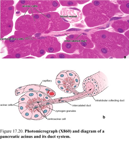
Figure 65. Diagram of a pancreatic acinus and its duct system
The exocrine component is a serous gland that synthesizes and secretes, into the duodenum, enzymes that are essential for digestion in the intestine.
The endocrine component synthesizes and secretes, into the blood, insulin and glucagons, hormones that regulate glucose, lipid and protein metabolism in the whole body.
Exocrine pancreas
The secretory units are acinar or tubuloacinar in shape and are formed by a simple epithelium of pyramidal serous cells. The serous secretory cells of the acinus produce the digestive enzyme precursors secreted by the pancreas. Pancreatic acini are unique among glandular acini, in that the initial duct that leads from the acinus, the intercalated duct, actually begins within the acinus. The duct cells located inside the acinus are referred to as centroacinar cells.
The acinar cells are characterized by acidophilic zymogen granules.
Zymogen granules contain a variety of digestive enzymes in an inactive form.
Pancreatic enzymes are capable of digesting most food substances.
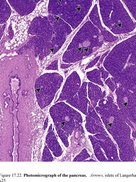
Figure 66. Photomicrograph of the pancreas; arrows islets of Langerhans
They include:
-trypsinogen, pepsinogen, and procarboxypeptidase digest proteins
-amylase digests carbohydrates
-lipase digests lipids
-deoxyribonuclease and ribonuclease digest nucleic acids.
The pancreatic digestive enzymes are activated only after they reach the lumen of the small intestine.
Duct system
The centroacinar cells are the beginning of the duct system of the exocrine pancreas.
Centroacinar cells are intercalated duct cells located in the acinus.
Centroacinar cells are continuous with the cells of the short intercalated duct that lies outside the acinus. The intercalated ducts are short and drain to intralobular collecting ducts. There are no striated (secretory) ducts in the pancreas.
The complex, branching network of intralobular ducts drains to the larger interlobular ducts, which are lined with a low columnar epithelium in which enteroendocrine cells and occasional goblet cells may be found. The interlobular ducts, in turn, drain directly into the main pancreatic duct. A second large duct, the ductus choledochus (accessory pancreatic duct) arises in the head of the pancreas.
The pancreas secretes about 1 liter per day. While the acini secrete a small volume of protein-rich fluid, the intercalated duct cells secrete a large volume of fluid rich in sodium and bicarbonate.
The bicarbonate serves to neutralize the acidity of the chyme that enters the duodenum from the stomach and to establish the optimum pH for the activity of the major pancreatic enzymes.
Hormonal control of exocrine secretion
Two hormones secreted by the enteroendocrine cells of the duodenum, secretin and CCK, also called pancreozymin, and are the principal regulators of the exocrine pancreas. The entry of the acidic chyme into the duodenum stimulates the release of these hormones into the blood. The coordinate action of the two hormones produces the secretion into the duodenum of a large volume of enzyme-rich, alkaline fluid.
Endocrine pancreas
The endocrine pancreas is a diffuse organ that secretes hormones that regulate blood glucose levels.
The islets of Langerhans, the endocrine component of the pancreas, are scattered throughout the organ. The islets constitute about 1-2% of the volume of the pancreas but are most numerous in the tail.
It is possible to identify three principal cell types designated A, B and D cells and three minor islet cell types designated PP, D-1 and EC cell.
Principal cell types of the islet
Figure 67. Diagram of an islet of Langerhans
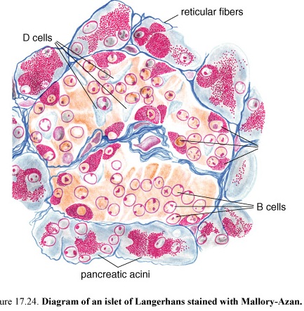
The B cells constitute about 70% of the total islet cells in humans and are generally located in its central portion.
They secrete insulin. Its principal effects are on the liver, skeletal muscle, and adipose tissue. Insulin stimulates
-uptake of glucose from the circulation
-utilization and storage of glucose by all cells
-phosphorylation of glucose on the cells
-synthesis of glycogen from the phosphorylated glucose
Absence or inadequate amounts of insulin lead to elevated blood glucose levels and the presence of glucose in the urine, a condition known as diabetes mellitus.
In addition, insulin stimulates glycerol synthesis in adipose cells and inhibits lipase activity in these cells.
The A cells constitute about 15-20% of the human islet population and are generally located peripherally in the islets. They secrete glucagons. Glucagon raises blood glucose.
Glucagon stimulates release of glucose into the blood-stream, and stimulates gluconeogenesis (synthesis of glucose from metabolites of amino acids) and glycogenolysis (breakdown of glycogen) in the liver.
Glucagon also stimulates proteolysis to promote gluconeogenesis, mobilizes fats from adipose cells, and stimulates hepatic lipase.
The D cells constitute about 5-10% of the total pancreatic endocrine tissue and are also located peripherally in the islets. D cells secrete somatostatin. The precise role of somatostatin in the islets is unclear, but it has been shown to inhibit both insulin and glucagon secretion.
The minor islet cells constitute about 5% of the islet tissue.
The PP cells secrete pancreatic polypeptide. It stimulates gastric chief cells, inhibits bile secretion and intestinal motility, inhibits pancreatic enzymes and bicarbonate secretion.
The D-1 cells secrete vasoactive intestinal peptide. Its principal effects are similar to those of glucagon plus these cells stimulate pancreatic exocrine secretion.
The EC cells secrete secretin, motilin and substance P. Secretin- acts locally to stimulate bicarbonate secretion in pancreatic fluid and pancreatic enzyme secretion, motilin-increases gastric and intestinal motility, substance P-unclear.
Liver
The liver is the largest mass of glandular tissue in the body and is also the largest internal organ. The liver has both exocrine and endocrine functions. The liver is an organ engaged in numerous metabolic conversions involving substrates brought to and from the digestive tube, pancreas and spleen. Some of the products of these metabolic conversions are carried in the exocrine secretion of the liver, called bile.
The endocrine secretions of the liver are released directly into the blood that supplies the liver parenchyma. These secretions include substances synthesized by the liver cells, i.e.,
-albumin
-prothrombin
-nonimmune α and β globulins
-numerous glycoproteins, including fibronectin
-glycogen, and glucose released by hydrolysis of glycogen
Blood supply to the liver
The liver receives blood that initially supplied the small intestine, pancreas and spleen.
The hepatic portal vein carries about 75% of the blood supply to the liver. The hepatic portal vein (hpv) carries venous blood from the digestive tube, pancreas and spleen into liver.
The hepatic artery, a branch of the celiac trunk, carries oxygenated blood to the liver.
Within the liver, the distributing branches of the hpv and hepatic artery, which supply the sinusoidal capillaries (sinusoids) that bathe the hepatocytes (epithelial cells of the liver) and the draining branches of the bile duct system course together in a relationship termed the portal triad.
Figure 68. Blood supply to the liver, portal triad

Liver lobules
There are three ways of describing the structure of the liver in terms of a functional unit: the “classic” lobule, the portal lobule, and the liver acinus.
Classic lobule
Figure 69. Diagram of a classic liver lobule
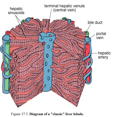

The classic lobule is the traditional description of the organization of the liver parenchyma. The classic hepatic lobule is a roughly hexagonal block of tissue.
The classic lobule consists of stacks of anastomosing plates of hepatic cells, separated by the anastomosing system of sinusoids that perfuse the cells with the mixed portal and arterial blood. At the center of the lobule is the terminal hepatic venule (central vein) into which the sinusoids drain. The plates of cells radiate from the central vein to the periphery of the lobule, as do the sinusoids. At the angles of the hexagon are the portal areas (portal canals), loose stromal connective tissue characterized by the presence of the portal triads. At the edges of the portal canal between the connective tissue stroma and hepatocytes is a small space called the space of Mall. This is one of the sites where lymph originates in
the liver.
Liver acinus
Figure70. Diagram of a classic liver acinus


The liver acinus described as diamond shaped, is the smallest functional unit in the hepatic parenchyma. The short axis of the acinus is defined by the terminal branches of the portal triad that lie along the border between two “classic” lobules. The long axis is a line drawn between the two central veins closest to the short axis.
The hepatocytes in each liver acinus are described as being arranged in three concentric elliptical zones surrounding the short axis. Thus,
Zone 1 is closest to the axis. Zone 2 is farthest from the axis. Zone 3 lies between zones 1 and 3.
The cells in zone 1 are the first to receive both nutrients and toxins in the blood and are the first to show morphologic changes following bile stasis. These cells are also the last to die and the first to regenerate.
The cells in zone 3, on the other hand, are the first to show ischemic necrosis. They are last to respond to toxic substances and to bile stasis
The cells in zone 3 have all characteristic between zones 1 and 3.
The liver acinus provides the best correlation among blood perfusion, metabolic activity, and liver pathology.
Portal lobule

The portal lobule emphasizes the exocrine functions of the liver. The morphologic axis of the portal lobule is the interlobular duct of the portal triad of the “classic” lobule. Its outer margins are imaginary lines between the three central veins that are closest to that portal triad. This defines a roughly triangular block of tissue that includes those portions of three classic lobules that secrete the bile that drains into its axial bile duct.
Blood vessels of the parenchyma
Figure 71. Diagram of the blood and bile flow in the liver

The blood vessels that occupy the portal canals are called interlobular vessels. Only the interlobular vessels that form the smallest portal triads send blood into the sinusoids. The larger interlobular vessels branch into distributing vessels that are located at the periphery of the lobule. These distributing vessels send inlet vessels to the sinusoids. In the sinusoids the blood flows centripetally toward the central vein. The central vein courses through the central axis of the classic liver lobule and empties into a sublobular vein. Several sublobular veins converge to form larger hepatic veins that empty into the inferior vena cava.
Sinusoids
Hepatic sinusoids are lined with a thin discontinuous endothelium. Hepatic sinusoids differ from other sinusoids in that a second cell type, the stellate sinusoidal macrophage or Kupffer cell is a regular part of the vessel lining.
Kupffer cells belong to the mononuclear phagocytic system. Processes of Kupffer cells often seem to span the sinusoidal lumen and may even partially occlude it. The Kupffer cells may be involved in the final breakdown of some damaged or senile red blood cells that reach the liver from the spleen.
Figure 72. Schematic diagram of a plate of hepatocytes interposed between hepatic sinusoids

Perisinusoidal space (space of Disse)
The Perisinusoidal space is the site of exchange of materials between blood and liver cells. The perisinusoidal space lies between the basal surfaces of the hepatocytes and the basal surfaces of the endothelial cells and Kupffer cells that line the sinusoids.
Irregular microvillous processes project into this space from the basal plasma membrane of the hepatocytes. Because of the large gaps in the endothelial layer and the absence of a continuous basal lamina, there is no significant barrier between the blood plasma in the sinusoid and the hepatocyte plasma membrane that forms the parenchymal border of the perisinusoidal space. A third cell type, the lypocyte or adipose cell (commonly called an Ito cell) is found in the perisinusoidal space. These cells have been shown to be the primary storage site for vitamin A, the vitamin A is transported from the liver to the retina, where it is used in the synthesis of visual pigments.
Hepatocytes
Hepatocytes make up the anastomosing cell plates of the liver lobule. Hepatocytes
are large polygonal cells that constitute about 80% of the cell population of the liver. Nuclei of hepatocytes are large and spherical and occupy the center of the cell. Liver cells are capable of considerable regeneration when liver substance is lost to hepatotoxic processes, disease, or surgery. The hepatocyte cytoplasm is generally acidophilic. Specific cytoplasmic components include:
-rER and free ribosomes
-numerous mitochondria
-glycogen
-lipid droplets
-small Golgi complexes (elements of the Golgi complex concentrated near the bile canaliculus are believed to be associated with “exocrine” secretion of bile.
-a lot of peroxisomes
Peroxisomes have specific oxidative functions in:
-gluconeogenesis
-metabolism of purines
-metabolism of alcohol
-metabolism of lipids
In humans catalase and D-amino acid oxidase, as well as alcohol dehydrogenase are found in peroxisomes.
The sER contains enzymes involved in degradation and conjugation of toxins and drugs as well as enzymes responsible for synthesizing cholesterol and the lipid portion of lipoproteins.
Certain drugs and hormones stimulate increased activity in the smooth ER.
Lysosomes
Lysosomes concentrated near the bile canaliculus correspond to the peribiliary dense bodies. They have varied contents including:
-pigment granules (lipofuscin)
-myelin figures
-partially digested cytoplasmic organelles
Hepatocyte lysosomes may also be a normal storage site for iron.
Biliary tree
The bile canaliculus is small canal formed by grooves in neighboring cells. The canaliculus contains short, irregular microvilli from the hepatocyte. The bile canaliculi form a ring about the hepatocyte and constitute a network that drains into small bile ducts, the canals of Hering, these in turn, drain into the bile duct of the portal canals. Bile canaliculi are formed by apposed grooves in the surface of adjacent hepatocytes. In three dimensions bile canaliculi may form a complete loop around four sides of the idealized six-sided hepatocytes. Bile flows in the canaliculi centrifugal. The flow of bile is in a direction opposite to the flow of blood, i.e. from the region of the central vein toward the portal canal. Near the portal canal, but still within the lobule, bile canaliculi join to form small ductules, the canals of Hering that are lined with cuboidal cells that are not hepatocytes.
Intra hepatic bile ducts
The ductules carry the bile to the interlobar bile ducts that form part of the portal triad. Interlobular duct join to form the right and left lobar ducts that in turn join at the hilum to form the common hepatic duct.
Extra hepatic bile ducts
Extra hepatic bile ducts carry the bile to the gall bladder and intestine.
Bile
About 90% of the bile salts a component of bile is reabsorbed and resecreted by the hepatocytes. Bile also consists of cholesterol, lecithin, bile pigments, water and electrolytes.
Gall bladder
The gall bladder concentrates and stores bile.
Mucosa
The empty or partially filled gall bladder has numerous deep mucosal folds. The mucosal surface consists of a simple columnar epithelium. The tall epithelial cells exhibit the following features:
-numerous, apical microvilli
-apical junctional complexes that join adjacent cells
-complex lateral plications
-concentrations of mitochondria in the apical and basal cytoplasm
Lamina propria
-Mucin-secreting glands
-large number of lymphocyte and plasma cells
Muscularis externa, adventitia and serosa
The smooth muscle bundles are some what randomly oriented. Contraction of the smooth muscle reduces the volume of the bladder, forcing its contents out through the cystic duct.
External to the muscularis externa is a thick layer of dense connective tissue.
Where the gall bladder is attached to the liver surface, this layer is referred to as the adventitia. The unattached surface is covered by a serosa of visceral peritoneum consisting of a layer of mesothelium and a thin layer of loose connective tissue.
Respiratory system
Figure 73. Diagram of respiratory passages
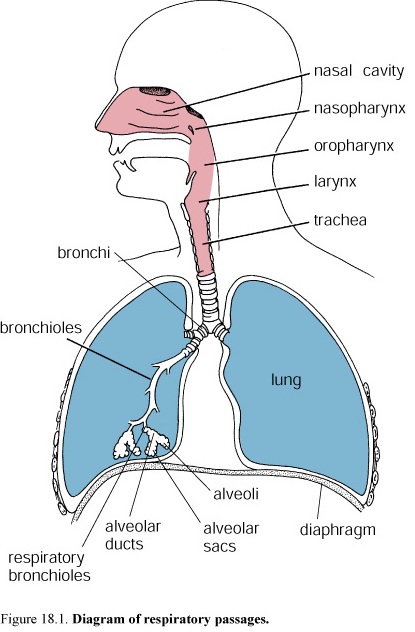
The respiratory system consists of the paired lungs and a series of air passages that lead to and from the lungs. Three principal functions are performed by this system, namely, air conduction, air filtration, and gas exchange (respiration). In addition, air passing through the larynx is used to produce speech, and air passing over the olfactory mucosa in the nasal cavities carries the stimuli for the sense of smell.
The Air Passages Consist of a Conducting Portion and a Respiratory Portion
The conducting portion of the respiratory system external to the lungs consists of
-Nasal cavities
-Nasopharynx and oropharynx
-Larynx
-Trachea
-Paired primary bronchi
Within the lungs are the internal bronchi, which undergo extensive branching to give rise to the distributing bronchioles. Collectively, the internal bronchi and the bronchioles constitute the bronchial tree.
The respiratory portion consists of
-Respiratory bronchioles
-Alveolar ducts
-Alveolar sacs
-Alveoli
Air passing through the respiratory passages must be conditioned before reaching the terminal respiratory units. Conditioning of the air occurs in the conducting portion of the system and consists of warming, moistening, and removal of particulate materials.
NASAL CAVITIES
The nasal cavities are paired chambers separated by a bony and cartilaginous septum. Each chamber is divided into three regions:
-Vestibule (nostril)
-Respiratory segment
-Olfactory segment
Vestibule of the Nasal Cavity
The vestibule communicates anteriorly with the external environment. It is lined with stratified squamous epithelium, and contains the hairs that filter out large particulate matter before it is carried in the airstream to the rest of the cavity. Sebaceous glands are also present, and their secretions assist in the entrapment of particulate matter.
Respiratory Segment of the Nasal Cavity
The respiratory segment constitutes most of the volume of the nasal cavities. It is lined by a ciliated, pseudostratified columnar epithelium.
It is composed of five cell types:
-Ciliated cells
-Goblet cells
-Brush cells, a general name for those cells in the respiratory tract that bear short, blunt microvilli
-Small granule cells, cells that resemble the basal cell but contain secretory granules
-Basal cells, stem cells from which the other cell types arise
The Mucosa of the Respiratory Segment Warms, Moistens, and Filters Inspired Air
The lamina propria of the respiratory segment has a rich, vascular network that includes a complex set of capillary loops. The lamina propria also contains mucous glands, many with serous demilunes. Their secretion supplements that of the goblet cells in the respiratory epithelium.
Olfactory Segment of the Nasal Cavity
Part of the dome of each nasal cavity form the olfactory segment and are lined with olfactory mucosa.
Figure 74. Diagram of the olfactory mucosa of the nasal cavity

The olfactory epithelium, is also pseudostratified, but it contains very different cell types. The olfactory epithelium is composed of the following cell types:
-Olfactory cells, bipolar neurons that span the thickness of the epithelium
-Supporting or sustentacular cells, columnar cells that provide mechanical and metabolic support to the olfactory cells
-Basal cells, stem cells from which new olfactory cells and supporting cells differentiate
-Brush cells, the same cell type that occurs in the respiratory epithelium
Lamina propria
This connective tissue contains numerous blood and lymphatic vessels.
The olfactory glands, a characteristic feature of the mucosa, are branched tubuloalveolar serous glands
Sinuses
Paranasal sinuses are air-filled spaces in the bones of the walls of the nasal cavity.
Pharynx
Figure 75. Diagram of the relationship of the pharynx to the respiratory and digestive system
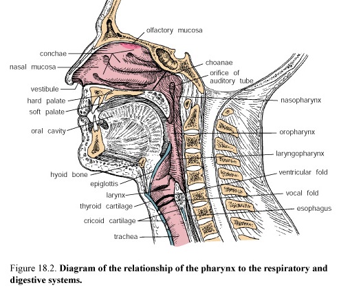
The pharynx connects the nasal and oral cavities to the larynx and esophagus. The pharynx is located posterior to the nasal and oral cavities and is divided regionally into the nasopharynx and oropharynx, respectively. The auditory (Eustachian) tubes connect the nasopharynx to each middle ear. Diffuse lymphatic tissue and lymphatic nodules are found in the wall of the nasopharynx. The concentration of such nodules in the posterior wall is called the pharyngeal tonsil.
Larynx
F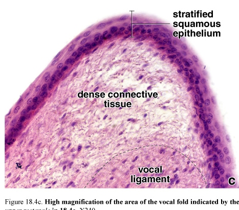 igure
76. The area of the vocal folds
igure
76. The area of the vocal folds
The passageway for air between the oropharynx and trachea is the larynx. It is a complex tubular segment of the respiratory system that is formed by irregularly shaped plates of hyaline and elastic cartilage.
Vocal Folds Control the Flow of Air Through the Larynx and Vibrate to Produce Sound
The vocal folds, also referred to as vocal cords, are two folds of mucosa that project into the lumen of the larynx. A supporting ligament and skeletal muscle, the vocalis muscle, is contained within each vocal fold-ligament. The ventricular folds located above the vocal folds are the "false vocal cords".
Stratified Squamous and Ciliated, Pseudostratified Columnar Epithelium Line the
The luminal surface of the vocal cords is covered with stratified squamous epithelium. The rest of the larynx is lined with the ciliated, pseudostratified columnar epithelium that characterizes the respiratory tract. The connective tissue of the larynx contains mixed mucoserous glands that secrete through ducts onto the laryngeal surface.
Trachea
Figure 77. Photomicrograph showing the relationship between the trachea and the esophagus at the base of the neck
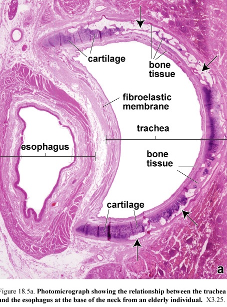
The trachea is a short tube. It serves as a conduit for air. The trachea extends from the larynx to about the middle of the thorax, where it divides into the two primary bronchi. The wall of the trachea consists of four layers: Mucosa, composed of ciliated, pseudostratified epithelium and an elastic fiber-rich lamina propria Submucosa composed of a slightly more dense connective tissue than the lamina propria. Cartilaginous layer composed of C-shaped hyaline cartilages. Adventitia, which binds the trachea to adjacent structures
1. Mucosa
Ciliated columnar cells, mucous (goblet) cells, and basal cells are the principal cell types in the tracheal epithelium. Brush cells are also present but in small numbers, as are the small granule cells.
Ciliated cells, the most numerous of the cell types. Cilia appear as short hair-like projections from the apical surface. The cilia provide a coordinated sweeping motion of the mucous coat.
Mucous cells are similar in appearance to the intestinal goblet cells and, thus, are often referred to by the same name. They accumulate mucinogen granules in their cytoplasm.
Brush cells are columnar cells that bear microvilli. The brush cell is regarded as a receptor cell.
Small granule cells are a representation in respiratory epithelium of the general class of enteroendocrine cells of the gut and gut derivatives. First type of small granule cell produces a catecholamine. A second cell type produces a polypeptide hormone.
Figure 79. Diagram of a brush cell(a) and small granule cell(b)
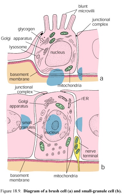
Basal cells serve as a reserve population by maintaining individual cell replacement in the epithelium.
Basement Membrane and Lamina Propria A Thick "Basement Membrane" Is Characteristic of Tracheal Epithelium Basement membrane appears as a glassy or homogeneous layer.
The lamina propria appears as a typical loose connective tissue. It is very cellular, containing numerous lymphocytes, many of which infiltrate the epithelium.
The boundary between mucosa and submucosa is defined by an elastic membrane.
2. Submucosa
In the trachea the submucosa is a relatively loose connective tissue similar in appearance to the lamina propria. Submucosal glands composed of mucus-secreting acini with serous demilunes are also present in the submucosa. The submucosal layer ends where its connective tissue fibers blend with the perichondrium of the cartilage layer.
3. Cartilaginous layer
The tracheal cartilages and trachealis muscle separate submucosa from adventitia.
A unique feature of the trachea is the presence of the C-shaped hyaline cartilages that are stacked on one another to form a supporting structure. Fibroelastic tissue and smooth muscle, the trachealis muscle, bridge the gap between the free ends of the C-shaped cartilages at the posterior border of the trachea, adjacent to the esophagus.
4. The adventitia
The outer layer, lies peripheral to the cartilage rings and trachealis muscle. It consists of loose connective tissue.
Bronchi
The trachea divides into two branches forming the primary or extrapulmonary bronchi (right and left bronchi). On entering the lungs the bronchi become the intrapulmonary bronchi, which branch immediately to give rise to the lobar bronchi (secondary bronchi).
The left and right lung is further divided into eight and ten bronchopulmonary segments.
Thus the lobar bronchi divide to give rise to segmental bronchi (tertiary bronchi); a segmental bronchus and the lung parenchyma that it supplies constitute a bronchopulmonary segment.
Bronchial Structure
The bronchi initially have the same general histologic structure as the trachea. At the point where the bronchi enter the lungs to become intrapulmonary bronchi, the structure of the bronchial wall changes. The cartilage rings are replaced by cartilage plates of irregular shape. Bronchi can be identified by their cartilage plates and a circular layer of smooth muscle. The second change observed in the wall of the intrapulmonary bronchus is the addition of smooth muscle to form a complete circumferential layer.
The wall of the bronchus consists of five layers.
Mucosa is composed of a pseudostratified epithelium having the same cellular composition as the trachea. The lamina propria is similar to that of the trachea but is reduced in amount in proportion to the diameter of the bronchi.
Muscularis is a continuous layer of smooth muscle in the larger bronchi. It may appear discontinuous in smaller bronchi. Contraction of the muscle maintains the appropriate diameter of the airway.
Submucosa remains as a relatively loose connective tissue. Glands are present as well as adipose tissue in the larger bronchi.
Cartilage layer consists of discontinuous cartilage plates that become reduced in size as the bronchial diameter diminishes.
Adventitia is dense connective tissue that is continuous with that of adjacent structures.
Figure 80. Divisions of the bronchial tree
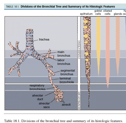
BRONCHIOLES
Bronchiolar Structure
Bronchioles are air-conducting ducts. The larger bronchioles represent branches of the segmental bronchi. These ducts branch repeatedly, giving rise to the smaller terminal bronchioles that also branch. They finally give rise to the respiratory bronchioles.
Cartilage and Glands Are Not Present in Bronchioles
The larger diameter bronchioles initially have a ciliated, pseudostratified columnar epithelium that gradually transforms to a simple ciliated columnar epithelium as the duct narrows. A relatively thick layer of smooth muscle is present in the wall of all bronchioles. Small bronchioles have a simple cuboidal epithelium. The smallest conducting bronchioles, the terminal bronchioles, are lined with a simple cuboidal epithelium in which Clara cells (see below) are found among the ciliated cells. A small amount of connective tissue underlies the epithelium, and a circumferential layer of smooth muscle underlies the connective tissue in the conducting portions.
Figure 81. Diagram of Clara cell between bronchial ciliated epithelial cells

Clara cells are nonciliated cells secrete a surface-active agent, a lipoprotein. The functional role of this agent is to prevent luminal adhesion should the wall of the airway fold on itself, particularly during expiration.
Respiratory Bronchioles Are the First Part of the Bronchial Tree That Allows Gas Exchange to Occur
Figure 82. Photomicrograph showing the respiratory portion of the bronchial tree. TB, terminal bronchiole; RB, respiratory bronchioles, AD, alveolar ducts; AS, alveolar sacs

Respiratory bronchioles constitute a transitional zone in the respiratory system concerned with both air conduction and gas exchange between air and blood. They have a narrow diameter and are lined by a cuboidal epithelium. Scattered, thin-walled outpocketings, alveoli, extend from the lumen of the respiratory bronchioles.
ALVEOLI
Alveoli Are the Site of Gas Exchange
Alveoli are the terminal air spaces of the respiratory system and are the actual site of gas exchange between the air and the blood. At some point, each alveolus is confluent with a respiratory bronchiole, by means of an alveolar duct, and an alveolar sac.
Alveolar ducts are elongate airways that have almost no walls, only alveoli, as their peripheral boundary with rings of smooth muscle in the knob-like interalveolar septa.
Figure 83. Photomicrograph showing an alveolar sac with adjacent alveoli.
AS, alveolar sac; A, alveoli

Alveolar sacs are spaces surrounded by clusters of alveoli. The surrounding alveoli open into these spaces. Alveoli are surrounded and separated from one another by a thin connective tissue layer that contains numerous blood capillaries. The tissue between adjacent alveolar air spaces is called the alveolar septum.
Alveolar Epithelium Is Composed of Type I and II Alveolar Cells and Occasional Brush Cells
Type I alveolar cells, also known as type I pneumocytes, are extremely thin squamous cells that line most of the surface of the alveoli.
Figure 84. Diagram of a type II alveolar cell
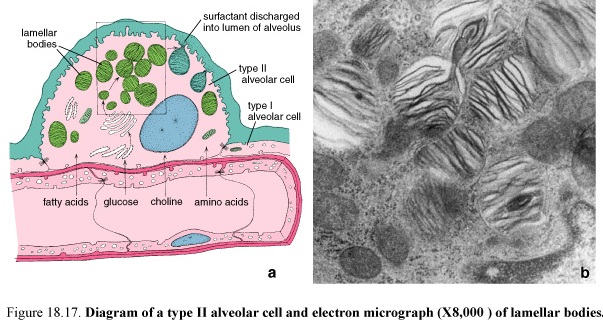
Type II alveolar cells are secretory cells. They are cuboidal cells interspersed among the type I cells. Their apical cytoplasm is filled with granules which rich in phospholipids, among which is the surface active agent surfactant. Without adequate secretions of surfactant, the alveoli would collapse on exhalation.
The Alveolar Septum Is the Site of the Air-Blood Barrier
Figure 85. Diagram of the alveolar septum
The components of the alveolar septum are
-Alveolar epithelial cells
-Basal lamina of the alveolar epithelium
-Basal lamina of the capillary endothelium
-Endothelial cells of the rich capillary network
-Other connective tissue elements, including fibroblasts, macrophages, collagen fibers, and elastic fibers
Most of the basal surface of the alveolar epithelium is in intimate association with the capillaries of the septum. Any one capillary may be shared by two or more alveoli.
The air-blood barrier refers to the cells and cell products across which gases must diffuse between the alveolar compartment and capillary compartment. The thinnest air-blood barrier consists of a monomolecular layer of surfactant, a type I epithelial cell and its basal lamina, and a capillary endothelial cell and its basal lamina. Often, these two basal laminae are fused.
Alveolar Macrophages Remove Inhaled Particulate Matter From the Air Spaces and Red Blood Cells From the Septum.
Urinary system
The urinary system consists of the paired kidneys, the paired ureters, which lead from the kidneys to the bladder, and the urethra, which leads from the bladder to the exterior of the body.
Kidneys Conserve Body Fluid and Electrolytes and Remove Metabolic Waste
The kidneys are highly vascular, large, reddish, bean-shaped organs.
The kidneys produce urine, initially an ultrafiltrate of the blood that is then modified by selective resorption and specific secretion by the cells of the kidney.
Kidneys Also Function as Endocrine Organs
The endocrine activities of the kidneys include
-Synthesis and secretion of erythropoietin, a growth factor regulating red blood cell formation
-Synthesis and secretion of renin, a hormone involved in control of blood pressure and blood volume
-Hydroxylation of vitamin D, a steroid prohormone, to produce its active form
KIDNEY STRUCTURE
Figure 86. Diagram of a kidney seen from behind

1. Capsule
The kidney surface is covered by a thin connective tissue capsule. The capsule consists of an outer layer of fibroblasts and collagen fibers and an inner layer of myofibroblasts. 2. Cortex and Medulla
Substance of kidney can be divided into two distinct regions:
-Cortex, the outer reddish brown-colored part
-Medulla, the much lighter-colored inner part
Approximately 90-95% of the blood passing through the kidney is in the cortex; 5-10% is in the medulla.
The Cortex Is Characterized by Renal Corpuscles and Their Tubules
The cortex consists of renal corpuscles, along with the convoluted and straight tubules of the nephron, the collecting tubules, and an extensive vascular supply. The renal corpuscles comprise the beginning segment of the nephron and contain a unique capillary network called glomerulus.
The surface of the kidney reveals a series of vertical striations that appear to emanate from the medulla. These are the medullary rays.
Each medullary ray contains straight collecting tubules and straight tubule components of the nephrons.
Figure 87.Diagram of adult human kidney
The Medulla Is Characterized by Straight Tubules, Collecting Ducts, and a Special Capillary Network
The straight tubular segments of the nephrons and the collecting ducts continue from the cortex into the medulla. They are accompanied by a capillary network, the vasa recta, that runs in parallel with the various tubules.
The tubules in the medulla, because of their arrangement and because of differences in length, collectively form a number of conical structures called pyramids. Usually 8— 12 pyramids may occur in the human kidney. The bases of the pyramids face the cortex and the apices face the renal sinus.
F igure
88. Light micrograph of human kidney showing the capsule (Cap) and
underlying cortex; outer layer of the capsule (OLC) and the inner
layer of the capsule (ILC)
igure
88. Light micrograph of human kidney showing the capsule (Cap) and
underlying cortex; outer layer of the capsule (OLC) and the inner
layer of the capsule (ILC)
The Renal Columns Represent Cortical Tissue Contained Within the Medulla
The caps of cortical tissue that lie over the pyramids form the renal columns.
The apical portion of each pyramid, which is known as the papilla, projects into a minor calyx, a cup-shaped structure that represents an extension of the renal pelvis. The tip of the papilla is perforated by the openings of the collecting ducts. The minor calyces are branches of the two or three major calyces that, in turn, are major divisions of the renal pelvis.
Kidney Lobes and Lobules
The Number of Lobes in a Kidney Equals the Number of Medullary Pyramids
The Lobule Consists of a Collecting Duct and All the Nephrons That It Drains
Nephron
The nephron is the basic functional and structural unit of the kidney.
In the human there are about: 2 million nephrons in each kidney. They are responsible for the production of urine and correspond to the secretory part of other glands. It is the collecting tubule that is responsible for the final concentration of the urine.
The renal corpuscle represents the beginning of the nephron. It consists of the glomerulus, a tuft of capillaries composed of 10-20 capillary loops, surrounded by a double-layered epithelial cup, the renal or Bowman's capsule. Bowman's capsule is the initial portion of the nephron where blood flowing through the glomerular capillaries undergoes a filtration process to produce the initial urine filtrate.
The glomerular capillaries are supplied by an afferent arteriole and are drained by an efferent arteriole that then branches, forming a new capillary network to supply the kidney tubules. This is then an arterial portal system. Where the afferent and efferent arterioles penetrate and exit from the parietal layer of Bowman's capsule is called the vascular pole. Opposite this site is the urinary pole of the renal corpuscle, where the proximal convoluted tubule begins.
Figure 89.Schematic diagram of a renal gromerulus and the structures associated at the vascular pole (top) and urinary pole (bottom)
Continuing from Bowman's capsule, the remaining segments of the nephron, namely, the tubular parts, are
-Proximal thick segment, consisting of the proximal convoluted tubule (pars convoluta) and the proximal straight tubule (pars recta)
-Thin segment, which constitutes the thin limb of the loop of Henle (the loop of Henle is the entire U-shaped portion of a nephron)
-Distal thick segment, consisting of the distal straight segment (pars recta) and the distal convoluted tubule (pars convoluta)
The distal convoluted tubule connects to the collecting tubule, often through a connecting tubule, thus forming the uriniferous tubule, i.e., the nephron plus collecting tubule.
Types of Nephrons
F igure
90.Diagram of two types of nephrons in the kidney and their
associated collecting ducts system
igure
90.Diagram of two types of nephrons in the kidney and their
associated collecting ducts system
The types of nephrons are based on where their renal corpuscles are located in the cortex:
•Cortical or subcapsular nephrons have their renal corpuscles located in the outer part of the cortex. They have short loops of Henle, extending only into the outer region of the pyramid.
•Juxtamedullary nephrons have their renal corpuscles in proximity to the base of a medullary pyramid. They have long loops of Henle and long thin segments that extend well into the inner region of the pyramid.
•Intermediate nephrons have their renal corpuscles in the midregion of the cortex. Their loops of Henle are of intermediate length.
Renal (Malpighian) Corpuscle
The Renal Corpuscle Contains the Filtration Apparatus of the Kidney
The renal corpuscle consists of the glomerular capillary tuft and the surrounding visceral and parietal epithelial layers of Bowman’s capsule. The filtration apparatus, enclosed by the parietal layer of Bowman’s capsule, consists of three components:
-endothelium of the glomerular capillaries has numerous fenestrations. -visceral layer of Bowman’s capsule -basal lamina lying between these two cellular elements
Figure 91.Schematic diagram of Mesangial cells and capillaries
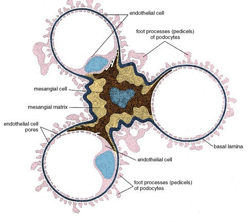
Podocytes Constitute the Visceral Layer of Bowman's Capsule
The podocytes, the cells of the inner or visceral layer of Bowman's capsule, extend processes around the glomerular capillaries. Each process, in turn, has numerous secondary processes called pedicels or foot processes. The elongated spaces between the interdigitating foot processes, called filtration slits, and allow the filtrate from the blood to enter the Bowman's space. The foot processes contain numerous microfilaments (actin) that are thought to have a role in regulating the size and patency of the filtration slits. The filtration slit membrane is thin membrane, spans the slits.
The thick basal lamina is the joint product of the endothelium and the podocytes. It is the principal component of the filtration barrier. It is called the glomerular basement membrane (GBM).
The Glomerular Basement Membrane Acts as a Physical Barrier and an Ion-Selective Filter
The components tend to localize in particular portions of the GBM:
The lamina rara interna, adjacent to the capillary endothelium
The lamina rara externa, adjacent to the podocyte processes
The lamina densa, the fused portion of the basal laminae, sandwiched between the laminae rarae
Type IV collagen is concentrated in the lamina densa, which is organized into a feltwork that acts as a physical filter. The GBM restricts the movement of particles, such as albumin or hemoglobin. The narrow slit pores formed by the pedicels and the filtration slit membranes also act as physical barriers to bulk flow and free diffusion.
The outer layer of Bowman's capsule, called the parietal layer, is a simple squamous epithelium. At the urinary pole of the renal corpuscle, it is continuous with the cuboidal epithelium of the proximal convoluted tubule.(figure 4)
The space between the visceral and parietal layers of Bowman's capsule is called the urinary or Bowman's space. It is the receptacle for the plasma filtrate produced by the filtration apparatus of the renal corpuscle.
Mesangium
The renal corpuscle contains an additional group of cells called mesangial cells. These cells and their extracellular matrix constitute the mesangium. Some Mesangial cells are located outside of the corpuscles along the vascular pole where they are also designated as lacis cells and form part of what is called the juxtaglomerular apparatus.
The functions of mesangial cells are:
-Mesangial cells are phagocytic; they can remove trapped residues and aggregated proteins from the GBM.
-Mesangial cells and their matrix provide structural support for the podocytes where the epithelial basement membrane is absent or incomplete.
-The cleaning of the GBM is believed to be the primary function of the mesangial cells.
Juxtaglomerular Apparatus
The Juxtaglomerular Apparatus Includes the Macula Densa, the Juxtaglomerular Cells, and the Extraglomerular Mesangial Cells
Lying directly adjacent to the afferent and efferent arterioles and adjacent to some extraglomerular mesangial cells at the vascular pole of the renal corpuscle is the terminal portion of the distal thick segment of the nephron. It contains, as part of its wall, cells that collectively are referred to as the macula densa. The cells of the macula densa are distinctive, in that they are narrower and usually taller than other distal tubule cells. The nuclei of these cells appear crowded, even to the extent that they appear partially superimposed over one another—thus, the name macula densa.
The smooth muscle cells of the adjacent afferent arteriole are modified. They contain secretory granules, and their nuclei are spherical. These smooth muscle cells are referred to as juxtaglomerular cells.
The Juxtaglomerular Apparatus Regulates Blood Pressure
The granules of the juxtaglomerular cells contain rennin, that is synthesized, stored, and released into the blood. In the blood, renin catalyzes the hydrolysis of circulating angiotensinogen to produce angiotensin. Then,
-Angiotensin II increases blood pressure.
-Angiotensin II stimulates the release of the hormone aldosterone from the zona glomerulosa of the adrenal gland.
-Aldosterone, in turn, increases distal tubular resorption of sodium and concomitant resorption of water, thereby raising blood volume and pressure.
-Angiotensin II is also a potent vasoconstrictor that has a regulatory role in the control of renal and systemic vascular resistance.
Renin secretion stimulates by decrease in NaCl concentration. The cells of the macula densa monitor the NaCl concentration in the afferent arteriole and regulate the release of renin by the juxtaglomerular cells.
KIDNEY TUBULE FUNCTION
As the glomerular filtrate passes through the uriniferous and collecting tubules of the kidney, it undergoes changes that involve both active and passive absorption, as well as secretion.
-Certain substances within the filtrate are reabsorbed, some partially, e.g., water, sodium, and bicarbonate and some completely, e.g., glucose.
-Other substances, e.g., creatinine and organic acids and bases, are added to the filtrate, i.e., the primary urine, by secretory activity of the tubule cells.
Thus, the volume of the filtrate is reduced substantially, and the urine is made hypertonic. The long loop of Henle and the collecting tubules that pass parallel to similarly arranged blood vessels, the vasa recta, serve as the basis the countercurrent multiplier mechanism that is instrumental in concentrating the urine and, thereby, making it hypertonic.
Proximal Thick Segment
The Proximal Thick Segment Is the Initial and Major Site of Reabsorption
The proximal convoluted tubule receives the primary filtrate from the urinary space of Bowman's capsule. The cuboidal cells of the tubule have the following features:
-A brush border, composed of numerous, relatively long microvilli
-A terminal bar apparatus, consisting of a narrow tight junction and a zonula adherens that seal off the intercellular space from the lumen of the tubule
-Large, flattened processes, plicae or folds, on the lateral surfaces of the cells, which alternate with similar processes of adjacent cells
-Extensive interdigitation of basal processes of adjacent cells
-Basal striations, consisting of elongate mitochondria concentrated in the basal processes and oriented normal to the basal surface
The Proximal Convoluted Tubule Reabsorbs 80% of the Primary Filtrate
Water and Electrolyte Reabsorption
The proximal convoluted tubule reabsorbs about 150 liters of fluid per day. As in the intestinal and gallbladder epithelia, this resorption of fluid is driven by active transport of Na+ into the lateral intercellular space. Here, the fluid is reabsorbed into the vessels of the peritubular capillary network.
The proximal tubule reabsorbs amino acids, sugars, and proteins.
Proteins and large peptides are reabsorbed by endocytosis in the proximal tubule
Proximal Straight Segment
The cells of the straight segment of the proximal tubule, i.e., the thick descending limb of the loop of Henle, are not as specialized for absorption as are those of the convoluted segment.
Thin Segment
The length of the thin segment of a nephron varies with its location in the cortex. The thin segment has four types of epithelial cells.
The cells possess few organelles. The specific functional roles of the four cell types in the thin segment are not yet clear.
Permeability of the Thin Segment
The descending portion of the thin segment is permeable, permitting free passage or equilibration of salt and water between the lumen of the nephron and the peritubular connective tissue.
Distal Thick Segment
The thick, straight, ascending segment of the distal tubule is the third part of the loop of Henle and includes both medullary and cortical portions, with the latter in the medullary rays. The straight segment of the distal tubule, like the ascending thin segment, transports ions from the tubular lumen to the interstitium. The large, cuboidal cells of the distal tubule have extensive basal-lateral plications and numerous large mitochondria associated with these basal folds. They have considerably fewer and less well-developed microvilli than proximal tubule cells.
The lateral margins of the cells are indistinct. The nucleus is located in the apical portion of the cell and sometimes, especially in the straight segment, causes the cell to bulge into the lumen.
The Distal Convoluted Tubule Exchanges Na+ for K+ Under Aldosterone Regulation
The distal convoluted tubule is short segment that responsible for:
-Reabsorption of Na+ and secretion of K+ in order to conserve Na+
-Continued reabsorption of bicarbonate ion
-Aldosterone secreted by the adrenal gland increases the reabsorption of Na+ and secretion of K+. This has the effect of increasing blood volume and blood pressure in response to increased blood Na+ concentration.
Antidiuretic hormone (ADH), can act on the terminal portion of the distal convoluted tubule to increase the permeability of the tubule to water, thereby producing a more concentrated urine. The action of this hormone is more significant in the collecting tubules and collecting ducts.
Collecting Tubules and Collecting Ducts
Both the collecting tubules and ducts are composed of a simple epithelium. The arched and cortical collecting tubules have flattened cells, somewhat squamous to cuboid in shape. The medullary collecting ducts have cuboidal cells, with a transition to columnar cells as the ducts increase in size.
Cytologically, two distinct types of cells are present in the collecting system:
Light cells, also called collecting duct or CD cells, are the principal cells of the system. They are cells with basal infoldings, true infoldings rather than processes that implicate with those of adjacent cells. They possess a single cilium and have relatively few short microvilli.
Dark cells, also called intercalated (1С) cells, occur in considerably smaller numbers. They have many mitochondria, and their cytoplasm appears more dense. Numerous vesicles are present in the apical cytoplasm.
INTERSTITIAL CELLS
The connective tissue of the kidney parenchyma, called interstitial tissue, surrounds the components of the nephrons, the ducts, and the blood and lymphatic vessels. In the cortex, two types of interstitial cells are recognized: cells that resemble fibroblasts, found between the basement membrane of the tubules and the adjacent peritubular capillaries, and occasional macrophages.
In the medulla, the principal interstitial cells resemble myofibroblasts. Prominent lipid droplets in the cytoplasm appear to increase and decrease in relationship to the diuretic state. Some experimental evidence suggests that these cells may secrete a hormone-like material that reduces blood pressure. In addition, it has been reported that prostaglandins and prostacyclin are synthesized in the interstitium.
HISTOPHYSIOLOGY OF THE KIDNEY
The Countercurrent Multiplier System Creates Hypertonic Urine
The ability to excrete a hypertonic urine depends or combination of three morphologic specializations:
-Loop of Henle
-Vasa recta, which form a loop paralleling the loop Henle
-Arrangement of the collecting ducts in the medulla
A Standing Gradient of Ion Concentration Produces Hypertonic Urine
The loop of Henle is the structure that allows the creation and maintenance in the medullary interstitium of a gradient of ion concentration that increases from the corticomedullary junction to the renal papilla. The concentration of NaCl in the interstitium gradually increases down the length of the loop of Henle and, consequently, through the thickness of the medulla from the corticomedullary junction to the papilla.
Collecting Ducts and Vasa Recta Are Countercurrent Exchangers
Vasa Recta
The efferent arterioles of the renal corpuscles of most of the cortex branch to form the capillary network that surrounds the tubular portions of the nephron in the cortex, the peritubular capillary network. The efferent arterioles of the juxtamedullary renal corpuscles form several unbranched arterioles that descend into the medullary pyramid. These arteriolae rectae make a hairpin turn deep in the medullary pyramid and ascend as the venulae rectae. Together, the descending arterioles and the ascending venules are called the vasa recta.
Countercurrent exchange occurs between the collecting system and vasa recta
Figure 92. Diagram showing movement of substances into and out of the nephron and collecting system

Because the ascending limb of the loop of Henle has a high level of transport activity and because it is impermeable to water, the modified filtrate that ultimately reaches the distal convoluted tubule is hypotonic. When ADH is present, the distal convoluted tubules, the collecting tubules and the collecting ducts are highly permeable to water. Within the cortex, the modified filtrate equilibrates to isotonicity in the distal convoluted tubule, partly by loss of water to interstitium and partly by addition of ions other than Na+ and Cl- to the filtrate. In the medulla, increasing amounts of water leave the filtrate as the collecting ducts pass through the increasingly hypertonic interstitium on their course to the papillae.
The vasa recta also form loops in the medulla parallel the loop of Henle. The vasa recta also form countercurrent exchange system in the following manner. As the arterial vessels descend through the medulla, the blood loses water to the interstitium and gains salt from the interstitium, so that at the tip of the loop, deep in the medulla, the blood essentially in equilibrium with the hypertonic interstitial fluid.
As the venous vessels ascend toward the corticomedullary junction, the process is reserved; the hypertonic blood loses salt to the interstitium and gains water from the interstitium. This passive countercurrent exchange of water and salt between the blood and the interstitium occurs without expenditure of energy by the endothelial cells.
Excretory passages
The urine that excreted at the tip of the papilla flows sequentially
-to the minor calyx of that papilla
-to a major calyx
-to the renal pelvis
-through the ureter to the urinary bladder
-through the urethra, where it is voided
All of these excretory passages except the urethra have the same general structure, namely, a mucosa, a muscularis, and an adventitia (or in some regions serosa)
Transitional epithelium
It lines all of the excretory passages. Transitional epithelium is essentially impermeable to salts and water. The apical surface of the cell is very irregular, with deep clefts penetrating into the apical region. Modified areas of plasma membrane called plaques are seen on the luminal surface of the cells.
Connective tissue and muscle
In the ureters and urethra there are usually two layers of smooth muscle beneath the lamina propria:
The inner layer is arranged in a loose spiral described as a longitudinal layer.
The outer layer is arranged in a tight spiral described as a circular layer
The smooth muscle of the urinary passages is mixed with connective tissue, so that it forms parallel bundles rather than pure muscular sheets. Peristaltic contractions of the smooth muscle move the urine from the minor calyces through the ureter to the bladder.
The smooth muscle of the bladder wall is less regularly arranged than that of the tubular portions of the excretory passages, and thus, there is more random mixing of muscle and collagen bundles. Contraction of the smooth muscle of the bladder muscle compresses the whole viscus and forces the urine into the urethra.
URETERS
The ureters conduct urine from the renal pelvis to the bladder and follow an oblique path through the wall of the bladder. Contraction of the smooth muscle of the bladder wall also compresses the openings of the ureters into the bladder. This is important in protecting the kidney from the spread of infection from the bladder and urethra.
The terminal portion of the ureters is a thick outer layer of longitudinal muscle.
URINARY BLADDER
The bladder is a distensible reservoir for urine; its size and shape change as it fills. The smooth muscle of the bladder wall forms an internal sphincter, a ring-like arrangement of muscle around the opening of the urethra. The triangular region defined by these three openings the trigone, is relatively smooth.
URETHRA
In the male, the urethra is about 20 cm long and serves as the terminal duct of both the urinary and genital systems. It has three distinct segments:
- Prostatic urethra extends for 3-4 cm from the neck of the bladder through the prostate gland
-Membranous urethra extends for about 1 cm from the apex of the prostate gland through the body wall
-Penile urethra extends for about 15cm through the length of the penis to open the body surface at the glans penis.
Male reproductive system
The male reproductive system consists of the testes, epididymides and genital ducts, accessory reproductive glands, and penis. The accessory glands include the seminal vesicles, prostate, and bulbourethral glands. The primary function of the testis is the production of sperm, the male gamete.
Figure 93. Schematic diagram demonstrating the components of the male reproductive system

TESTIS
Figure 94. Diagram and photomicrograph of a sagittal section of the human testis

The Testes Produce Sperm and Androgens
Androgens, especially testosterone, are steroid hormones secreted by the interstitial cells in the testes. They influence embryonic development, sexual maturation, and reproductive function.
The Testes Have an Unusually Thick Connective Tissue Capsule, the Tunica Albuginea
A thick fibrous connective tissue capsule, the albuginea, covers each testis. The inner part of this capsule is the tunica vasculosa, a loose connective tissue layer that contains blood vessels. Each testis is divided into approximately 250 lobules by incomplete connective tissue septa that project from the capsule.
Along the posterior surface of the testis, the albuginea thickens and projects inward as the mediastinum testis.
Each Lobule Contains Several Highly Convoluted Seminiferous Tubules
Each lobule contains 1—4 seminiferous tubules, which produce the sperm, and a connective tissue stroma in which the interstitial or Leydig cells are contained. Each tubule within the lobule forms a convoluted loop. The ends of the loop in the vicinity of the mediastinum are called the tubuli recti or straight tubule and are continuous with the rete testis anastomosing channel system within the mediastinum.
The Seminiferous Tubules Consist of a Seminiferous Epithelium Surrounded by a Tunica Propria
The seminiferous epithelium is a complex stratified epithelium composed of two basic populations of cells:
-Spermatogenic cells, which regularly replicate and differentiate into mature sperm
-Sertoli cells, also known as supporting or sustentacular cells.
Sertoli cells
Figure 95. Schematic drawing of the Sertoli cell and its relationship to the adjacent spermatogenic cells
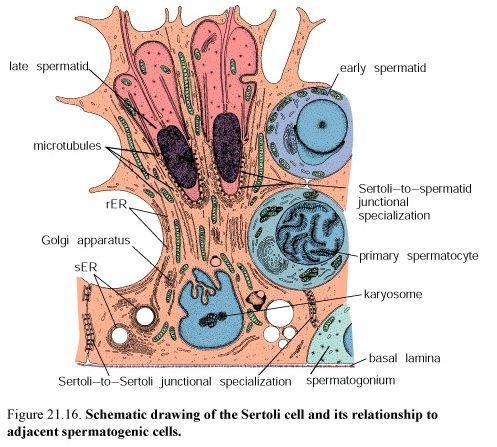
The Sertoli cells are columnar cells with complex apical and lateral processes that surround the adjacent spermatogenic cells and fill the spaces between them. The Sertoli cells give structural organization to the tubules as they extend through the full thickness of the seminiferous epithelium.
The Sertoli –Sertoli junctional complex divides the seminiferous epithelium into basal and luminal compartments.
Spermatogonia and early primary spermatocytes are restricted to the basal compartment, i.e., between the Sertoli-Sertoli junctions and the basal lamina. More mature spermatocytes and spermatids are restricted to the luminal side of the Sertoli-Sertoli junctions. The function of Sertoli cells is in the exchange of metabolic substrates and wastes between the developing spermatogenic cells and the circulatory system. In addition, Sertoli cells phagocytized and break down any spermatogenic cells that fail to differentiate completely.
The Sertoli-Sertoli junctional complex is the site of the blood-testis barrier.
Plasma proteins and circulating antibodies are excluded from the lumen of the seminiferous tubules. The exocrine secretory products of the Sertoli cells, particularly androgen-binding protein (ABP), can become highly concentrated in the lumen. Most importantly, the barrier isolates the genetically and, therefore, antigenically different haploid germ cells (secondary spermatocytes, spermatids, and sperm) from the immune system of the adult male. Antigens produced by or specific to the sperm are prevented from reaching the systemic circulation.
The tunica propria also called the lamina propria, is a multilayered connective tissue that lacks typical fibroblasts. In man, it consists of three to five layers of myoid cells and collagen fibrils external to the basal lamina of the seminiferous epithelium. Rhythmic contractions of the myoid cells create peristaltic waves that help move spermatozoa and testicular fluid through the seminiferous tubules to the excurrent duct system.
Leydig Cells
Leydig cells are large, polygonal, acidophilic cells that typically contain lipid droplets. The enzymes necessary for the synthesis of testosterone from cholesterol are associated with the sER. Leydig cells secrete testosterone.
INTRATESTICULAR DUCTS
Straight Tubules, the Tubuli Recti, Join the seminiferous Tubules with the Rete Testis of the mediastinum
At the end of each seminiferous tubule there is an abrupt transition to the tubuli recti or straight tubules. This short terminal section of the seminiferous tubule is lined only by Sertoli cells. Near their termination, the tubuli recti narrow, and their lining changes to a simple cuboidal epithelium.
The straight tubules empty into the rete testis, a complex series of interconnecting channels within the highly vascular connective tissue of the mediastinum. A simple cuboidal or low columnar epithelium lines the channels of the rete testis. These cells have a single apical cilium and few apical microvilli.
EXCURRENT DUCT SYSTEM
Efferent Ductules
The Efferent Ductules Connect the Rete Testis to the Epididymis
In man, approximately 20 efferent ductules (ductuli efferentes) connect the channels of the rete testis at the superior end of the mediastinum to the proximal portion of the ductus epididymis. They are highly coiled and form 6-10 conical masses.
The efferent ductules are lined with a pseudostratified columnar epithelium. Interspersed among the columnar cells are a few basal cells and intraepithelial lymphocytes. The tall columnar cells are generally ciliated.
The smooth muscle forms a layer in which the cells are arrayed as a circular sheath in the wall of the ductule. Transport of the sperm in the efferent ductules is effected largely by both ciliary action and contraction of this fibromuscular layer.
Ductus Epididymis
F igure
96. Photomicrograph of the human ductus epididymis
igure
96. Photomicrograph of the human ductus epididymis
The Ductus Epididymis Is a Highly Coiled Tube in Which Sperm Undergo Further Maturation
The epididymis lies along the posterior surface of testis. It consists of the ductus epididymis and its associated vascularized connective tissue, smooth muscle, and a fibrous connective tissue tunic. The ductus epididymis is a highly coiled tube. It is divided a head (caput), a body (corpus), and a tail (cauda). Sperm mature during their passage through the epididymis. This maturation, like their earlier differentiation, is androgen dependent. During this process, changes occur in the sperm plasma membrane, including the addition to the glycocalyx of glycoproteins secreted by the epididymal epithelial cells. The ductus epididymis is lined with a pseudostratified columnar epithelium. It contains principal cells (tall) and basal cells (short). Numerous, long microvilli called stereocilia extend from the luminal surface of the principal cells. Basal cells are the stem cells of the duct epithelium.
Epididymal Cells Function in Both Absorption and Secretion
The Smooth Muscle Coat
In the head of the epididymis and most of the body, the smooth muscle coat consists of a thin layer of circular smooth muscle. In the tail, inner and outer longitudinal layers are added. These three layers are then continuous with the three smooth muscle layers of the ductus deferens, the next component of the excurrent duct system.
Ductus deferens
The ductus deferens (vas deferens), the terminal portion of the excurrent system, ends in the prostatic urethra
The ductus deferens is a direct continuation of the epididymis. It ascends along the posterior border of the testis, close to the testicular vessels nerves. The vas deferens then descends to the pelvis, to the level of the bladder, where it connects to the prostatic urethra. The distal end of the ductus deferens enlarges to form the ampulla. It is joined there by the duct of the seminal vesicle continues through the prostate gland to the urethra as ejaculatory duct. The ampulla has taller, branched mucosal folds. There is no muscularis layer in the wall of the ejaculatory tract.
The ductus deferens is lined with a pseudostratified columnar epithelium. The tall columnar cells also have long microvilli that extend into the lumen. The rounded basal cells rest on the basal lamina. The lumen of the duct does not appear smooth. It appears to be thrown into deep longitudinal folds throughout most of its length, probably due to contraction.
Accessory sex glands
Seminal vesicles
The paired seminal vesicles secrete the fluid that is rich in fructose. The seminal vesicles are paired, elongate tubular glands that have a muscular and fibrous coat. The pseudostratified columnar epithelium contains tall, nonciliated columnar cells and short round cells. Prostaglandins are synthesized in large amounts in the seminal vesicles. Contraction of the smooth muscle coat of the seminal vesicles during ejaculation discharges their secretion into the ejaculatory ducts and helps to flush sperm out of the urethra.
Prostate Gland
Figure 98. Schematic drawings of the zones of the human prostate gland

The Prostate, the Largest Accessory Sex Gland, Secretes Acid Phosphatase, Fibrinolysin, and Citric Acid
The prostate consists of a collection of 30—50 tubuloalveolar glands that surround the proximal urethra. The glands are arranged in three concentric layers: a mucosal layer, a submucosal layer, and the most peripheral layer containing the main prostatic glands. The mucosal glands secrete directly into the urethra; the other two layers have ducts that open into the prostatic sinuses located on either side of the crest of the posterior wall of the urethra.
The epithelium is generally columnar but may have patches that are cuboidal, squamous, or pseudostratified. It secretes protein enzymes. The secretions are pumped into the urethra at the time of ejaculation by contraction of the fibromuscular tissue of the prostate. The epithelium of the prostate is also depended on testosterone for normal morphology and function.
Bulbourethral Glands
The Bulbourethral Glands Secrete Preseminal Fluid
The paired bulbourethral glands are pea-sized structures located in the urogenital diaphragm. The glands are compound tubuloalveolar glands that structurally resemble mucus secretory glands. It is lined by simple columnar epithelium.
Erotic stimulation causes release of the secretion, which constitutes the major portion of the preseminal fluid and probably serves as a lubricant of the penile urethra.
Semen
Semen contains fluids and sperm from the testis and secretory products from the epididymis, vas deferens, pros seminal vesicles, and bulbourethral glands.
The volume of the average ejaculate of semen is 3 mL. This normally contains up to 100 million sperm mL, of which it is estimated that 20% are morphologic abnormal and nearly 25% are immotile.
Penis
Figure 99. Photomicrograph of the histologic section of the penis

The penis is the termination of both the urinary system and the reproductive excurrent duct system in the male. The urethra, which originates at the bladder and extends through the penis, carries both semen and urine to the exterior.
Erection of the penis involves the filling of the vascular sinuses of the corpora cavernosa and spongiosum
The penis consists principally of two dorsal masses of erectile tissue, the corpora cavernosa, and a ventral mass erectile tissue, the corpus spongiosum, in which the urethra is embedded. A dense fibroelastic layer, the tunica albuginea, binds the three together and forms a capsule around each one. The corpora cavernosa are lined with vascular endothelium.
The skin of the penis is thin and loosely attached to the underlying loose connective tissue except at the glans, where it is very thin and tightly attached. There is a thin layer of smooth muscle that is continuous with the dartos layer of the scrotum.
The Female Reproductive System
The Female Reproductive System Consists of Internal Sex Organs and External Genital Structures
The internal organs are the ovaries, oviducts, uterus, and vagina.
The external genitalia include the mons pubis, labia majora and minora, clitoris, vestibule and opening of the vagina, and external urethral orifice and mammary glands.
Figure 100. Schematic drawing of the female internal sex organs
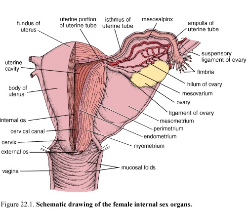
OVARY
Production of Gametes and Production of Steroid Hormones Are the Two Major Functions of the Ovary
Two major groups of steroid hormones are secreted by the ovaries. They are the estrogens and the progestogens.
Estrogens promote growth and maturation of internal and external sex organs and are responsible for the typical female characteristics.
Progestogens prepare the internal sex organs, mainly the uterus, for pregnancy by promoting secretory changes in the endometrium. Both hormones play an important role in the menstrual cycle by preparing the uterus for implantation of a fertilized ovum.
Ovarian Structure
The ovaries are paired, almond-shaped, pinkish-white structures. The surface of the ovary is covered by tunica albuginea. Each ovary is attached to the mesovarium. The ovary is composed of a cortex and a medulla.
-Medulla or medullary region located in the central portion of the ovary contains loose connective tissue, a mass of relatively large contorted blood vessels, lymphatic vessels, and nerves.
Figure 101. Schematic drawing of a section through the ovary

-Cortex or cortical region found in the peripheral portion of the ovary surrounding the medulla contains the ovarian follicles embedded in a richly cellular connective tissue. Scattered smooth muscle fibers are present in the stroma around the follicles.
Follicle Development
Three basic types of ovarian follicles can be identified, based on developmental state:
1. Primordial follicles
2. Growing follicles (primary and secondary (or antral) follicles)
3. Mature or Graafian follicles

The Primordial Follicle Is the Earliest Stage of Follicular Development
Primordial follicles first appear in the ovaries during the third month of fetal development. Early growth of the primordial follicles is independent of gonadotropin stimulation. The primordial follicles are found in the stroma of the cortex just beneath the tunica albuginea. A single layer of squamous follicular cells surrounds the oocyte.

The Primary Follicle Is the First Stage of the Growing Follicle
Initially, the oocyte enlarges and the surrounding flattened follicular cells proliferate and become cuboidal. As the oocyte grows, a homogeneous, acidophilic layer called the zona pellucida appears between the oocyte and the adjacent follicular cells. Follicular cells undergo stratification to form the granulosa layer of the primary follicle.
Connective Tissue Cells Form the Theca Layers of the Primary Follicle
-The theca interna is the inner, highly vascularized layer of cuboidal secretory cells. Cells of the theca interna possess a large number of luteinizing hormone (LH) receptors.
-The theca externa is the outer layer of connective tissue cells.
Maturation of the Oocyte Occurs in the Primary Follicle
The oocytes exhibit specialized secretory vesicles known as cortical granules. They are located just beneath the plasma membrane.
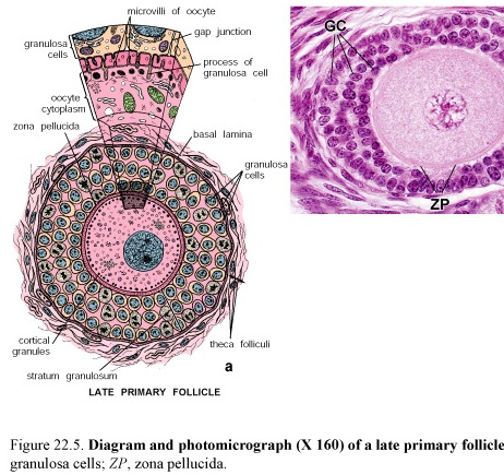
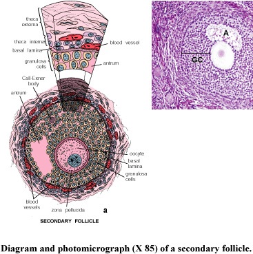
The Secondary Follicle Is Characterized by a Fluid-Containing Antrum
Liquor folliculi includes estrogen. In the secondary follicle begins to form a single cavity called the antrum. The follicle is now identified as a secondary or antral follicle. The granulosa cells form the cumulus oophorus that projects into the antrum. The cells of the cumulus oophorus that immediately surround the oocyte and remain with it at ovulation are referred to as the corona radiata.
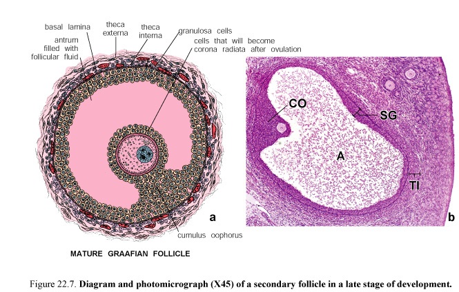
The Graafian Follicle Contains the Mature Secondary Oocyte
The mature follicle, also known as a Graafian follicle, has a diameter of 10 mm or more. The stratum granulosum appears to become thinner as the antrum increases in size. The oocyte and cumulus cells are gradually loosened from the rest of the granulosa cells in preparation for ovulation. The cumulus cells immediately surrounding the oocyte now form a single layer of cells of the corona radiata.
The granulosa and thecal cells then undergo luteinization and produce progesterone.
Ovulation
Figure 102. Ovulation

Ovulation is the process by which an oocyte is released from the Graafian follicle.
A combination of hormonal changes and enzymatic effects is responsible for the actual release of the secondary oocyte at the middle of the menstrual cycle. The factors include
-Increase in the volume and pressure of the follicular fluid
-Enzymatic proteolysis of the follicular wall by activated plasminogen
Just before ovulation, blood flow stops in a small area of the ovarian surface which known as the stigma. Last one becomes elevated and then ruptures. The oocyte is forcefully expelled from the ruptured follicle.
The oocyte is then transported into the abdominal ostium of the oviduct.
FERTILIZATION
Fertilization normally occurs in the ampulla of the oviduct. Several spermatozoa may penetrate the zona pellucida, but only one spermatozoon completes the fertilization process.
Corpus Luteum
The Collapsed Follicle Undergoes Reorganization into the Corpus Luteum After Ovulation
At ovulation, the follicular wall, composed of the remaining granulosa and thecal cells, is transformed into the corpus luteum (yellow body) or luteal gland. At first, bleeding from the capillaries in the theca interna into the follicular lumen leads to the formation of the corpus hemorrhagicum with a central clot. Connective tissue from the stroma then invades the former follicular cavity. These luteal cells become filled with lipid droplets which give them a yellow appearance. As the corpus luteum begins its formation, blood and lymphatic vessels from the theca interna rapidly grow into the granulosa layer. This highly vascularized structure located in the cortex of the ovary secretes progesterone. If fertilization and implantation do occur, the corpus luteum increases in size to form the corpus luteum of pregnancy. It remains active 12 weeks. If fertilization and implantation do not occur, the corpus luteum remains active only for 14 days; in this case it is called the corpus luteum of menstruation.
The corpus luteum degenerates and undergoes a slow involution after pregnancy or menstruation. A white scar, the corpus albicans, is formed as intercellular hyaline material.
Atresia
Most Ovarian Follicles Are Lost by Atresia
Very few of the ovarian follicles that begin their differentiation in the embryonic ovary are destined to complete their maturation. Most of the follicles degenerate and disappear through a process called follicular atresia.
The oocyte undergoes typical changes associated with degeneration and autolysis.
Blood Supply to the Ovaries Comes From Two Different Sources: Ovarian and Uterine Arteries
OVIDUCT
The oviducts are paired tubes that extend bilaterally from the uterus toward the ovaries. The oviducts, also commonly referred to as the uterine or Fallopian tubes, transmit the ova from the ovary to the uterus and provide the necessary environment for fertilization and for initial development of the conceptus to the morula stage.
The oviduct wall consists of next layers.
-The serosa or peritoneum consists of mesothelium and a thin layer of connective tissue.
-The muscularis, throughout most of its length, is organized into an inner, relatively thick circular layer and an outer, thinner longitudinal layer.
-The mucosa exhibits relatively thin longitudinal folds that project into the lumen of the oviduct throughout its length.
There is, however, no submucosa. The mucosal lining is simple columnar epithelium composed of two kinds of cells, ciliated and nonciliated. Nonciliated cells are secretory cells that produce the oviductal fluid that provides nutritive material for the ovum.
Oviduct Transport
Bidirectional Transport Occurs in the Oviduct
The oviduct demonstrates active movements just before ovulation as the fimbriae become closely apposed to the ovary and localize over the region of the ovarian surface where rupture will occur. As the egg is released, the ciliated cells in the infundibulum sweep it toward the opening of the oviduct and, thus, prevent it from passing into the peritoneal cavity. The egg is transported along the oviduct by peristaltic contractions.
Uterus
Figure 103. Schematic diagram illustrating the blood supply to the endometrium of the uterus
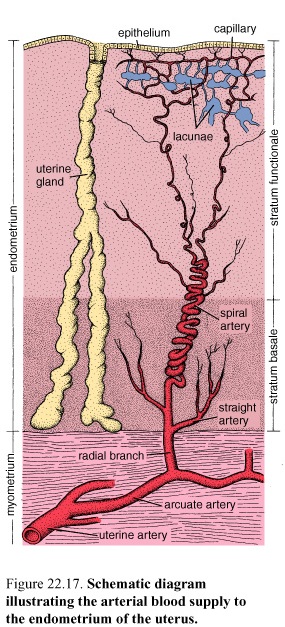
The human uterus is a hollow, pear-shaped organ. Its lumen is continuous with the oviducts and the vagina. The uterine wall is composed of three layers. From the lumen outward they are
-Endometrium, the mucosa of the uterus.
-Myometrium, the thick muscular layer. It is continuous with the muscle layer of the oviduct and vagina. The smooth muscle fibers also extend into the ligaments connected to the uterus.
-Perimetrium, the external serous layer or visceral peritoneal covering of the uterus. The perimetrium consists of a mesothelium and a thin layer of loose connective tissue. Underneath the mesothelium is a layer of elastic tissue. The perimetrium covers the entire posterior surface of the uterus but only part of the anterior surface. The remaining part of the anterior surface consists of connective tissue or adventitia.
The myometrium and the endometrium both undergo cyclic changes each month to prepare the uterus for implantation of an embryo. These changes constitute the menstrual cycle. If an embryo implants, the cycle stops, and both layers undergo considerable growth and differentiation during pregnancy.
The Myometrium Forms a Structural and Functional Syncytium
The myometrium is the thickest layer of the uterine wall. It is composed of three indistinctly defined layers of smooth muscle:
- The middle muscle layer contains numerous large blood vessels (venous plexuses) and lymphatics and is called the stratum vasculare. It is the thickest layer and has smooth muscle bundles described as oriented circular or spiral pattern interlaced with each other.
-The smooth muscle bundles in the inner and outer layers are described as predominantly oriented parallel to the long axis of the uterus.
The Endometrium Proliferates and Then Degenerates During a Menstrual Cycle
The endometrium undergoes cyclic changes each month that prepare it for the implantation of the embryo. Changes in the secretory activity of the endometrium during the cycle are correlated with the maturation of the ovarian follicles. The discharge of tissue and blood from the vagina, which usually continues for a period of 3-5 days, is referred to as menstruation or menstrual flow.
The endometrium consists of two layers or zones that differ in structure and function:
• Stratum functionale or functional layer: This layer is the thick part of the endometrium, which is sloughed off at menstruation.
• Stratum basale or basal layer: This layer is retainer during menstruation.
The Stratum Functionale Is the Layer That Proliferates and Degenerates During the Menstrual Cycle
It is lined with a simple columnar epithelium with a mixture of secretory and ciliated cells. The surface epithelium invaginates into the underlying lamina propria, the endometrial stroma forming uterine glands. These glands are simple tubular. No submucosa separates the endometrium from the myometrium.
The Vasculature of the Endometrium Also Proliferate and Degenerates in Each Menstrual Cycle
The endometrium contains a unique system of blood vessels. The main branch of the radial artery continues upward and becomes highly coiled. It is called the spiral artery.
Cervix
The Endometrium of the Cervix Differs From the Rest of the Uterus
The cervical mucosa contains large, branched glands. The amount and properties of the mucus secreted by the gland cells vary during the menstrual cycle under the influence of the ovarian hormones.
Proliferative, Secretory, and Menstrual Phases Are the Defined Cyclic Changes of the Endometrium
The menstrual cycle is convenient to describe the cycle as having three successive phases:
-Proliferative phase, occurring concurrently with follicular maturation and influenced by ovarian estrogen secretion
-Secretory phase, coinciding with the functional activity of the corpus luteum and primarily influenced by progesterone secretion
-Menstrual phase, commencing as hormone production by the ovary declines with the degeneration of the corpus luteum
Figure 104. Relationship of morphological and physiological events that occur in the menstrual cycle
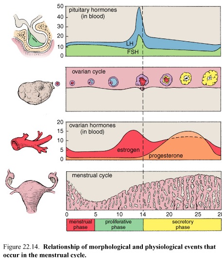
It must be emphasized that the phases are part of a continuous process and that there is no abrupt change from one to the next.
The Proliferative Phase of the Menstrual Cycle Is Regulated by Estrogens
At the end of the menstrual phase, the endometrium consists of a thin band of connective tissue, about 1 mm thick, containing the basal portions of the uterine glands and the lower portions of the spiral arteries. This layer is the stratum basale; the layer that was sloughed off was the stratum functionale. Under the influence of estrogens, the proliferative phase is initiated. Cells in the stratum basale proliferate rapidly, and the following changes can be seen:
-Epithelial cells in the basal portion of the glands reconstitute the glands and migrate to cover the denuded endometrial surface.
-Spiral arteries lengthen as the endometrium is reestablished; these arteries are only slightly coiled and do not extend into the upper third of the endometrium.
The proliferative phase continues for 1 day after ovulation, which occurs at about day 14 of a 28-day cycle. At the end of this phase, the endometrium has reached a thickness about 3 mm. The glands have narrow lumina and are relatively straight.
The Secretory Phase of the Menstrual cycle is Under the Control of Progesterone
Under the influence of progesterone, dramatic change occur in the stratum functionale, beginning a day or two after ovulation. The endometrium becomes edematous and may eventually reach a thickness of 5-6 mm. The glands enlarge and become corkscrew shaped, and their lumina become sacculated as they fill with secretory products. The mucoid fluid being produced by the gland epithelium is rich in nutrients, particularly glycogen. The growth seen at this stage results from hypertrophy of the epithelial cells, an increase in vascularity, and edema of the endometrium. The spiral arteries lengthen and become more coiled. They extend nearly to the surface of the endometrium.
The sequential influence of estrogens and progesterone on the stromal cells makes them capable of undergoing transformation into decidual cells.
The Menstrual Phase Results From a Decline in the Ovarian Secretion of Progesterone and Estrogen
The corpus luteum remains active in hormone production for only about 10 days if fertilization does not occur. As the hormone levels rapidly decline, changes occur in the blood supply to the stratum functionale. Initially, periodic contraction of the walls of the spiral arteries, lasting for several hours, cause the stratum functionale to become ischemic, the glands stop secreting, and the endometrium shrinks in height as the stroma becomes less edematous. When spiral arteries close off, blood flows into the stratum basale but not into the stratum functionale. The desquamation continues until only the stratum basale remains. As noted, this is a cyclic process.
In the absence of fertilization, a cessation of bleeding would accompany the growth and maturation of new ovarian follicles. The epithelial cells would rapidly proliferate and migrate to restore the surface epithelium as the proliferative phase of the next cycle begins.
If Fertilization and Implantation Occur, a Gravid Phase Replaces the Menstrual Phase of the Cycle
Vagina
The Vagina Is a Fibromuscular Tube Lined by Nonkeratinized Stratified Squamous Epithelium
The vagina is a fibromuscular sheath. The vaginal wall consists of an
-Inner mucosal layer
-Intermediate muscular layer
-Outer adventitial layer
The mucosa of the vagina has numerous transverse folds or rugae and is lined with stratified nonkeratinized squamous epithelium.
The Vaginal Mucosa Contains No Glands
The vaginal surface is lubricated by mucus produced by the cervical glands. The epithelium undergoes cyclic changes during the menstrual cycle. Under the influence of estrogens, during the follicular phase, the epithelial cells synthesize and accumulate glycogen as they migrate toward the surface.
The lamina propria exhibits two distinct regions. The outer region is a highly cellular loose connective tissue. The deeper region, adjacent to the muscular layer, is denser and may be considered a submucosa. Numerous elastic fibers are present here. Many lymphocytes and leukocytes are found in the lamina propria.
The vaginal muscularis is organized in two, sometimes indistinct, intermingling smooth muscle layers, an outer longitudinal layer and an inner circular layer.
The vaginal adventitia is organized into an inner dense connective tissue layer, adjacent to the muscularis, and an outer loose connective tissue layer that blends with the adventitia of the surrounding structures.
MAMMARY GLANDS
Figure 105. Schematic drawing of the human breast as seen during lactation

The mammary glands or breasts are a distinguishing feature of mammals. Multiple glands develop along paired epidermal thickenings, called the mammary ridges (milk lines.
In the female, the mammary glands undergo further development under hormonal influence. They are also influenced by changes in the ovarian hormone levels during each menstrual cycle. The actual initiation of milk secretion is induced by prolactin secreted by the adenohypophysis. The ejection of the milk from the breast is stimulated by oxytocin released from the neurohypophysis. With the change in the hormonal environment at menopause, the glandular component of the breast regresses or involutes and is replaced by fat and connective tissue.
Mammary Glands Are Modified Apocrine Sweat Glands That Develop Under the Influence of Sex Hormones
The inactive adult mammary gland is composed of 15-20 irregular lobes of branched tubuloalveolar glands. The lobes, separated by fibrous bands of connective tissue, radiate from the mammary papilla or nipple and are further subdivided into numerous lobules. Abundant adipose tissue is present in the dense connective tissue of the interlobular spaces. The intralobular connective tissue is much less dense and contains little fat.
The epidermis of the adult nipple and areola is highly pigmented and somewhat wrinkled and has long dermal papillae invading into its deep surface. During pregnancy, the areola becomes larger, and the degree of pigmentation increases further. Deep to the areola and nipple, bundles of smooth muscle fibers are arranged radially and circumferentially in the dense connective tissue and longitudinally along the lactiferous ducts. These muscle fibers allow the nipple to become erect in response to various stimuli.
The areola contains sebaceous glands, sweat glands, and modified mammary glands. These glands, which are described as having a structure intermediate between sweat glands and true mammary glands, produce small elevations on the surface of the areola. Numerous sensory nerve endings are present in the nipple. The areola contains fewer sensory nerve endings.
The tubuloalveolar glands, derived from modified sweat glands in the epidermis, lie in the subcutaneous tissue. Each gland ends in a lactiferous duct that opens through a constricted orifice onto the nipple. Beneath the areola, the pigmented area surrounding the nipple, each duct has a dilated portion, the lactiferous sinus. Near their openings, the lactiferous ducts are lined with stratified squamous epithelium.
The Morphology of the Secretory Portion of the Mammary Gland Varies With the Menstrual Cycle
In the inactive gland, the glandular component is sparse and consists chiefly of duct elements. During the menstrual cycle, the inactive breast undergoes slight cyclic changes. Early in the cycle, the ducts appear as cords with little or no lumen. Under estrogen stimulation, at about the time of ovulation, the secretory cells increase in height, lumina appear in the ducts as small amounts of secretions accumulate, and fluid accumulates in the connective tissue.
Mammary Glands Undergo Dramatic Proliferation and Development During Pregnancy
The mammary glands exhibit a number of changes in preparation for lactation. The changes in the glandular tissue are accompanied by decreases in the amount of connective tissue and adipose tissue. Plasma cells, lymphocytes, and eosinophils infiltrate the fibrous component of the connective tissue as the breast develops. As the cells proliferate by mitotic division, the ducts branch and alveoli begin to develop. In the later stages of pregnancy, alveolar development becomes more prominent. The actual proliferation of the stromal cells declines and subsequent enlargement of the breast occurs through hypertrophy of the secretory cells and accumulation of secretory product in the alveoli.
Both Merocrine and Apocrine Secretion Are Involved in Production of Milk
The secreting cells contain large lipid droplets and secretory granules depending on the secretory state. The secretory cells produce two distinct products that are released by different mechanisms:
Merocrine secretion: The protein component of the milk is synthesized in the rER, packaged into membrane-limited secretory granules for transport in the Golgi apparatus, and released from the cell by fusion of the granule's limiting membrane with the plasma membrane.
Apocrine secretion: The fatty or lipid component of the milk arises as lipid droplets free in the cytoplasm. The lipid coalesces to form large droplets that pass to the apical region of the cell and project into the lumen of the acinus. The droplets are invested with an envelope of plasma membrane as they are released. A thin layer of cytoplasm is trapped between the plasma membrane and lipid droplet and is released with the lipid, but the cytoplasmic loss in this process is minimal.
The secretion released in the first few days after childbirth is known as colostrum. This premilk is an alkaline, yellowish secretion with a higher protein, vitamin A, sodium, and chloride content and a lower lipid, carbohydrate, and potassium content than milk. It contains considerable amounts of antibodies that provide the newborn with some degree of passive immunity. As these wandering cells decrease in number after parturition, the production of colostrum stops, and lipid-rich milk is produced.
Hormonal Regulation of the Mammary Gland
The initial growth and development of the mammary gland at puberty occur under the influence of estrogens and progesterone being produced by the maturing ovary. During pregnancy, the corpus luteum and placenta continuously produce estrogens and progesterone. Estrogen present in the circulation stimulates proliferation of the lactiferous duct components, and progesterone stimulates growth of alveoli. It is now believed that the growth of the mammary glands is also dependent on the presence of prolactin, produced by the adenohypophysis; hCS, produced by the placenta; and adrenal glucocorticoids.
Lactation Is Under the Neurohormonal Control of the Adenohypophysis and Hypothalamus
Although estrogen and progesterone are essential for the physical development of the breast during pregnancy, both of these hormones also have an overriding suppressive effect on prolactin and hCS, the levels of which increase as pregnancy progresses. Immediately after birth, however, the sudden loss of estrogen and progesterone secretion from the placenta and corpus luteum allows the lactogenic effect of prolactin to assume its natural role. Production of milk also requires adequate secretion of growth hormone, adrenal glucocorticoids, and parathyroid hormones.
The act of suckling during breast-feeding initiates sensory impulses from receptors in the nipple to the hypo-thalamus. The impulses inhibit the release of prolactin-inhibiting factor, and prolactin is then released from the adenohypophysis. The sensory impulses also cause the release of oxytocin in the neurohypophysis. The oxytocin stimulates the myoepithelial cells that surround the base of the alveolar secretory cells and the base of the cells in the larger ducts, causing them to contract and eject the milk from the alveoli and the ducts. In the absence of suckling, secretion of milk ceases, and the mammary glands begin to regress. The glandular tissue then returns to an inactive condition.
Literature
Human Histology. Inderbir Singh, - 2002. - C.294.
General histology. Shapovalova E. – 2004. – C. 152.
Histology and atlas. Michael H. Ross at al. – 1995. – C. 823.
Histology for the first year medical students by Dr.Adel Zohdy. – 2000. – C. 286.
Basic Histology. Nai Sarak at al. – 2003. – C. 470.