

Human_Histology
.pdf228Textbook of Human Histology
Muscle
The seminal vesicles consist of a thin intermediate layer of smooth muscles. The muscle layer contains outer longitudinal and inner circular fibers.
Connective Tissue
The outer covering of loose connective tissue forms the adventitial layer containing blood vessels and nerves.
Function
The seminal vesicles produce a thick secretion that forms the bulk of semen. The secretion contains fructose which provides nutrition to spermatozoa. It also contains amino acids, proteins, prostaglandins, ascorbic acid, and citric acid. This secretion is expelled during ejaculation by contraction of the smooth muscle of the vesicle.
Clinical Correlation
semenanalysis absence of frustose suggests congenital absence of seminal vesicle or portion of the ductal system or both.
PROSTATE
The prostate is the largest accessory sex gland. It surrounds the beginning of urethra and is the shape of a chestnut. It is made up of 30 to 50 compound tubulo-alveolar glands that are embedded in a framework of fibromuscular tissue. The glandular part of the prostate is poorly developed at birth. It undergoes considerable proliferation at puberty, and degenerates in old age.
In sections, the glandular tissue is seen in the form of numerous follicles that are lined by columnar epithelium (Plate 18.6). The epithelium is thrown into numerous folds (along with some underlying connective tissue). The follicles drain into 12 to 20 excretory ducts that open into the prostatic urethra. The ducts are lined by a double layered epithelium. The superficial (luminal) layer is columnar, and the deeper layer is cuboidal.
Small rounded masses of uniform or lamellated structure are found within the lumen of the follicles. They are called amyloid bodies or corpora amylacea. These are more abundant in older individuals. These consist of condensed glycoprotein. They are often calcified.
The fibromuscular tissue forms a conspicuous feature of sections of the prostate. It contains collagen fibers and smooth muscle. Within the gland the fibromuscular tissue forms septa that separate the glandular elements. These septa are continuous with a fibrous capsule that surrounds the prostate. The capsule contains numerous veins and parasympathetic ganglion cells.
On the basis of differences in the size and nature of the glands the prostate can be divided into an outer (or peripheral) zone, and an internal zone. An innermost zone lying immediately around the prostatic urethra is also described (Fig. 18.7).
The glands in the outer zone are the main prostatic glands. They open into long ducts that join the urethra. The internal (or submucous) glands have short ducts. The innermost (or mucous) glands open directly into the urethra. The internal and innermost zones together form the central zone.
The prostate is traversed by the prostatic urethra. The gland is also traversed by the ejaculatory ducts.
Function
The prostate produces a secretion that forms a considerable part of semen. The secretion is rich in enzymes (acid phosphatase, amylase, protease) and in citric acid.
The prostate also produces substances called prostaglandins that have numerous actions.
Clinical Correlation
Benign nodular hyperplasia of prostate: Non-neoplastic tumorlike enlargement of the prostate, is a very common condition becomes increasingly more frequent above the age of 50 years and its incidence approaches 75–80% in men above 80 years. The central zone commonly undergoes benign hypertrophy in old persons. Enlargement of the prostate can compress the urethra leading to problems in passing urine.
Carcinoma of prostate: Cancer of the prostate is the second most common form of cancer in males, followed in frequency by its prevalence increases with increasing age so that more than 50% of men 80 years old have asymptomatic (latent) carcinoma of the prostate. Many a times, carcinoma of the prostate is small and detected as microscopic foci in a prostate removed for benign enlargement of prostate or found incidentally at autopsy. The peripheral zone is often the site of carcinoma.
PENIS
The penis is the erectile copulatory organ in males. It consists of a root that is fixed to the perineum, and of a free part that is called the body or corpus. A transverse section through the free part of the penis is shown in Figure 18.8. The penis is covered all round by thin skin that is attached loosely to underlying tissue.
The substance of the penis is made up of three masses of erectile tissue, two dorsal and one ventral. The dorsal masses are the right and left corpora cavernosa, while the ventral mass is the corpus spongiosum.
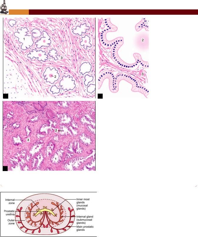
PLATE 18.6:
B
(
Fig. 18.7: Arrangement of glandular tissue in prostate
(Schematic representation)
Chapter 18 Male Reproductive System 229
(
& &
;&
'& :
(&
*& :
Each corpus cavernosum is surrounded by a dense sheath containing collagen fibers, elastic fibers and some smooth muscle. In the midline the sheaths of the right and left corpora cavernosa fuse to form a median septum. The corpora cavernosa lie side by side and are separated only by a median fibrous septum.
The corpus spongiosum is placed in the midline ventral to the corpora cavernosa. The corpus spongiosum is also surrounded by a sheath, but this sheath is much thinner than that around the corpora cavernosa. An additional sheath surrounds both the corpora cavernosa and the

230 Textbook of Human Histology
Fig. 18.8: Transverse section of penis (Schematic representation)
corpus spongiosum. The corpus spongiosum is traversed by the penile urethra throughout its length.
The tip of urethra at glans penis is lined by stratified squamous non-keratinized epithelium. Many small mucous glands of Littre are scattered along the length of urethra that secrete mucus and have lubricating functions.
Many sensory nerve endings are present in the penis, particularly on the glans.
% &
, *
Numerous septa arising from the connective tissue sheath extend into the corpora cavernosa and into the corpus spongiosum, and form a network. The spaces of the network are lined by endothelium. The spaces are in communication with arteries and veins. They are normally empty.
!! ! pressure. This results in enlargement and rigidity of the organ. The process of erection involves the corpora cavernosa more than the corpus spongiosum, rigidity of the former! sheath. The corpus spongiosum does not become so rigid
Contd...
Contd...
as its sheath is elastic, and the vascular spaces within it are smaller. As a result, the penile urethra remains patent during# $
Blood to the corpora cavernosa is supplied mainly by the deep arteries of the penis. These arteries give off branches that follow a spiral course before opening into the cavernous spaces. They are called helicrine arteries and have an unusual structure. The circular muscle in their media is very thick so that the vessels can be completely occluded. The tunica intima shows longitudinal thickenings.
Erection is produced by complete relaxation of smooth muscle both in the walls of arteries and in the septa. Helicrine arteries are connected to veins by arteriovenous anastomoses. Normally, the anastomoses are patent. Their closure (caused by parasympathetic nerves) causes cavernous!! ! !! ! # %
sure in them compresses the veins that lie just deep to the& ! out of the spaces. At the end of erection smooth muscle!! $ ! Contraction of muscle in the trabeculae gradually forces blood out of the spaces.

Female Reproductive System
The female reproductive system includes (Fig. 19.1)
A pair of ovaries
A pair of uterine tubes
Uterus
Vagina
External genitalia
Mammary glands
THE OVARIES
The ovaries are the female gonads, responsible for the formation of ova. They also produce hormones (estrogen and progesterone) that are responsible for the development of the female secondary sex characters, and produce marked cyclical changes in the uterine endometrium. Each ovary is an oval structure about 3 cm in long diameter.
General Structure
Each ovary consists of the following parts (Fig. 19.2 and Plate 19.1):
Germinal epithelium: The free surface of ovary is covered by a single layer of cubical cells that constitute the germinal epithelium. This epithelium is continuous with the mesothelium lining the peritoneum, and represents a modification of the latter.
Fig. 19.1: Parts of female reproductive system (Schematic representation)
Note: The term germinal epithelium is a misnomer. The epithelium does not produce germ cells. The cells of this epithelium bear microvilli, and contain numerous mitochondria. They become larger in pregnancy.
Tunica albuginea: The germinal epithelium rests on a connective tissue layer called the tunica albuginea. The tunica albuginea of the ovary is much thinner, and less dense, than that of the testis
Cortex: Deep to the tunica albuginea the cortex has a stroma made up of reticular fibers and numerous fusiform cells that resemble mesenchymal cells. Scattered in this stroma there are ovarian follicles at various stages of development. Each follicle contains a developing ovum
Medulla: The medulla consists of connective tissue in which numerous blood vessels (mostly veins) are seen. Elastic fibers and smooth muscle are also present. The hilum of the ovary is the site for entry of blood vessels and lymphatics. It is continuous with the medulla. The hilum also contains some remnants of the mesonephric ducts; and hilus cells that are similar to interstitial cells of the testis.
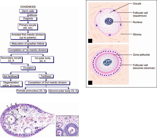
OOGENESIS
The process of formation of ovum from the stem cells is called oogenesis (Flowchart 19.1). The process of oogenesis consists of following stages:
Oogonia: The stem cells from which ova are derived are called oogonia. These are large round cells present in the cortex of the ovary. Oogonia are derived (in fetal life) from primordial germ cells that are formed in the region of the yolk sac, and migrate into the developing ovary. They increase in number by mitosis.
All oogonia to be used throughout the life of a woman are produced at a very early stage (before birth) and do not multiply thereafter. At birth the number of oogonia in an ovary is about one million. Many oogonia formed in this way degenerate, the process starting before birth and progressing throughout life, so that the number of oogonia becomes less and less with increasing age.

232 Textbook of Human Histology
Fig. 19.2: Histological structure of ovary showing follicles at various stages of development (Schematic representation)
Primary oocyte: An oogonium enlarges to form a primary oocyte. The primary oocyte contains the diploid number of chromosomes, i.e. 46. It undergoes the first meiotic division to form two daughter cells each of which has 23 chromosomes.
Secondary oocyte: The cytoplasm of the primary oocyte is not equally divided. Most of it goes to one daughter
Embryological Considerations
At the time of birth all primary oocytes are in the prophase
! " #$" % &
' *
+
+, +
to complete the second meiotic division and degenerates !
, " %
-
$ &
cell that is large and is called the secondary oocyte. The second daughter cell has hardly any cytoplasm, and forms the first polar body.
Ovum: The secondary oocyte now undergoes the second meiotic division, the daughter cells being again unequal in size. The larger daughter cell produced as a result of this division is the mature ovum. The smaller daughter cell (which has hardly any cytoplasm) is the second polar body. Thus, one primary oocyte ultimately gives rise to only one ovum.
Formation of Ovarian Follicles
Ovarian follicles (or Graafian follicles) consists of a developing ova surrounded by follicular (granulosa) cells. The development and maturation of an ovarian follicle passes through four stages—the process is called folliculogenesis (Figs. 19.2 and 19.3).
Primordial follicle: Some cells of the stroma become flattened and surround an oocyte (Figs. 19.3 and 19.4A). These stromal cells are now called follicular cells. The oocyte (20–25 μm) and the flat surrounding cells form a primordial follicle. Primordial follicles are the smallest and simplest in structure located at the periphery of the cortex. Numerous primordial follicles are present in the ovary at birth. They undergo further development only at puberty.
Primary follicle: The first indication that a primordial follicle is beginning to undergo further development is that the flattened follicular cells become columnar
Chapter 19 Female Reproductive System 233
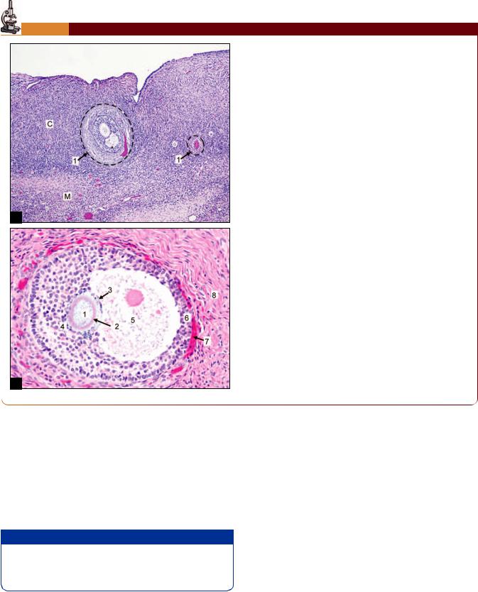
PLATE 19.1: Ovary
A
B
The surface is covered by a cuboidal epithelium. Deep to the tutes the tunica albuginea
The substance of the ovary has an outer cortex in which follicles of various sizes are present; and an inner medulla vessels
Just deep to the tunica albuginea many primordial follicles each of which contains a developing ovum surrounded by
Large follicles have a follicular cavity surrounded by several layers of follicular cells
oophoricus
The capsule consists of an inner cellular part (the theca! "# #
Key
C. Cortex
M.Medulla
1.Ovarian follicle
2.Zona pellucida
3.Cumulus oophoricus
4.Discus proligerus
5.Antrum folliculi
6.Membrana granulosa
7.Capsule of follicle
8.Stroma
(Figs. 19.3 and 19.4B). Follicles at this stage of development are called primary follicles. The outermost layer of the follicular cells rest on a well-defined basement membrane which separates it from the ovarian stroma. A homogeneous membrane, the zona pellucida, appears between the follicular cells and the oocyte (which enlarges in size 50–80 μm in diameter. With the appearance of the zona pellucida the follicle is now referred to as a multilaminar primary follicle.
+ ,
./0 ,1
Secondaryfollicle:Thefollicularcellsproliferatetoform several layers of cells that constitute the membrana granulosa. The cells are now called granulosa cells. The oocyte enlarges and reaches its maximum size (125 μm). This is a secondary follicle (Figs. 19.3A and B).
So far the granulosa cells are in the form of a compact mass. However, the cells to one side of the ovum soon partially separate from one another so that a follicular cavity (or antrum folliculi) appears between them. It is with the appearance of this cavity that a true follicle (= small sac) can be said to have been formed. The follicular cavity is filled by a fluid, the liquor folliculi. With the formation of follicular cavity the size of the follicle increases.
234 Textbook of Human Histology
Flowchart 19.1: Stages of oogenesis
A
B
Figs. 19.4A and B: $/& . 3 $9& Primary follicle (Schematic repesentation)
|
|
|
|
A |
|
B |
|
|
Figs. 19.3A and B: '$/& . |
$0 |
|
& 2 3 !2 3 %2 43 2 3 "2 3 52 3 62
3 72 3 82 3 2 3
2 3 !2 3 %2 3 2
3 "2$9& '$ &
2 3 !2 3 %2 3 2 3 "2 follicles (Schematic representation)
Graafian follicle: With the further development, the follicular cavity rapidly increases in size. As a result, the wall of the follicle (formed by the granulosa cells) becomes relatively thin. The graffian follicle now measures about 10 mm or more in diameter and is seen bulging out of the cortex (Fig. 19.2). The oocyte now lies eccentrically in the follicle surrounded by some
granulosa cells that are given the name of cumulus oophoricus (or cumulus oophorus, or cumulus ovaricus). The inner most layer of cumulus oophorus that lies directly adjacent to the zona pellucida is called corona radiata. The granulosa cells that attach the oocyte to the wall of the follicle constitute the discus proligerus (Fig. 19.5).
As the follicle expands the stromal cells surrounding the membrana granulosa become condensed to form a covering called the theca interna (theca = cover). The cells of the theca interna later secrete a hormone called estrogen, and they are then called the cells of the thecal gland.
Outside the theca interna some fibrous tissues become condensed to form another covering for the follicle. This is the theca externa. The theca interna and externa are collectively called the theca folliculi.
Ovulation
The ovarian follicle is at first very small compared to the thickness of the ovarian cortex. As the follicle enlarges it becomes so big that it not only reaches the surface of the ovary, but forms a bulging in this situation. As a result the stroma and the theca on this side of the follicle are stretched and become very thin (Fig. 19.6).

Chapter 19 Female Reproductive System 235
Fig. 19.6: Relationship of a growing ovarian follicle to the ovary
(Schematic representation)
Fig. 19.5: Mature ovarian follicle (Schematic representation)
Fig. 19.8: : ; 4 with yellow granules (Schematic representation)
Fig. 19.7: Structure of ovum at the time of ovulation
(Schematic representation)
An avascular area (stigma) appears over the most convex point of the follicle. At the same time the cells of the cumulus oophoricus become loosened by accumulation of fluid between them. The follicle ultimately ruptures and the ovum is shed from the ovary. The shedding of the ovum is called ovulation. The “ovum” that is shed from the ovary is not fully mature. It is really a secondary oocyte surrounded by zona pellucida and corona radiata
(Fig. 19.7).
Fate of the Ovum
The ovary is closely embraced by the fimbriated end of the uterine tube. Therefore, the ovum is easily carried into the tube partly by the follicular fluid discharged from the follicle and partly by the activity of ciliated cells lining the tube. The ovum slowly travels through the tube
toward the uterus, taking three to four days to do so. If sexual intercourse takes place at about this time, the spermatozoa deposited in the vagina swim into the uterus and into the uterine tube. One of these spermatozoa may fertilize the ovum. If this happens, the fertilized ovum begins to develop into an embryo. It travels to the uterus and gets implanted in its wall. On the other hand, if the ovum (secondary oocyte) is not fertilized it dies in 12 to 24 hours. It passes through the uterus into the vagina and is discharged.
Corpus Luteum
The corpus luteum is an important structure. It secretes a hormone, progesterone. The corpus luteum is derived from the ovarian follicle, after the latter has ruptured to shed the ovum, as follows (Figs. 19.8 and 19.9).
Corpus hemorrhagicum: When the follicle ruptures its wall collapses and becomes folded. Sudden reduction in pressure caused by rupture of the follicle results in bleeding into the follicle. The follicle filled with blood is called the corpus hemorrhagicum. At this stage, the follicular cells are small and rounded.

236 Textbook of Human Histology
Fig. 19.9: : $ & (Schematic representation)
Corpus luteum: The cells now enlarge rapidly. As they increase in size their walls press against those of neighboring cells so that the cells acquire a polyhedral shape (Figs. 19.8 and 19.9).
Their cytoplasm becomes filled with a yellow pigment called lutein. They are now called luteal cells. The presence of this yellow pigment gives the structure a yellow color, and that is why it is called the corpus luteum (= yellow body).
Some cells of the theca interna also enlarge and contribute to the corpus luteum. The cells of the corpus luteum contain abundant smooth ER and considerable amount of lipids.
The corpus luteum secretes progesterone. This secre-
tion has to be poured into blood like secretions of endocrine glands. All endocrine glands are richly supplied with blood vessels for this purpose. The ovarian follicle itself has no blood vessels, but the surrounding theca interna is full of them. When the corpus luteum is forming, blood vessels from the theca interna invade it and provide it with a rich blood supply.
The subsequent fate of the corpus luteum depends on whether the ovum is fertilized or not.
If the ovum is not fertilized, the corpus luteum persists for about 14 days. During this period it secretes progesterone. It remains relatively small and is called the corpus luteum of menstruation. At the end of its functional life, it degenerates and becomes converted into a mass of fibrous tissue called the corpus albicans (= white body) (Fig. 19.10).
If the ovum is fertilized and pregnancy results, the corpus luteum persists for three to four months. It is larger than the corpus luteum of menstruation, and is called the corpus luteum of pregnancy. The progesterone secreted by it is essential for the maintenance of pregnancy in the first few months. After the fourth month, the corpus luteum is no longer needed, as the placenta begins to secrete progesterone.
Fate of Ovarian Follicles
The series of changes that begin with the formation of an ovarian follicle, and end with the degeneration of the corpus luteum constitute what is called an ovarian cycle.
In each ovarian cycle one follicle reaches maturity, sheds an ovum, and becomes a corpus luteum. At the same time, several other follicles also begin to develop, but do not reach maturity. It is interesting to note that, contrary to what one might expect, these follicles do not persist into the next ovarian cycle, but undergo degeneration. The ovum and granulosa cells of each follicle disappear. The cells of the theca interna, however, proliferate to form the interstitial glands, also called the corpora atretica. These glands are believed to secrete estrogens. After a period of activity, each gland becomes a mass of scar tissue indistinguishable from the corpus albicans formed from the corpus luteum.
The cortex of an ovary (taken from a woman in the reproductive period) can show ovarian follicles (at various stages of maturation), corpora lutea, corpora albicantes, and corpora atretica.
The changes taking place during the ovarian cycle are greatly influenced by certain hormones produced by the hypophysis cerebri. The hormones produced by the theca interna and by the corpus luteum in turn influence other parts of the female reproductive system, notably the uterus, resulting in a cycle of changes referred to as the uterine cycle or menstrual cycle.
UTERINE TUBES
Uterine tubes are paired muscular tubes and are also called fallopian tubes. Each uterine tube has a medial or uterine end, attached to (and opening into) the uterus, and a lateral end that opens into the peritoneal cavity near the ovary. The tube has (from medial to lateral side)
A uterine part that passes through the thick uterine wall
A relatively narrow, thick walled part called the isthmus
A thin-walled dilated part called the ampulla
Funnel-shaped infundibulum. It is prolonged into a number of finger like processes or fimbriae.

Chapter 19 Female Reproductive System 237
Fig. 19.10: Comparison of fate of ovarian follicles that shed an ovum and of those that do not (Schematic representation)
The wall of the uterine tube consists of following layers from within outwards.
Mucous Membrane
The mucous membrane shows numerous branching folds that almost fill the lumen of the tube (Plate 19.2). These folds are most conspicuous in the ampulla. Each fold has a highly cellular core of connective tissue. It is lined by columnar epithelium that rests on a basement membrane. Some of the lining cells are ciliated: ciliary action helps to move ova toward the uterus.
Other cells are secretory in nature and are also called as peg cells. They contain secretory granules and are not ciliated. Their surface shows microvilli. A third variety of intercalary cells is also described.
Muscle Coat
The muscle coat has an inner circular layer and an outer longitudinal layer of smooth muscle. An additional inner longitudinal layer may also be present. The circular layer is thickest in the uterine part of the tube. The circular muscle is thickest in the isthmus. The pattern of mucosal folds is also different in this region. There is some evidence that the isthmus may have some control on the passage of a fertilized ovum through it.
Serosa
It consists of mesothelium supported by connective tissue.
Functions
The uterine tube conveys ova, shed by the ovary, to the uterus. Ova enter the tube at its fimbriated end. Spermatozoa enter the uterine tube through the vagina and uterus. Fertilization normally takes place in the ampulla. When fertilization occurs, the fertilized ovum travels toward the uterus through the tube. Secretions present in the tubes provide nutrition, oxygen and other requirements for ova and spermatozoa passing through the tube.
Clinical Correlation
Ectopic Tubal Pregnancy
The term ectopic tubal pregnancy is used for implantation of a fertilized ovum in the uterine tube. Though ectopic pregnancy may rarely occur in the uterine horn, cornu, ovary and abdominal cavity, tubal pregnancy is by far the most common form of ectopic gestation. The most frequent site of tubal pregnancy is the ampullary portion and the least common is interstitial pregnancy. Ectopic tubal pregnancy is a potentially hazardous problem because of rupture which is followed by intraperitoneal hemorrhage.