

Human_Histology
.pdf278 Textbook of Human Histology
Contd...
As there are three types of cones, responding to the colors red, green, and blue we can distinguish three cor responding types of cone bipolars (red cone bipolar, green cone bipolar, blue cone bipolar).
When a photoreceptor (rod or cone) is exposed to light it releases neurotransmitter at its synapse with the bipolar cell. Some bipolars respond to neurotransmitter by depolarization (and secretion of neurotransmitter at their synapses with ganglion cells). These are called ON bipolars as they are “switched on” by light. Other bipolars respond to release of neurotransmitter by hyperpolariza tion. In other words they are “switched off” by light and are called OFFbipolars.
On the basis of structural characteristics, and the synap ses established by them, cone bipolars are divided into three types: midget, blue cone, and diffuse.
A midget bipolar establishes synapses with a single cone (which may be red or green sensitive). Some midget bipolars synapse with indented areas on cone pedicles forming triads (Fig. 21.11). These are ON bipolars. Other midget bipolars establish “flat” synapses with the cone pedicle (and are also referred to as flatbipolars). These are OFFbipolars.
A blue cone bipolar connects to one blue cone, and establishes triads. It may be of the ON or OFF variety.
Diffuse cone bipolars establish synapses with several cone pedicles. They are not color specific.
Axons of rod bipolar neurons synapse with up to four gang lion cells, but those of one midget bipolar neuron synapse with only one (midget) ganglion cell, and with amacrine neurons.
Ganglion Cells
We have seen that the dendrites of ganglion cells synapse with axons of bipolar cells, and also with processes of ama crine cells. The axons arising from ganglion cells constitute the fibers of the optic nerve.
Ganglion cells are of two main types. Those that synapse with only one bipolar neuron are monosynaptic, while those that synapse with many bipolar neurons are polysynaptic. Monosynaptic ganglion cells are also called midget ganglion cells. Each of them synapses with one midget bipolar neuron. We have seen that midget bipolars in turn receive impulses from a single cone. This arrangement is usual in the central region of the retina, and allows high resolution of vision to be attained.
Polysynaptic ganglion cells are of various types. Some of them synapse only with rod bipolars (rod ganglion cells).
Others have very wide dendritic ramifications that may synapse with several hundred bipolar neurons (diffuse ganglion cells). This arrangement allows for summation of stimuli received through very large numbers of photo receptors facilitating vision in poor light. On physiological grounds ganglion cells are also classified as “ON” or “OFF” cells.
Contd...
Contd...
Horizontal Neurons
Horizontal neurons establish numerous connections between photoreceptors (Fig. 21.17). Some of them are excitatory, while others are inhibitory. In this way these neurons play a role in integrating the activity of photorecepors located in adjacent parts of the retina. As they participate in synapses between photoreceptors and bipolar neurons horizontal neurons may regulate synaptic transmission between these cells.
Amacrine Neurons
Different types of amacrine neurons are recognized depend ing upon the pattern of branching. We have seen that the processes of amacrine neurons enter the internal plexiform layer where they may synapse with axons of several bipolar cells, and with the dendrites of several ganglion cells (Fig. 21.18). They also synapse with other amacrine cells. At many places an amacrine process synapsing with a ganglion cell is accompanied by a bipolar cell axon. The two are referred to as a dyad.
Internal plexiform cells (present in the internal plexi form layer) represent a third variety of horizontally oriented neurons in the retina.
Fig. 21.17: Connections of a cone horizontal neuron (Schematic representation)
Fig. 21.18: Connections of an amacrine neuron (Schematic representation)
Contd...

Chapter 21 Special Senses: Eye 279
Contd... |
Contd |
|
Apart from integration of impulses from rods and cones horizontal, amacrine and internal plexiform cells act as “gates” that can modulate passage of inputs from rods and cones to ganglion cells. In this connection it is to be noted that processes of amacrine neurons are interposed between processes of bipolar cells and ganglion cells, while processes of horizontal cells are interposed between photoreceptors and bipolar cells.
Some Further Remarks about Connections of Retinal Neurons
Whiletherearewelloverahundredmillionphotoreceptors in each retina there are only about one million ganglion cells, each giving origin to one fiber of the optic nerve.
(The bipolar cells are intermediate in number between photoreceptors and ganglion cells). In passing from the photoreceptors to the ganglion cells there has, therefore, to be considerable convergence of impulses (Fig. 21.19).
Each ganglion cell would be influenced by impulses originating in several photoreceptors. On functional con siderations it would be expected that such convergence would be most marked near the periphery of the retina; and that it would involve the rods much more than the cones. It has been estimated that in the peripheral parts of the retina one ganglion cell may be connected to as many as 300 rods or to 10 cones. Convergence leads to summation of impulses arising in many photoreceptors and allows vision even in very dim light. It would also be expected that convergence would be minimal in the macula, and absent in the foveola, to allow maximal resolution.
The second highly important fact about intraretinal con nections is the presence of numerous arrangements for interaction of adjacent regions of the retina as follows:
Firstly, cone pedicles establish numerous contacts with other cone pedicles and with adjacent rod spherules.
Except in the fovea, most photoreceptors are connec ted to more than one bipolar cell. In turn each bipolar cell is usually connected to more than one ganglion cell.
The vertically arranged elements of the retina (photo receptors, bipolar cells, ganglion cells) are intimately interconnected to adjacent elements through hori zontal neurons and amacrine neurons.
Mechanism of Firing of Bipolar Neurons
When no light falls on the retina photoreceptors are depolarized. Exposure to light causes hyperpolarization.
When a photoreceptor is depolarized it releases inhibitor at its junction with a bipolar neuron. This prevents the bipolar neuron from firing. Release of inhibitor is controlled by voltage gated calcium channels.
Hyperpolarization of photoreceptor, caused by exposure to light, leads to closure of Ca++ gates and release of inhibitor is stopped. This causes the bipolar neuron to fire. As explained earlier, this description applies to ONbipolars.
Rhodopsin, present in photoreceptors, is a complex of a protein opsin and cis-retinal that is sensitive to light. When exposed to light cisretinal is transformed to trans retinal. This leads to decrease in concentration of cyclic GMP that in turn leads to closure of sodium channels. Closure of sodium channels results in hyperpolarization of photoreceptor (Flowchart 21.1).
Flowchart. 21.1: Mechanism of firing of bipolar neurons
Fig. 21.19: How impulses arising in several photoreceptors concentrate on one ganglion cell (Schematic representation)
Contd...

Chapter
22
Special Senses: Ear
INTRODUCTION
Ear is the peripheral sense organ concerned with hearing and equilibrium. Anatomically speaking, the ear is made up of three main parts called the external ear, the middle ear, and the internal ear. The external and middle ears are concerned exclusively with hearing. The internal ear has a cochlear part concerned with hearing; and a vesti bular part which provides information to the brain regard ing the position and movements of the head.
The main parts of the ear are shown in Figure 22.1.
External Ear
The external ear consists of auricle or pinna and external acoustic meatus (external auditory canal).
The part of the ear that is seen on the surface of the body (i.e. the part that the lay person calls the ear) is ana tomically speaking, the auricle or pinna. Leading inwards from the auricle there is a tube called the external acoustic meatus.
The inner end of the external acoustic meatus is closed by a thin membranous diaphragm called the tympanic membrane. This membrane separates the external acous tic meatus from the middle ear.
Middle Ear
The middle ear is a small space placed deep within the petrous part of the temporal bone. It is also called the tympanum.
Medially, the middle ear is closely related to parts of the internal ear. It is lined with mucous membrane.
The cavity of the middle ear is continuous with that of the nasopharynx through a passage called the auditory tube. Within the cavity of the middle ear there are three small bones or ossicles: the malleus, the incus, and the stapes. They form a chain that is attached on one side to the tympanic membrane, and at the other end to a part of the internal ear.
Internal Ear
The internal ear is in the form of a complex system of cavities lying within the petrous temporal bone. It has sense organs for both hearing and balance. It has a central part called the vestibule. Continuous with the front of the vestibule there is a spiral shaped cavity called the cochlea. Posteriorly, the vestibule is continuous with three semi circular canals.
Fig. 22.1: The main parts of the ear (Schematic representation)

Chapter 22 Special Senses: Ear 281
Plate 22.1: Pinna
The pinna has a core of elastic cartilage covered on both sides by true skin in which hair follicles and sweat glands are seen.
Key
1.Elastic cartilage
2.Hair follicle
3.Sweat gland
4.Stratified squamous keratinized epithelium
Pinna (As seen in drawing)
THE EXTERNAL EAR
The Auricle (Pinna)
The auricle consists of a thin plate of elastic cartilage covered on both sides by true skin (Plate 22.1). The skin is closely adherent to the cartilage on its lateral surface while it is comparatively loose on medial surface. Epithe lium is squamous keratinising. Hair follicles, sebaceous glands, and sweat glands are present in the skin, adipose tissue is present only in lobule.
Clinical Correlation
Grafts in rhinoplasty: The conchal cartilage is frequently used to correct depressed nasal bridge. The composite grafts of the skin and cartilage can be used for repair of defects of ala of nose.
Grafts in tympanoplasty: Tragal and conchal cartilage and perichondrium and fat from lobule are often used during tympanoplasty operations.
The External Acoustic Meatus
EAC is usually divided into 2 parts: (1) cartilaginous and
(2) bony. Its outer onethird (8 mm) is cartilaginous and its inner twothirds (16 mm) is bony.
Cartilaginous EAC: It is a continuation of the cartilage that forms the framework of the pinna. The skin of the cartilaginous canal is thick and contains hair follicles, ceruminous and pilosebaceous glands that secrete wax.
Note: The ceruminous glands secrete the wax of the ear. They are modified sweat glands lined by a columnar, cuboidal or squamous epithelium.
Clinical Correlation
Fissures of Santorini: Transverse slits in the floor of carti laginous EAC called “fissures of Santorini” provide passages for infections and neoplasms to and from the surrounding soft tissue (especially parotid gland). The parotid and mastoid infec tions can manifest in the EAC.
Hair follicles are present only in the outer cartilaginous canal and therefore furuncles (staphylococcal infection of hair folli cles) are seen only in the cartilaginous EAC.
Dimensions: External auditory canal (EAC) measures about 24 mm and extends from the concha to the tympanic membrane.
Bony EAC: It is mainly formed by the tympanic portion of temporal bone but roof is formed by the squamous part of the temporal bone.

282Textbook of Human Histology
Skin of the bony EAC is thin and continuous over the tympanic membrane skin is devoid of subcuta neous layer, hair follicles and ceruminous glands.
Isthmus: Approximately 6 mm lateral to tympanic membrane, bony EAC has a narrowing called the isthmus.
Clinical Correlation
Foreign body impacted medial to bony isthmus of EAC are diffi cult to remove.
Foramen of Huschke: In children and occasionally in adults, anteroinferior bony EAC may have a deficiency that is called foramen of Huschke. Foramen of Huschke permits spread of infections to and from EAC and parotid.
Note: The skin of EAC has a unique selfcleansing mechanism. This migratory process continues from the medial to lateral side. The sloughed epithelium is extruded out as a component of cerumen.
The Tympanic Membrane
Dimensions: Its dimensions are: 9–10 mm height and 8–9 mm width. It is 0.1 mm thick.
Position: Tympanic membrane (TM) is a partition wall between the EAC and the middle ear. It is positioned obliquely. It forms angle of 55° with deep EAC.
Structure: Tympanic membrane consists of the follow ing three layers:
Outer epithelial layer: It is continuous with the EAC skin.
Middle fibrous layer: The middle layer is made up of fibrous tissue, which is lined on the outside by skin (continuous with that of the external acoustic meatus), and on the inside by mucous membrane of the tympanic cavity.
The fibrous layer contains collagen fibers and some elastic fibers. The fibers are arranged in two layers. In the outer layer they are placed radially, while in the inner layer they run circularly.
Inner mucosal layer: The mucous membrane is lined by an epithelium which may be cuboidal or squa mous. It is said that the mucosa over the upper part of the tympanic membrane may have patches of ciliated columnar epithelium, but this is not borne out by EM studies.
Otoscopy: Normal tympanic membrane is shiny and pearlygrey in color. Its transparency varies from person to person.
MIDDLE EAR
The Tympanic Cavity
also covers the ossicles. The lining epithelium varies from region to region. Typically it is cuboidal or squamous. At places it may be ciliated columnar. The ossicles of the middle ear consist of compact bone, but do not have marrow cavities.
The Auditory Tube (Eustachian Tube)
It is a channel connecting the tympanic cavity with the nasopharynx. The length of eustachian tube (ET) is 36 mm. Its lateral third is bony and medial 2/3 (i.e. 24 mm) is fibrocartilaginous.
The bone or cartilage is covered by mucous membrane which is lined by ciliated columnar epithelium. Near the pharyngeal end of the tube the epithelium becomes pseudostratified columnar. Goblet cells and tubuloalveolar mucous glands are also present. A substantial collection of lymphoid tissue, present at the pharyngeal end, forms the tubal tonsil.
THE INTERNAL EAR
Introduction
The internal ear is in the form of a complex system of cavities within the petrous temporal bone. Because of the complex shape of these intercommunicating cavities the internal ear is also called the labyrinth.
It consists of a bony labyrinth contained within the petrous part of temporal bone.
Note: The basic structure of the labyrinth is best understood by looking at a transverse section through a relatively simple part of it, e.g. a semicircular canal (Fig. 22.2). The space bounded by bone is bony labyrinth. Its wall is made up of bone that is more dense than the surrounding bone. Its inner surface is lined by periosteum.
Lying within the bony labyrinth there is a system of ducts which constitute the membranous labyrinth. The space
The walls of the tympanic cavity are formed by bone which is lined by mucous membrane. The mucous membrane
Fig. 22.2: Basic structure of internal ear as seen in a section through a semicircular canal (Schematic representation)

Fig. 22.3: Bony labyrinth as seen from the lateral side
(Schematic representation)
within the membranous labyrinth is filled by a fluid called the endolymph. The space between the membranous labyrinth and the bony labyrinth is filled by another fluid called the perilymph.
Bony Labyrinth
The bony labyrinth consists of three parts:
Vestibule
Semicircular canals
The bony cochlea
Vestibule
Vestibule is the central part (Fig. 22.3). It is continuous anteriorly with the cochlea; and posteriorly with three semicircular canals.
Semicircular Canal
There are three semicircular canals (SCCs): lateral (hori zontal), posterior and superior (anterior). Each canal occu pies 2/3rd of a circle and has a diameter of 0.8 mm. They lie in planes at right angles to one another. Each canal has two ends: ampullated and nonampullated. All the three ampullated ends and nonampullated end of lateral SCC open independently and directly into the vestibule.
Note: The nonampullated ends of posterior and superior canals join and form a crus commune (4 mm length), which then opens into the medial part of vestibule. So, the three SCCs open into the vestibule by five openings.
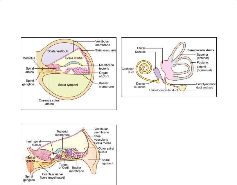
The Bony Cochlea
The cochlea has a striking resemblance to a snail shell. It is basically a tube that is coiled on itself for two and three fourth turns. The “turns” rest on a solid core of bone called the modiolus.
Chapter 22 Special Senses: Ear 283
of the cochlea. A structure that is “inferior” in the upper part of the canal becomes “superior” in the lower part. For descriptive convenience the structures lying next to the modiolus are described as “inner” and those away from it as “outer”. These terms as used here are not equivalents of “medial” and “lateral” as normallyused. The words “superior“ and “inferior” indicate relationships as they exist in the lowest (or basal) turn of the cochlea.
In sections, through the middle of the cochlea the coch lear canal is cut up six times as shown in Plate 22.2. The cochlear canal is partially divided into two parts by a bony lamina that projects outwards from the modiolus. This bony projection is called the spiral lamina. Passing from the tip of the spiral lamina to the opposite wall of the canal there is the basilar membrane.
The spiral lamina and the basilar membrane together divide the cochlear canal into three parts—scala vestibuli, scala tympani, and scala media (membranous cochlea). The lower most channel is the scala tympani. When traced proximally, the scala tympani opens in the medial wall of the middle ear through an aperture the fenestra cochleae (round window), which is closed by the secondary tympanic membrane.
The part of the cochlear canal above the basilar mem brane is further divided into two parts by an obliquely placed vestibular membrane (of Reissner). The part above the vestibular membrane is the scala vestibuli. When traced proximally it becomes continuous with the vesti bule (Fig 22.4). At the apex of the cochlea the scala vesti buli becomes continuous with the scala tympani called helicotrema. Both scala vestibuli and scala tympani are filled with perilymph. The triangular space between the basilar and vestibular membranes is called the duct of the cochlea. This duct represents the membranous laby rinth of the cochlea and contains endolymph.
The vestibular membrane consists of a basal lamina lined on either side by squamous cells. Some of the cells show an ultrastructure indicative of a fluid transport function. The cells of the membrane form a barrier to the flow of ions between endolymph and perilymph so that these two fluids have different concentrations of electrolytes.
The basilar membrane is divisible into two parts. The part supporting the organ of Corti is the zona arcuata. The part lateral to the zona arcuata is the zona pectinata. The zona arcuata is made up of a single layer of delicate filaments of collagen. The zona pectinata is made up of three layers of fibers.
Note: Aqueduct of cochlea: The scala tympani is connected
Note: Because of the spiral nature of the cochlea the mutual relationships of the structures within it differ in different parts
with the subarachnoid space through the aqueduct of cochlea. It is thought to regulate perilymph and pressure in bony labyrinth.

284 Textbook of Human Histology
Plate 22.2: Cochlea
A |
B |
Cochlea. A. As seen in drawing (low magnification); B. As seen in drawing (magnified view)
Key |
|
|
|
1. |
Petrous temporal bone |
9. |
Basilar membrane |
2. |
Modiolus |
10. |
Membrana tectoria |
3. |
Canal for passage of cochlear nerve fibers |
11. |
Organ of Corti |
4. |
Spiral ganglion |
12. |
Spiral lamina |
5. |
Scala vestibuli |
13. |
Spiral ligament |
6. |
Scala media |
14. |
Stria vascularis |
7.Scala tympani
8.Vestibular membrane
A low power view to show the general structure of the cochlea:
The cochlea is embedded in the petrous temporal bone
It is in the form of a spiral canal and is, therefore, cut up six times
Thecone-shapedmassofbonesurroundedbytheseturnsofthecochleaiscalledthemodioluswhichcontainsacanalthroughwhich fibers of the cochlear nerve pass
A mass of neurons belonging to the spiral ganglion lies to the inner side of each turn of the cochlea
Thepartstobeidentifiedineachturnofthecochleaarethescalavestibuli,scalamedia,thescalatympani,thevestibularmembrane, the basilar membrane, the membrana tectoria, and the organ of Corti, and the spiral lamina
Outer wall of the cochlear turn is the spiral ligament and it is lined by avascularized epithelium (stria vascularis).
Membranous Labyrinth
Membranous labyrinth consists of cochlear duct, utricle, saccule, three semicircular ducts, and endolymphatic duct and sac.
The parts of the membranous labyrinth are shown in Figure 22.5. Within each semicircular canal the mem branous labyrinth is represented by a semicircular duct. The part of the membranous labyrinth present in the cochlea is called the duct of the cochlea (membranous
cochlea or scala media). The part of the membranous labyrinth that lies within the vestibule is in the form of two distinct membranous sacs called the saccule and the utricle.
Utricle: The utricle, which is oblong and irregular, has anteriorly upward slope at an approximate angle of 30°. It lies in the posterior part of bony vestibule and receives the five openings of the three semicircular ducts. The utricle (4.33 mm2) is bigger than saccule (2.4 mm2)
Chapter 22 Special Senses: Ear 285
Fig. 22.4: Structure of cochlear canal to show scala vestibuli and tympani (Schematic representation)
Fig. 22.6: Structure of the organ of Corti (Schematic representation)
and lies superior to saccule. The utricle is connected with the saccule through utriculosaccular duct. Its sen sory epithelium, which is called macula, is concerned with linear acceleration and deceleration.
Saccule: The saccule lies anterior to the utricle opposite the stapes footplate in the bony vestibule. Its sensory epithelium, macula responds to linear acceleration and deceleration. The saccule is connected to the cochlea
through the thin reunion duct.
The wall of the membranous labyrinth is trilaminar. The outer layer is fibrous and is covered with perilymphatic cells. The middle layer is vascular. The inner layer is epi thelial, the lining cells being squamous or cuboidal. Some of the cells (called dark cells) have an ultrastructure indicative of active ionic transport. They probably control the ionic composition of endolymph.
Fig. 22.5: Parts of the membranous labyrinth
(Schematic representation)
Inner Ear Fluids
Perilymph fills the space between bony and membranous labyrinth while endolymph fills the entire membranous labyrinth.
Perilymph
It resembles extracellular fluid and is rich in sodium ions. The aqueduct of cochlea provides communication between scala tympani and subarachnoid space. Perilymph perco lates through the arachnoid type connective tissue present in the aqueduct of cochlea.
Source: There are two theories:
Filtrate of blood serum from the capillaries of spiral ligament.
CSF reaching labyrinth via aqueduct of cochlea.
Endolymph
It resembles intracellular fluid and is rich in potas sium ions. Protein and glucose contents are less than in perilymph.
Source: They are believed to be following:
Stria vascularis
Dark cells of utricle and ampullated ends of semi circular ducts.
Absorption: There are following two opinions regarding the absorption of endolymph:
Endolymphatic sac: The longitudinal flow theory believes that from cochlear duct endolymph reaches saccule, utricle and endolymphatic duct and is then absorbed by endolymphatic sac.
Stria vascularis: The radial flow theory believes that endolymph is secreted as well as absorbed by the stria vascularis.

286Textbook of Human Histology
The Spiral Organ of Corti
This sensory organ of the hearing is situated on the basilar membrane. It is spread like a ribbon along the entire length of basilar membrane (Figs. 22.4 and 22.6).
The spiral organ of Corti is so called because (like other structures in the cochlea) it extends in a spiral manner through the turns of the cochlea. It is made up of epithelial cells that are arranged in a complicated manner. The cells are divisible into the true receptor cells or hair cells, and supporting elements which are given different names depending on their location. The cells of the spiral organ are covered from above by a gelatinous mass called the membrana tectoria. It consists of delicate fibers embedded in a gelatinous matrix. This material is probably secreted by cells lining the vestibular lip of the limbus lamina spiralis.
In Figure 22.7 a tunnel can be seen called the tunnel of Corti, which is situated between the inner and outer rod cells and contains a fluid called cortilymph. The base of the tunnel lies over the basilar membrane.
To the internal side of the inner rod cells there is a single row of inner hair cells. The inner hair cell is supported by tall cells lining the tympanic lip of the internal spiral sulcus.
On the outer side of each external rod cell there are three or four outer hair cells. The outer hair cells do not lie directly on the basilar membrane, but are supported by the phalangeal cells (of Dieters) which rest on the basilar membrane. To the outer side of the outer hair cells and the phalangeal cells, there are tall supporting cells (cells of Hensen). Still more externally the outer spiral sulcus is lined by cubical cells (cells of Claudius).
A narrow space the cuniculum externum intervenes between the outermost hair cells and the cells of Hensen.
A third space, the cuniculum medium (or space of Nuel) lies between the outer rod cell and the outer hair cells. The spaces are filled with perilymph (or cortilymph).
We will now examine some of the structures mentioned above in greater detail.
Rod Cells (Fig. 22.7)
Each rod cell (or pillar cell) has a broad base (or footplate, or crus) that rests on the basilar membrane; an elongated middle part (rod or scapus); and an expanded upper end called the head or caput.
The bases of the rod cells are greatly expanded and contain their nuclei. The bases of the inner and outer rod cells meet each other forming the base of the tunnel of Corti. The heads of these cells also meet at the apex of the tunnel. Here a convex prominence on the head of the outer rod cell fits into a concavity on the head of the inner rod cell. The uppermost parts of the heads are expanded
Fig. 22.7: The cells in the organ of Corti
(Schematic representation)
into horizontal plates called the phalangeal processes. These processes join similar processes of neighboring cells to form a continuous membrane called the reticular lamina.
The Hair Cells
These important receptor cells of hearing transduce sound energy into electrical energy. The hair cells are so called because their free “upper” or apical ends bear a number of “hair”. The hair are really stereocilia. Each cell is columnar or piriform. The hair cells are distinctly shorter than the rod cells. Their apices are at the level of the reticular lamina. Their lower ends (or bases) do not reach the basilar membrane. They rest on phalangeal cells. The plasma membrane at the base of each hair cell forms numerous synaptic contacts with the terminations of the peripheral processes of neurons in the spiral ganglion. Some efferent terminals are also present. The apical surface of each hair cell is thickened to form a cuticular plate the edges of which are attached to neighboring cells.
There are two types of hair cells—inner and outer. Differences between inner and outer hair cells are given in Table 22.1.
Inner hair cells: Inner hair cells (IHCs) form a single row and are richly supplied by afferent cochlear fibers. These are flaskshaped cells and relatively short (Fig. 22.8). They are very important in the transmission of auditory impulses. Their nerve fibers are mainly afferent.
Outer hair cells: Outer hair cells (OHCs) are arranged in three or four rows and mainly receive efferent inner vation from the olivary complex. These are long cylin drical cells which modulate the function of inner hair cells (Fig. 22.9). Their nerve fibers are mainly efferent. The lower end of each outer hair cell fits into a depression on the upper end of a phalangeal cell, but the inner hair cells do not have such a relationship. The “hair” of the

Chapter 22 Special Senses: Ear 287
Table 22.1: Difference between inner hair cells (IHCs) and outer hair cells (OHCs)
|
Inner hair cells |
Outer hair cells |
||
Cells numbers |
3500 |
|
|
12000 |
Rows |
One |
Three or four |
||
Shape |
Flask |
Cylindrical |
||
Nerve supply |
Mainly afferent fibers |
Mainly efferent fibers |
||
Development |
Early |
Late |
||
Function |
Transmit auditory stimuli |
Modulate function of inner hair cells |
||
Ototoxicity |
More resistant |
More sensitive and easily damaged |
||
High intensity noise |
More resistant |
More sensitive and easily damaged |
||
|
|
|
|
|
Generation of otoacoustic emissions |
No |
Yes |
||
|
|
|
|
|
|
|
|
|
|
|
|
|
|
|
Fig. 22.8: Structure of the inner hair cell (Schematic representation)
outer hair cells are somewhat longer and more slender than those on inner hair cells. They are arranged as a shallow “U” rather than a “V”. Occasionally, the outer hair cells may have more than three rows of hair, and the rows may assume the shape of a “W” (instead of a “V”).
Added Information
With the EM the “hair” of hair cells are seen to be similar to microvilli. Each hair has a covering of plasma membrane within which there is a core of microfilaments. Each hair is cylindrical over most of its length, but it is much narrowed at its base. The hair can, therefore, bend easily at this site.
The hair on each hair cell are arranged in a definite manner. When viewed from “above” they are seen to be
Contd...
Fig. 22.9: Structure of the outer hair cell (Schematic representation)
Contd...
arranged in the form of the letter “V” or “U”. Each limb of the “V” has three rows of hairs. The hairs in the three rows are of unequal height being tallest in the “outer” row, inter mediate in the middle row, and shortest in the “inner” row. The “V” formed by the hairs of various hair cells are all in alignment, the apex of the “V” pointing toward the “outer” wall of the cochlear canal. At the point corresponding to the apex of the “V” there is a centriole lying just under the apical cell membrane, but a true kinocilium is not present (unlike hair cells of ampullary crests).
The above description applies to both inner and outer hair cells.
The direction of the “V” is of functional importance. Like hair cells of the maculae and cristae, those of the cochlea
Contd...