

Human_Histology
.pdf198 Textbook of Human Histology
Clinical Correlation |
Serosa |
The serosal layer has a lining of mesothelium resting onconnective tissue. The fundus and lower surface of body
Entamoeba histolytica
!" # # $" %
& ! |
|
|
|
" |
|
! |
|
|
portal |
||
hypertension |
! |
||
"! " ' "
( " % ! ) !* + #hepatic coma
EXTRAHEPATIC BILIARY APPARATUS
The extrahepatic biliary apparatus consists of the gallbladder and the extrahepatic bile ducts.
THE GALLBLADDER
Gallbladder is a muscular sac situated on the visceral surface of liver in the fossa for gallbladder. The gallbladder stores and concentrates bile. This bile is discharged into the duodenum when required.
The wall of the gallbladder is made up of:
A mucous membrane
A fibromuscular coat
A serous layer that covers part of the organ (Plate 16.3)
Mucous Membrane
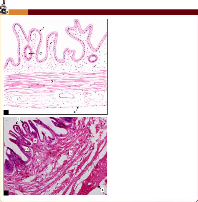
The mucous membrane of the gallbladder is lined by a tall columnar epithelium with a striated border. The mucosa is highly folded. In sections, the folds may look like villi.
Note: Because of the resemblance to villi, students sometimes
mistake sections of the gallbladder for those of the intestines.
The two are easily distinguished if it is remembered that there are no goblet cells in the epithelium of the gallbladder.
Fibromuscular Coat
The fibromuscular coat is made up mainly of connective tissue containing the usual elements. Smooth muscle fibers are present and run in various directions.
of gallbladder is covered by serosa, whereas the upper surface is attached to the fossa for gallbladder by means of connective tissue (adventitia).
Cells of Gallbladder
With the EM the lining cells of the gallbladder are seen to have irregular microvilli on their luminal surfaces. Near the lumen the lateral margins of the cells are united by prominent junctional complexes. More basally the lateral margins are separated by enlarged intercellular spaces into which complex folds of plasma membrane extend. Numerous blood capillaries are present near the bases of the cells.
These features indicate that bile is concentrated by absorption of water at the luminal surface of the cell. This water is poured out of the cell into basal intercellular spaces from where it passes into blood. Absorption of salt and water from bile into blood is facilitated by presence of Na+ and K+ ATPases in cell membranes of cells lining the gallbladder.
Clinical Correlation
" ," " -" "
THE EXTRAHEPATIC DUCTS
These are the right, left and common hepatic ducts; the cystic duct; and the bile duct. All of them have a common structure. They have a mucosa surrounded by a wall made up of connective tissue, in which some smooth muscle may be present.
The mucosa is lined by a tall columnar epithelium with a striated border.
Hepatopancreatic Duct
At its lower end the bile duct is joined by the main pancreatic duct, the two usually forming a common hepatopancreatic duct (or ampulla) that opens into the duodenum at the summit of the major duodenal papilla.
The mucosa of the hepatopancreatic duct is highly folded. These folds are believed to constitute a valvular mechanism that prevents duodenal contents from entering the bile and pancreatic ducts.
Chapter 16 Hepatobiliary System and Pancreas 199
PLATE 16.3: Gallbladder
3
- -
"6
7 8
Note: 9 /
:
A
Key
! ; 3
# <% =' 6
" "
B
Gallbladder. A. As seen in drawing; B. Photomicrograph
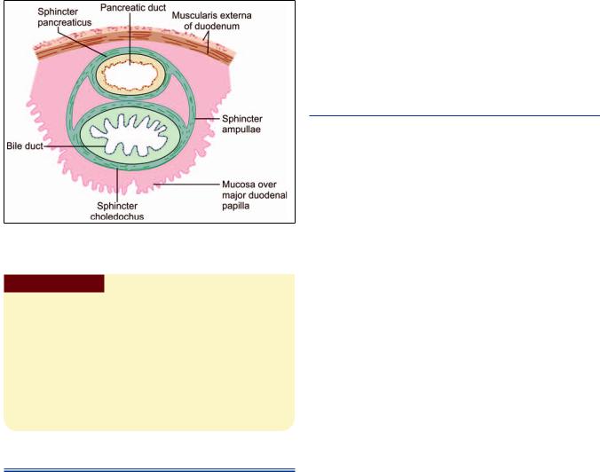
Sphincter of Oddi
Well developed smooth muscle is present in the region of the lower end of the bile duct. This muscle forms the sphincter of Oddi.
From a functional point of view this sphincter consists of three separate parts. The sphincter choledochus surrounds the lower end of the bile duct. It is always present, and
its contraction is responsible for filling of the gallbladder. A less developed sphincter pancreaticus surrounds the terminal part of the main pancreatic duct (Fig. 16.9).
A third sphincter surrounds the hepatopancreatic duct (or ampulla) and often forms a ring round the lower ends of both the bile and pancreatic ducts. This is the sphincter ampullae. The sphincter ampullae and the sphincter pancreaticus are often missing.
200 Textbook of Human Histology
Fig. 16.9: Section through the major duodenal papilla to show the components of the sphincter of Oddi (Schematic representation)
Clinical Correlation
. * " |
" |
|
|
! "" /$ * ! " + 0 "
|
/ 0 |
|
|
" " "$ biliary colic
THE PANCREAS
The pancreas extends from the concavity of the duodenum, on the right to the spleen and on the left in the posterior abdominal wall retroperitoneally. The pancreas is covered by connective tissue that forms a capsule for it. Septa arising from the capsule extend into the gland dividing it into lobules. Each pancreatic islet is surrounded by a network of reticular fibers.
It is a gland that is partly exocrine, and partly endocrine, the main bulk of the gland being constituted by its exocrine part (Plate 16.4).
The exocrine pancreas secretes enzymes that play a very important role in the digestion of carbohydrates, proteins and fats. After digestion, and absorption through the gut, these products are carried to the liver through the portal vein.
The endocrine pancreas produces two very important hormones, insulin, and glucagon. These two hormones are also carried through the portal vein to the liver where
they have a profound influence on the metabolism of carbohydrates, proteins and fats.
The functions of the exocrine and endocrine parts of the pancreas are thus linked. The linkage between the two parts is also seen in their common embryonic derivation from the endodermal lining of the gut.
THE EXOCRINE PANCREAS
The exocrine pancreas is in the form of compound tubuloalveolar serous gland.
Note: Its general structure is very similar to that of the parotid gland, but the two are easily distinguished because of the presence in the pancreas of endocrine elements.
Capsule
A delicate capsule surrounds the pancreas. Septa extend from the capsule into the gland and divide it into lobules.
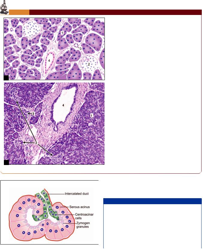
Pancreatic Acini
The secretory elements of the exocrine pancreas are long and tubular (but they are usually described as acini as they appear rounded or oval in sections). Their lumen is small (Fig. 16.10).
Secretory Cells
The cells lining the alveoli appear triangular in section, and have spherical nuclei located basally. In sections stained with hematoxylin and eosin the cytoplasm is highly basophilic (blue) particularly in the basal part. With suitable fixation and staining numerous secretory (or zymogen) granules can be demonstrated in the cytoplasm, specially in the apical part of the cell. These granules are eosinophilic. They decrease considerably after the cell has poured out its secretion.
With the EM the cells lining the alveoli show features that are typical of secretory cells. Their basal cytoplasm is packed with rough endoplasmic reticulum (this being responsible for the basophilia of this region). A welldeveloped Golgi complex is present in the supranuclear part of the cell. Numerous secretory granules (membrane bound, and filled with enzymes) occupy the greater part of the cytoplasm (except the most basal part).
Centroacinar Cells
In addition to secretory cells, the alveoli of the exocrine pancreas contain centroacinar cells that are so called because they appear to be located near the center of the acinus. These cells really belong to the intercalated ducts (see below) that are invaginated into the secretory
Chapter 16 Hepatobiliary System and Pancreas 201
PLATE 16.4: Pancreas
A
B
Pancreas. A. As seen in drawing; B. Photomicrograph
Fig. 16.10: Pancreatic serous acinus
|
|
|
|
|
> ? |
|
|
6 3 > ?
@
3 3A/ acini
,
) @3) < 3
Key
! 6
# ) <
% )
' )
elements. Some cell bodies of autonomic neurons, and undifferentiated cells are also present in relation to the secretory elements.
Added Information
The secretory cells produce two types of secretion:One of these is watery and rich in bicarbonate. Bicar-
bonate is probably added to pancreatic secretion by cells lining the ducts. It helps to neutralize the acid contents entering the duodenum from the stomach. Production of this secretion is stimulated mainly by the hormone secretin liberated by the duodenal mucosa.
The other secretion is thicker and contains numerous enzymes(includingtrypsinogen,chymotrypsinogen,amylase,
Contd...

202 Textbook of Human Histology
Contd...
lipases, etc.). The production of this secretion is stimulated mainly by the hormone cholecystokinin (pancreozymin) liberated by endocrine cells in the duodenal mucosa.
Secretion by cells of the exocrine pancreas, and the amines produced either in the gastrointestinal mucosa or in pancreatic islets. (These include gastrin, vasoactive intestinal polypeptide, and pancreatic polypeptide). Secretion is also
Theenzymes are synthesized in the rough endoplasmic
reticulum. From here they pass to the Golgi complex where they are surrounded by membranes, and are released into the cytoplasm as secretory granules. The granules move to the luminal surface of the cell where the secretions are poured out by exocytosis. Within the cell the enzymes are in an inactive form. They become active only after mixing with present in the epithelium lining the duodenum.
Duct System
Secretions produced in the acini are poured into intercalated ducts (also called intralobular ducts). These ducts are invaginated deeply into the secretory elements (Fig. 16.10). As a result of this invagination, the interacalated ducts are not conspicuous in sections.
From the intercalated ducts the secretions pass into larger, interlobular ducts. They finally pass into the duodenum through the main pancreatic duct and the accessory pancreatic duct. The cells lining the pancreatic ducts control the bicarbonate and water content of pancreatic secretion. These actions are under hormonal and neural control. The walls of the larger ducts are formed mainly of fibrous tissue. They are lined by a columnar epithelium.
The terminal part of the main pancreatic duct is surrounded by a sphincter. A similar sphincter may also be present around the terminal part of the accessory pancreatic duct.
THE ENDOCRINE PANCREAS
The endocrine pancreas is in the form of numerous rounded collections of cells that are embedded within the exocrine part. These collections of cells are called the pancreatic islets, or the islets of Langerhans. The human pancreas has about one million islets. They are most numerous in the tail of the pancreas.
Each islet is separated from the surrounding alveoli by a thin layer of reticular tissue. The islets are very richly supplied with blood through a dense capillary plexus. The
intervals between the capillaries are occupied by cells arranged in groups or as cords. In ordinary preparations stained with hematoxylin and eosin, all the cells appear similar, but with the use of special procedures three main types of cells can be distinguished.
Alpha Cells (A-Cells)
In islets of the human pancreas the alpha cells (or A-cells) tend to be arranged toward the periphery (or cortex) of the islets. They form about 20% of the islet cells. They contain smaller granules that stain brightly with acid fuchsin. They do not stain with aldehyde fuchsin.
When seen with electron microscopy, the granules of alpha cells (A2) appear to be round or ovoid with high electron density.
The alpha cells secrete the hormone glucagon.
Beta Cells (B-Cells)
The beta cells (or B-cells) tend to lie near the center (or medulla) of the islet. About 70% of the cells of islet are of this type. The beta cells contain granules (larger than alpha cells) that can be stained with aldehyde fuchsin. When seen with electron microscopy, the granules of beta cells are fewer, larger, and of less electron density than those of alpha cells.
The beta cells secrete the hormone insulin.
Delta Cells (D-Cells)
Delta cells (or D-cells), like alpha cells, are also peripherally placed. The delta cells (also called type III cells) stain black with silver salts (i.e. they are argyrophile). They resemble alpha cells in having granules that stain with acid fuchsin; and are, therefore, sometimes called A1 cells in distinction to the glucagon producing cells that are designated A2. The two can be distinguished by the fact that A2 cells are not argyrophile.
When seen with electron microscopy, the granules of delta cells (A1) appear to be round or ovoid with low electron density.
The delta cells probably produce the hormones gastrin and somatostatin. Somatostatin inhibits the secretion of glucagon by alpha cells, and (to a lesser extent) that of insulin by beta cells.
Added Information
Apart from the three main types of cells described above some other types are also present in the islets of Langerhans. These are the PP cells containing pancreatic polypeptide (and located mainly in the head and neck of the pancreas), and D1 cells (or type IV cells) probably containing vasoactive intestinal polypeptide (or a similar amine). A few cells secreting serotonin, motilin and substance P are also present.

BLOOD SUPPLY
The gland is richly supplied with blood vessels that run through the connective tissue. The capillary network is most dense in the islets. Here the endothelial lining is fenestrated providing intimate contact of islet cells and circulating blood.
Lymphatics are also present in the pancreas.
NERVE SUPPLY
The connective tissue of the pancreas also serves as a pathway for nerve fibers, both myelinated and unmyelinated. Groups of neurons are also present.
Pancreatic islets are richly innervated by autonomic nerves. Noradrenaline and acetyl choline released at nerve endings influence secretion by islet cells.
Chapter 16 Hepatobiliary System and Pancreas 203
Clinical Correlation
Acute pancreatitis: " ! 1 2$! " 34 54$" serum amylase ! # 63
783
Chronic pancreatitis: chronic relapsing
pancreatitis " |
|
|
|
* |
|
9 |
! * % |
|
( " /
The Urinary System
The urinary organs are:A pair of kidneysA pair of uretersThe urinary bladderThe urethra.
Urine production, and the control of its composition, is exclusively the function of the kidneys. The urinary bladder is responsible for storage of urine until it is voided. The ureter and urethra are simple passages for transport of urine.
Functions
The urinary organs are responsible for the production, storage, and passing of urine. Many harmful waste products (that result from metabolism) are removed from blood through urine. These include urea and creatinine that are end products of protein metabolism.
Many drugs, or their breakdown products, are also excreted in urine.
Considerable amount of water is excreted through urine. The quantity is strictly controlled being greatest when there is heavy intake of water, and least when intake is low or when there is substantial water loss in some other way (for example by perspiration in hot weather). This enables the water content of plasma and tissues to remain fairly constant.
Note: In diseased conditions urine can contain glucose (as in diabetes mellitus), or proteins (in kidney disease), the excretion of which is normally prevented.
THE KIDNEYS
Each kidney has a characteristic bean-like shape. A thin layer of fibrous tissue, which constitutes the capsule, intimately covers kidney tissue. The capsule of a healthy kidney can be easily stripped off, but it becomes adherent in some diseases.
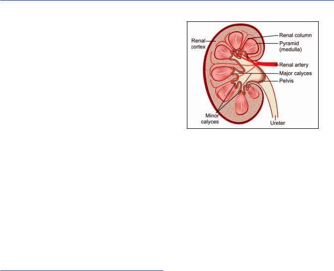
The kidney has a convex lateral margin; and a concavity on the medial side that is called the hilum. The hilum leads into a space called the renal sinus. The renal sinus is occupied by the upper expanded part of the ureter called the renal pelvis.
Fig. 17.1: Some features seen in a coronal section through the kidney (Schematic representation)
Within the renal sinus the pelvis divides into two (or three) parts called major calyces. Each major calyx divides into a number of minor calyces (Fig. 17.1). The end of each minor calyx is shaped like a cup. A projection of kidney tissue, called a papilla fits into the cup.
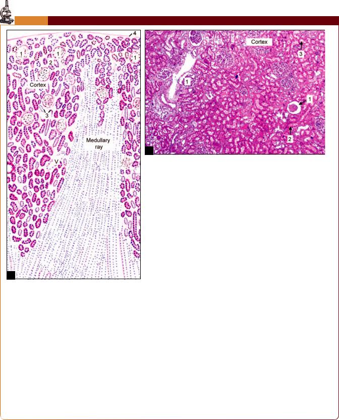
Kidney tissue consists of an outer part called the cortex, and an inner part called the medulla (Fig. 17.1 and Plate 17.1).
Medulla
The medulla is made up of triangular areas of renal tissue that are called the renal pyramids (Fig. 17.1). Each pyramid has a base directed toward the cortex; and an apex (or papilla) that is directed toward the renal pelvis, and fits into a minor calyx. Pyramids show striations that pass radially toward the apex.
Cortex
The renal cortex consists of the following:
Tissue lying between the bases of the pyramids and the surface of the kidney, forming the cortical arches or cortical lobules. This part of the cortex shows light and dark striations. The light lines are called medullary rays (Plate 17.1).
Chapter 17 The Urinary System 205
PLATE 17.1:
Key
. /
0
1 &
2
3 3 !
4 4
!
The kidney is covered by a capsule
Deep to the capsule there is the cortex
Deep to the cortex there is the medulla of the kidney
In the cortex we see circular structures called renal corpuscles surrounding which there are tubules cut in various shapes
! cuboidal epithelium with brush border
" # # $ % $ & ! ! epithelium
PCT are more in number than DCT
% ! # #$#$# # ' ( # ! ! # ! ' (thin segments) are lined by simple squamous epithelium
Note: + % ! % , % ! #-! # ! ## !
206 Textbook of Human Histology
Fig. 17.2: Parts of a nephron. A collecting duct is also shown (Schematic representation)
Tissue lying between adjacent pyramids is also a part of the cortex. This part constitutes the renal columns.
In this way each pyramid comes to be surrounded by a “shell” of cortex. The pyramid and the cortex around it constitute a lobe of the kidney. This lobulation is obvious in the fetal kidney.
"
" #$
Most of the interstitial space in the renal cortex is occupied by blood vessels and lymphatics. In the medulla the interstitium is composed mainly of a matrix containing proteins cells are present.
It has been held that interstitial cells produce prostaglandins, but it now appears that prostaglandins are produced by epithelial cells of collecting ducts.
The Uriniferous Tubules
From a functional point of view the kidney may be regarded as a collection of numerous uriniferous tubules that are specialized for the excretion of urine. Each uriniferous tubule consists of an excretory part called the nephron, and of a collecting tubule. The collecting tubules draining different nephrons join to form larger tubules called the papillary ducts (of Bellini), each of which opens into a minor calyx at the apex of a renal papilla. Each kidney contains one to two million nephrons.
Urinary tubules are held together by scanty connective tissue. Blood vessels, lymphatics and nerves lie in this connective tissue.
Fig. 17.3: Basic structure of a renal corpuscle
(Schematic representation)
Nephron
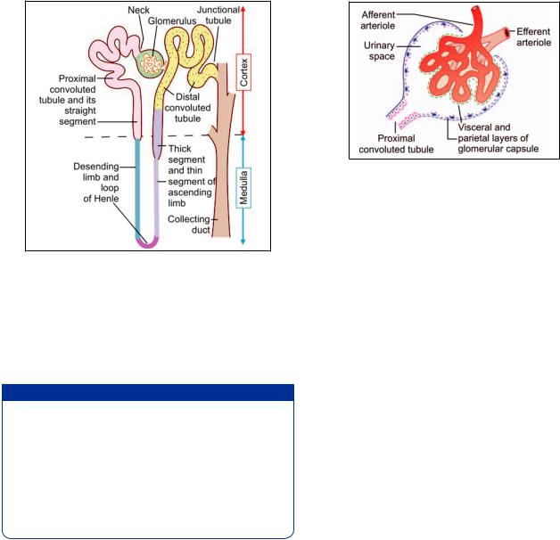
Nephron is the structural and functional unit of kidney and there are about 1–4 million nephrons in each kidney. The nephron consists of a renal corpuscle (or Malpighian corpuscle), and a long complicated renal tubule. Renal tubule is made up of three parts:
The proximal convoluted tubuleLoop of Henle
The distal convoluted tubule (Fig. 17.2)
Renal corpuscle is situated in the cortex of the kidney either near the periphery or near the medulla. Based on the situation of renal corpuscle, the nephrons are classified into two types:
Cortical nephrons or superficial nephrons (which have their corpuscles in the outer cortex).
Juxtamedullary nephrons (which have their corpuscles in the inner cortex near medulla or corticomedullary junction).
Renal corpuscles, and (the greater parts of) the proximal and distal convoluted tubules are located in the cortex of the kidney. The loops of Henle and the collecting ducts lie in the medullary rays and in the substance of the pyramids.
The Renal Corpuscle
The renal corpuscle is a rounded structure consisting of (a) a rounded tuft of blood capillaries called the glomerulus; and (b) a cup-like, double layered covering for the glomerulus called the glomerular capsule (or Bowman’s capsule) (Fig. 17.3). The glomerular capsule represents the cup-shaped blind beginning of the renal tubule. Between the two layers of the capsule there is a urinary space that is continuous with the lumen of the renal tubule.
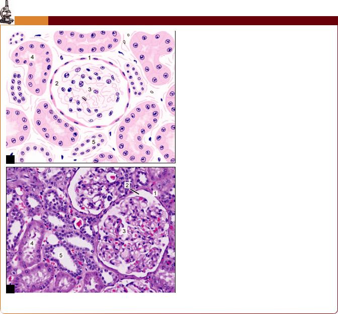
Glomerulus
The glomerulus is a rounded tuft of anastomosing capillaries (Plate 17.2). Blood enters the tuft through an afferent arteriole and leaves it through an efferent arteriole
Chapter 17 The Urinary System 207
PLATE 17.2: % & ! '
In the high power view of renal cortex large renal corpuscles ,
: a rounded glomerulus, and an outer wall, the glomerular 5 % 6
A urinary space between the glomerulus and the capsule is seen
# !! %
& # #
#
Key
. 5 % 60 7 !1 8
29 &
% !
(Note that the efferent vessel is an arteriole, and not a venule. It again breaks up into capillaries). The afferent and efferent arterioles lie close together at a point that is referred to as the vascular pole of the renal corpuscle.
The Mesangium
On entering the glomerulus the afferent arteriole divides (usually) into five branches, each branch leading into an independent capillary network. The glomerular circulation can, therefore, be divided into a number of lobules or segments.
Glomerular capillaries are supported by the mesangium that is made up of mesangial cells surrounded by a non-cellular mesangial matrix. The mesangium forms a mesentery-like fold over the capillary loop. Mesangial cells give off processes that run through the matrix. Mesangium intervenes between the capillaries of the glomerular segments.
Mesangial cells contain filaments similar to myosin. They bear angiotensin II receptors. It is believed that stimulation by angiotensin causes the fibrils to contract. In this way mesangial cells may play a role in controlling blood