

Human_Histology
.pdf168 Textbook of Human Histology
Fig. 14.8: Arrangement of cells in a taste bud
(Schematic representation)
The cells present in taste buds are elongated and are vertically orientated, those toward the periphery being curved like crescents (Fig. 14.8). Each cell has a central broader part containing the nucleus, and tapering ends. The cells are of two basic types. Some of them are receptor cells or gustatory cells. Endings of afferent nerves end in relation to them. Other cells perform a supporting function and are called supporting cells.
Note: However, it is by no means easy to distinguish between receptor and supporter cells, the essential difference being the presence of innervation. Early observers using the light microscope found hairs at the tips of some cells and concluded that these were the receptor cells. However, this has not been confirmed by EM studies. The latter have shown that the “hair” seen with the light microscope are microvilli that are more common on supporting cells rather on receptors. Two types of receptor cells can be distinguished on the basis of the vacuoles present in them.
Added Information
Supporting cells are probably of three types. Some of them that lie at the periphery of the taste bud form a sheath for it. Those near the center of the bud are truly supporting. They! " ! # of the taste bud. Microvilli are often present at the tips of these cells. A third variety of supporting cell is seen in the basal part of the bud. These basal cells multiply and produce new supporting and receptor cells to replace those that are worn out. This may be correlated with the fact that cells of taste buds have a short life and are continuously being replaced.
Recognition of Various Tastes by Tongue
It has been held that taste buds in different parts of the tongue may respond best to particular modalities of taste. However, it is now known that the same taste bud can respond to different types of taste (sweet, sour, salt, and bitter) and that taste is a complicated sensation depending
upon the overall pattern of responses from taste buds all over the tongue. With this reservation in mind, we may note that sweet taste is best appreciated at the tip of the tongue, salt by the area just behind the tip and along the lateral border, and bitter taste by circumvallate papillae.
Clinical Correlation
Fissured Tongue
It is a genetically-determined condition characterized by numerous small furrows or grooves on the dorsum of the tongue.
Hairy Tongue
ted.These “hairs” (papillae) are stained black, brown or yellowish-
SALIVARY GLANDS
There are two main groups of salivary glands—major and minor. The major salivary glands are the three paired glands: parotid, submandibular, and sublingual. The parotid glands are located laterally to the mandibular ramus and its main duct drains into the oral cavity opposite the second maxillary molar. The submandibular glands are present in the floor of the mouth, superior to digastric muscles. The sublingual gland lie anterior to submandibular glands. The ducts of submandibular and sublingual glands empty in the floor of the mouth.
The minor salivary glands are numerous and are widely distributed in the mucosa of oral cavity. Some of the minor salivary glands are the Von Ebner’s gland in tongue, buccal glands in cheeks and labial glands in lips.
The secretions of these glands help to keep the mouth moist, and provide a protective and lubricant coat of mucous. Some enzymes (amylase, lysozyme), and immunoglobulin IgA are also present in the secretions.
Structural Organization
Basically, a salivary gland consists of stroma, parenchyma and a duct system which carries the secretions into the oral cavity.
Stroma
The stroma consists of connective tissue capsule and septa.
Numerous septa arise from the capsule and enter the parenchyma of the gland, dividing the gland into numerous lobules.
These septa bring the blood vessels and nerves into the gland. Large ducts of the glands are also present in it.
Chapter 14 Digestive System: Oral Cavity and Related Structures 169
PLATE 14.4: Parotid Gland
/ / features are:
( gen granules and are darkly stained
$ $
$
$
A
Key
$
! 3
# |
$ |
|
|
|
$ |
|
|
3 |
) $
+ .
- 5
B
Parotid gland. A. As seen in drawing; B. Photomicrograph
Parenchyma
Parenchyma has two components: the secretory part and conducting part (Plate 14.3 to 14.7).
Secretory Part
Salivary glands are compound tubuloalveolar glands (racemose glands). Their secretory elements (also referred to as end pieces or as the portio terminalis) may be rounded (acini), pear shaped (alveoli), tubular, or a mixture of these (tubuloacinar, tubuloalveolar). The secretory elements lead into a series of ducts through which their secretions are poured into the oral cavity.
In sections through salivary glands we see a large number of closely packed acini with ducts scattered between them. These elements are supported by connective tissue that also divides the glands into lobules, and forms capsules around them. Blood vessels, lymphatics and
nerves run in the connective tissue that may at places contain some adipose tissue.
The acini are made up of either serous or mucous cells. A salivary gland may have only one type of acini or there may be a mix of both serous and mucous acini, these are called mixed acini. A secretory unit, or gland, with only one type of cell (serous or mucous) is said to be homocrine. If it contains more than one variety of cells it is said to be
heterocrine.
The structure of mucous and serous acini has already been described and compared in Chapter 4. It would be worthwhile if your revise that chapter now.
Myoepithelial Cells
Myoepithelial cells are present in relation to acini and intercalated ducts of salivary glands. They may also be seen in relation to larger ducts (intralobular and extralobular).

170 Textbook of Human Histology
PLATE 14.5: Submandibular Gland
The submandibular gland is a mixed salivary gland, predominantly
3 & in the form of a crescent and called as demilunes
3 /
A
Key
$! 3# 6) 6
B + $
- 5
A.As seen in drawing; B. Photomicrograph
|
These cells lie between the epithelial cells and their base- |
|
ment membrane. The myoepithelial cells located on acini |
|
are often branched (stellate) and may form “baskets” around |
|
the acini (Fig. 14.9)). Those located on the ducts are fusi- |
|
form and run longitudinally along them. |
|
Conducting part: |
|
Duct System |
|
Secretions produced in acini pass along a system of ducts, |
|
different parts of which have differing structure. The |
|
smallest ducts are called intercalated ducts. These are lined |
|
by cuboidal or flattened cells. Intercalated ducts open into |
|
striated ducts lined by columnar cells. They are so called |
|
because the basal parts of the cells show vertical striations. |
|
Both intercalated and striated ducts are intralobular ducts. |
Fig. 14.9: Surface view of an acinus showing myoepithelial cell. Its |
Striated ducts open into excretory ducts (interlobular)that |
|
|
processes form a basket around the acinus (Schematic representation) are lined by simple columnar epithelium. |
|

Chapter 14 Digestive System: Oral Cavity and Related Structures 171
PLATE 14.6:
|
$/ |
|
% / 4 |
|
|
|
The serous acini are darkly stained and have rounded |
|
nucleus placed near the center of the cell |
|
/ > |
|
|
|
? |
|
|
A
Key
3! 6
# 3) 6
B
A.As seen in drawing; B. Photomicrograph
Clinical Correlation
Sialorrhea (Ptyalism): rhea or ptyalism.
Xerostomia: ! Sialadenitis: denitis.
Tumors of Salivary Glands
Pleomorphic adenoma (mixed salivary tumor): It is the most common tumor of major and minor salivary gland. It is characterized by pleomorphic or mixed appearance in which there are epithelial elements present in a matrix of mucoid, myxoid, and chondroid tissue.
Contd...
Mucoepidermoid carcinoma: It is the most common malignant salivary gland tumor. The tumor is composed of combination of 4 types of cells: mucin-producing, squamous, intermediate, and clear cells. Well-differentiated tumors have predominance of mucinous cells, while poorly differentiated have more solid
Added Information
Cells of Salivary Glands
Serous Cells
Serous cells are usually arranged in the form of rounded acini. As a result each cell is roughly pyramidal having
Contd... |
Contd... |

172 Textbook of Human Histology
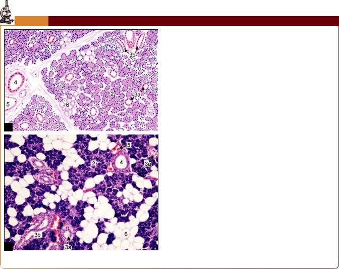
PLATE 14.7: Sublingual Gland
The sublingual gland is predominantly a mucous gland
3
A
B
Sublingual gland. A. As seen in drawing; B. Photomicrograph
Contd...
Fig. 14.10: Some features of serous cell in a salivary gland (Schematic representation)
Key
$
! 6
# 3
) 3
+ @
- :
a broad base (toward the basement membrane) and a narrow apex (toward the lumen) (Fig. 14.10). Some microvilli and pinocytotic vesicles are seen at the apex of the cell. The lumen of the acinus often extends for some distance between adjacent cells: these extensions are called intercellular secretory canaliculi. Deep to these canaliculi the cell membranes of adjoining cells are united by tight junctions. Deep to these junctions, the lateral cell margins show folds that interdigitate with those of adjoining cells. The apical cytoplasm contains secretory granules that are small, homogeneous, and electron dense. The cytoplasm also contains a prominent Golgi complex and abundant rough endoplasmic reticulum, both features indicating considerable synthetic activity. $ % ! %
present.
Contd...

Chapter 14 Digestive System: Oral Cavity and Related Structures 173
Contd... |
Contd... |
|
|
|
|
|
|
|
Fig. 14.11: Some features of mucous cells in salivary glands (Schematic representation)
Mucous Cells
Mucous cells are usually arranged in the form of tubular secretory elements (Fig. 14.11). Crescents present in relation to them are located at the ends of the tubules. The cells lining mucous cells tend to be columnar rather than! ! Rough endoplasmic reticulum and Golgi complex are similar to those in serous cells, but microvilli, foldings of plasma membrane, and intercellular canaliculi are not usually seen.
Seromucous Cells
From the point of view of ultrastructure many cells of salivary glands are intermediate between serous and mucous cells. They are referred to as seromucous cells. Most of& ! parotid and submandibular glands are really seromucous.
The secretions of all types of salivary secretory cells contain protein-carbohydrate complexes. Their concentration is lowest in cases of serous cells, very high in mucous cells, and with widely differing concentrations in seromucous cells.
We have seen that in the submandibular glands mucous acini are often capped by serous demilunes. The serous cells of a demilune drain into the lumen of the acinus through" & mucous cells.
With the EM myoepithelial cells are seen to contain the' ! " *! + + the cell. Cilia are present on some myoepithelial cells. It has been suggested that the cilia may subserve a sensory or chemoreceptor function.
Myoepithelial cells are contractile, their contraction helping to squeeze out secretion from acini.
The cells lining the striated ducts show an interesting ultrastructure (Fig. 14.12). Their basal striations are seen to
Contd...
Fig. 14.12: Electron microscopic structure of a cell from striated duct (Schematic representation)
be due to the presence of numerous deep infoldings of the basal parts of the cell membranes. Numerous elongated mitochondria are present in the intervals between the folds. Similar cells are also present scattered in the epithelium of the excretory ducts. These cells are believed to play a role in regulating the water and electrolyte content of saliva to make it hypotonic. Immunoglobulin A, produced by plasma cells lying subjacent to the epithelium, passes into saliva through the cells lining the striated ducts.
Innervation of Salivary Glands
Secretion by salivary glands is under hormonal as well as neural control. A local hormone plasmakinin formed by! / " " ! innervated by autonomic nerves, both parasympathetic (cholinergic) and sympathetic (adrenergic). Parasympathetic nerves travel to secretory elements along ducts, while sympathetic nerves travel along arteries. Synaptic contacts between nerve terminals and effector cells form neuroeffector junctions.
Two types of junction, epilemmal and hypolemmal, are present. At epilemmal junctions the nerve terminal is separated from the secretory or effector cell by the basal lamina. At hypolemmal junctions the nerve terminal pierces the basal lamina and comes into direct contact with the effector cell. Nerve impulses reaching one effector cell spread to others through intercellular contacts. Classically, salivary secretion has been attributed to parasympathetic stimulation. While this is true, it is believed that sympathetic nerves can also excite secretion either directly, or by vasodilation. Autonomic nerves not only stimulate secretion, but also appear to determine its viscosity and other characteristics. Autonomic nerve terminals are also seen on myoepithelial cells and on cells lining the ducts of salivary! / + by cells lining the ducts. Salivary glands are sensitive to pain, and must therefore have a sensory innervation as well.

Digestive System: Esophagus, Stomach, and Intestines
The gastrointestinal tract (GIT) or alimentary canal is a long muscular tube that begins at the oral cavity and ends in the anus. Different parts of the tract are specialized to perform different functions, and hence structural modifications are seen in various parts of the GIT.
The esophagus and anal canal are merely transport passages. The part of the alimentary canal from the stomach to the rectum is the proper digestive tract, responsible for digestion and absorption of food. Reabsorption of secreted fluids is an important function of the large intestine.
GENERAL STRUCTURE OF GIT
The structure of the alimentary canal, from the esophagus up to the anal canal, shows several features that are common to all these parts. We shall consider these common features before examining the structure of individual parts of the canal.
The walls of the oral cavity and pharynx are partly bony, and partly muscular. From the upper end of the esophagus up to the lower end of the anal canal the alimentary canal has the form of a fibromuscular tube. The wall of the tube is made up of the following layers (from inner to outer side) (Fig. 15.1).
The innermost layer is the mucous membrane that is made up of:
A lining epithelium
A layer of connective tissue, the lamina propria, that supports the epithelium
Fig. 15.1: Layers of the gut (Schematic representation)
A thin layer of smooth muscle called the muscularis mucosae.
The mucous membrane rests on a layer of loose areolar tissue called the submucosa.
The gut wall derives its main strength and form because of a thick layer of muscle (muscularis externa) that surrounds the submucosa.
Covering the muscularis externa there is a serous layer or (alternatively) an adventitial layer.
Primarily, it is the mucosa in which changes are seen in the alimentary tract; the other layers remain almost the same.
The Mucosa
The Lining Epithelium
The lining epithelium is columnar all over the gut; except in the esophagus, and in the lower part of the anal canal, where it is stratified squamous. This stratified squamous epithelium has a protective function in these situations. The cells of the more typical columnar epithelium are either absorptive or secretory.
The epithelium of the gut presents an extensive absorptive surface. The factors contributing to the extent of the surface are as follows:
The considerable length of the alimentary canal, and specially that of the small intestine.
The presence of numerous folds involving the entire thickness of the mucous membrane. These folds can be seen by naked eye. The submucosa extends into the folds.
At numerous places the epithelium dips into the lamina propria forming crypts (see below).
In the small intestine the mucosa bears numerous finger-like processes that project into the lumen. These processes are called villi. Each villus has a surface lining of epithelium and a core formed by an extension of the connective tissue of the lamina propria. The luminal surfaces of the epithelial cells bear numerous microvilli.
Chapter 15 Digestive System: Esophagus, Stomach, and Intestines 175
The epithelium of the gut also performs a very important secretory function. The secretory cells are arranged in the form of numerous glands as follows:
Some glands are unicellular, the secretory cells being scattered among the cells of the lining epithelium.
In many situations, the epithelium dips into the lamina propria forming simple tubular glands (These are the crypts referred to above).
In other situations (e.g. in the esophagus, duodenum) there are compound tubuloalveolar glands lying in the submucosa. They open into the lumen of the gut through ducts traversing the mucosa.
Finally, there are the pancreas and the liver that form distinct organs lying outside the gut wall. They pour their secretions into the lumen of the gut through large ducts (In this respect, these glands are similar to the salivary glands). The liver and pancreas, and most of the salivary glands, are derivatives of the epithelial lining of the gut. Embryologically, this epithelium is derived from endoderm.
The Lamina Propria
The lamina propria is made up of collagen and reticular fibers embedded in a glycosaminoglycan matrix. Some fibroblasts, blood capillaries, lymph vessels, and nerves are seen in this layer. In the small intestine the lamina propria forms the core of each villus. It surrounds and supports glandular elements and the overlying epithelium.
Prominent aggregations of lymphoid tissue (as well as scattered lymphocytes) are present in the lamina propria. Some of this lymphoid tissue extends into the submucosa and is called as gut associated lymphoid tissue (GALT).
The Muscularis Mucosae
This is a thin layer of smooth muscle that separates the connective tissue of the lamina propria from the submucosa. It consists of an inner layer in which the muscle fibers are arranged circularly (around the lumen) and an outer layer in which the muscle fibers run longitudinally. The muscularis mucosae extends into mucosal folds, but not into villi. Contractions of the muscularis mucosae are important for the local mixing of intestinal contents.
The Submucosa
This layer of loose areolar tissue connects the mucosa to the muscularis externa. Its looseness permits some mobility of the mucosa over the muscle. Numerous blood vessels, lymphatics and nerve fibers traverse the submucosa. Smaller branches arising from them enter the mucous membrane.
The Muscularis Externa
Over the greater part of the gut the muscularis externa consists of smooth muscle. The only exception is the upper part of the esophagus where this layer contains striated muscle fibers. Some striated muscle fibers are also closely associated with the wall of the anal canal.
The muscle layer consists (typically) of an inner layer of circularly arranged muscle fibers, and an outer longitudinal layer.
Both layers really consist of spirally arranged fasciculi, the turns of the spiral being compact in the circular layer, and elongated in the longitudinal layer.
The arrangement of muscle fibers shows some variation from region to region. In the stomach an additional oblique layer is present. In the colon the longitudinal fibers are gathered to form prominent bundles called the taenia coli.
Localized thickenings of circular muscle fibers form sphincters that can occlude the lumen of the gut. For example, the pyloric sphincter is present around the pyloric end of the stomach, and the internal anal sphincter surrounds the anal canal. A functional sphincter is seen at the junction of the esophagus with the stomach. A valvular arrangement at the ileocaecal junction (ileocaecal valve) prevents regurgitation of caecal contents into the ileum.
The Serous and Adventitial Layers
Covering the muscle coat, there is the serous layer which is the outermost layer of the alimentary canal. This layer is merely the visceral peritoneum that covers most parts of the gastrointestinal tract. In some places where a peritoneal covering is absent (e.g. over part of the esophagus) the muscle coat is covered by an adventitia made up of connective tissue.
Nerve Plexuses
The gut is richly supplied with nerves. A number of nerve plexuses are present as follows:
The myenteric plexus (of Auerbach) lies between the circular and longitudinal coats of muscularis externa.
The submucosal plexus (of Meissner) lies in the submucosa (near its junction with the circular muscle layer).
A third plexus is present near the muscularis mucosae. The nerve fibers in these plexuses are both afferent and
efferent. The efferent fibers supply smooth muscle and glands. Postganglionic neurons meant for these structures lie amongst the nerve fibers forming these plexuses.

176 Textbook of Human Histology
THE ESOPHAGUS |
The Submucosa |
The esophagus is a long muscular tube beginning at the end of cricoid cartilage and opens into the cardiac end of stomach. It conducts chewed food (bolus) and liquids to stomach.
MICROSCOPIC FEATURES
The wall of esophagus has the usual four layers viz., mucosa, submucosa, muscularis externa and an external adventitia (Fig. 15.2 and Plate 15.1). The esophagus does not have a serous covering except over a short length near its lower end.
The Mucosa
The mucous membrane of the esophagus shows several longitudinal folds that disappear when the tube is distended.
The mucosa is lined by stratified squamous epithelium, which is normally not keratinized.
Occasional melanocytes and endocrine cells are present. A columnar epithelium, similar to that lining the cardiac end of the stomach, may extend for some distance into the abdominal part of the esophagus.
Finger-like processes (or papillae) of the connective tissue of the lamina propria project into the epithelial layer (just like dermal papillae). This helps to prevent separation of epithelium from underlying connective tissue.
At the upper and lower ends of the esophagus some tubuloalveolar mucous glands are present in the lamina propria.
The muscularis mucosae is absent or poorly developed in the upper part of the esophagus. It is distinct in the lower part of the esophagus, and is thickest near the esophagogastric junction. It consists chiefly of longitudinal muscular fibers, but a few circular fibers are also present.
Fig. 15.2: Transverse section of esophagus showing all the four layers of wall. The lumen of esophagus is star shaped (Schematic representation)
The only special feature of the submucosa is the presence of compound tubuloalveolar mucous glands. Small aggregations of lymphoid tissue may be present in the submucosa, specially near the lower end. Some plasma cells and macrophages are also present.
The Muscularis Externa
The muscle layer consists of the usual circular and longitudinal layers. However, it is unusual in that the muscle fibers are partly striated and partly smooth. In the upper one-third (or so) of the esophagus the muscle fibers are entirely of the striated variety, while in the lower one-third all the fibers are of the smooth variety. Both types of fibers are present in the middle one-third of the esophagus.
Added Information
cardioesophagealjunction. However, the circular muscle is not thicker here than elsewhere in the esophagus, and its role as a sphincter is not generally accepted. However, a physiological sphincter does appear to exist. The anatomical factors that could account for this sphincteric action are not agreed upon.
The Adventitia
The muscle layer of the esophagus is surrounded by dense fibrous tissue that forms an adventitial coat for the esophagus. The lowest part of the esophagus is intraabdominal and has a covering of peritoneum.
Pathological Correlation
Achalasia (Cardiospasm): Achalasia of the esophagus is a neuromuscular dysfunction due to which the cardiac sphincter fails to relax during swallowing and results in progressive dysphagia and dilatation of the esophagus (mega-esophagus).
Barrett’s esophagus: This is a condition in which, following esophagus is replaced by columnar epithelium (columnar metaplasia). The condition is seen more commonly in later age disease.
THE STOMACH
Stomach is a muscular bag that receives food bolus from esophagus. The food passes through the esophagus and enters the stomach where it is converted into a thick paste known as chyme. Anatomaically, stomach is divided into four regions: Cardia, fundus, body, and pylorus
(Fig. 15.3).

Chapter 15 Digestive System: Esophagus, Stomach, and Intestines 177
PLATE 15.1: Esophagus
|
|
|
|
|
|
|
|
B |
|
|
|
|
|
Esophagus. A. As seen in drawing |
A |
|
|
|
[to be provided by author]; B. Photomicrograph |
!
! "
! # #
! " $%
! #& ' & # '
Note: " ('
Key
)' * "
+'
,' *
-' . "
/' * &
'
"' 0
1' 2
* ' *
Histologically fundus and body of stomach have a similar structure.
MICROSCOPIC FEATURES
The wall of the stomach has the four basic layers a mucous membrane, a submucosa, a muscularis externa, and a serous layer. The mucous membrane and the muscularis externa have some special features that are described below.
The Mucous Membrane
As seen with the naked eye the mucous membrane shows numerous folds (or rugae) that disappear when the
Fig. 15.3: Anatomical regions of stomach (Schematic representation) stomach is distended.