

Учебники / ABC of Ear, Nose and Throat 5 ed Ludman 2007
.pdfSore Throats |
51 |
|
|
|
|
Figure 11.1 Acute tonsillitis.
Indications for tonsillectomy
Indications for tonsillectomy have become more stringent recently, with a reduction in numbers as risk/benefit analysis has developed (Fig. 11.1). There is also a better understanding of the natural history, at least in children with relatively mild symptoms who are likely to improve over 3 years. Tonsillectomy is reasonable if the infection is due to true tonsillitis, is severe enough to preclude normal activity and has been recurring on and off for at least a year, with at least five episodes. The benefits must be balanced against a small but significant risk of complications, particularly haemorrhage, which had an incidence of 2–8% in one national audit.
The incidence of surgery diminishes with age, particularly over 30. There is a distinct group of adults with low-grade continuing sore throat symptoms, punctuated by occasional acute episodes due to chronic tonsil sepsis, who may be helped by tonsillectomy.
Sore throats with cervical adenopathy
Severe sore throat with marked neck lymphadenopathy in young people, particularly with no history of recurrent sore throat, may be due to infectious mononucleosis, and the monospot test should be checked in these cases. Management is supportive, but severe cases may need admission for intravenous rehydration. Antibiotics, and sometimes steroids, are usually given in hospital despite the viral cause, as secondary bacterial infection, sometimes with anaerobes, is common. Epstein–Barr virus is the most common cause, although cytomegalovirus, toxoplasmosis, rubella and human immunodeficiency virus (HIV) are also implicated. During recovery, patients should be warned to avoid contact sports for at least 6 weeks because of a risk of damage to an enlarged liver and spleen, and abnormal liver function should be monitored until recovery is complete.
Complications of throat sepsis
Quinsy
Peritonsillar abscess (quinsy) presents as a unilateral erythematous swelling lateral to the tonsil, and in a patient with systemic upset is the commonest septic complication. Restricted mouth opening is
usual. Traditional management by incision in the unanaesthetized patient (topical anaesthesia being potentially dangerous because of the risk of aspiration of pus) has been replaced by aspiration with a large-bore hypodermic needle. Antibiotics, usually penicillin V, with or without metronidazole are administered.
Parapharyngeal abscess
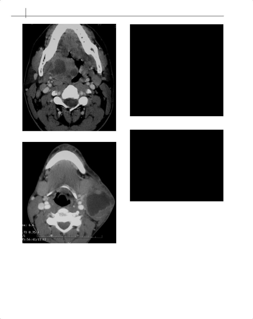
Deep neck space infection secondary to tonsil, or sometimes dental, sepsis is less common, but needs management in hospital, sometimes with airway protection by intubation or tracheostomy before surgical incision and drainage and infusion of intravenous antibiotics (see Fig. 11.2). The commonest variety is parapharyngeal abscess presenting in a severely ill patient with marked unilateral, tender, often red, neck swelling.
Acute retropharyngeal abscess
This is uncommon and usually secondary to tonsil or adenoid sepsis, sometimes in the immune compromised (Fig. 11.3). Airway protection before incision and drainage, which may be peroral, is essential. Tuberculosis is a rare cause for chronic retropharyngeal abscess nowadays in Western countries, but needs to be excluded.
Sore throat with acute airway distress
Acute upper airway distress or obstruction is most commonly inflammatory (Fig. 11.4), usually due to bacterial infection with Haemophilus influenzae. Vaccination programmes have reduced the incidence of this once common paediatric illness, usually manifesting as acute epiglottitis, but it is still seen quite commonly in adults where the infection tends to affect the whole supraglottic region. Mild cases in cooperative adults may be investigated by nasopharyngeal endoscopy, but there is a risk, particularly in children, of precipitating complete airway obstruction, and interference of any kind should be avoided – even the use of a tongue depressor or attempts at imaging. Large doses of intravenous broad-spectrum antibiotics, usually a third generation cephalosporin, with intravenous steroids are the management of choice and will usually result in improvement if not complete resolution within 48–72 hours.
The life-threatening nature of this disease cannot be over-empha- sized. Rapid deterioration, even in apparently stable patients, may occur to precipitate critical airway obstruction.
Sore throat with subacute airway obstruction
In adults, subacute stridor may be caused by neoplasia, most commonly in the subglottic region (see Chapter 19), or rarely due to bilateral vocal cord palsy from neurological disease, or may be a complication of thyroid neoplasia or surgery.
Sore throat with a chronic upper airway obstruction
Low-grade upper airway obstruction in adults may be due to neoplasms or rarer conditions such as amyloidosis, sarcoid or vascu-
52 ABC of Ear, Nose and Throat
(a)
(a)
(b)
Figure 11.3 (a) Lateral soft tissue of neck showing widening of retropharyngeal space and (b) CT scan of the same patient showing abscess cavity in retropharyngeal space.
(b)
Figure 11.2 (a) Tonsillar and parapharyngeal abscess and (b) large neck abscess secondary to tonsillitis.
litis, notably Wegener’s granuloma. Patients present with noisy breathing or hoarseness and may be distressed or cyanosed. They should be referred early for specialist care. Examination with the flexible nasolaryngoscope is the first step, followed up by endoscopy and biopsy under general anaesthesia when appropriate (Fig. 11.5).
Pharyngitis
Pharyngitis may be acute and is most often caused by viruses, rhinoviruses, influenza A and B, herpes simplex and zoster and other infections involving pharyngeal lymphoid tissue. The symptoms of pharyngitis may not correlate with the clinical picture on inspection.
Chronic pharyngitis may be specific or non-specific. Non-spe- cific pharyngitis is more difficult to define and diagnose. Patients usually complain of long-standing discomfort in the throat, pain or catching on swallowing, and sometimes earache. The observation of red patches on the posterior pharyngeal wall is not a reliable indication for a firm diagnosis, nor are there any helpful laboratory tests. There are a number of sources of infection of the lymphoid tissue of

Sore Throats |
53 |
|
|
|
|
Figure 11.4 Rare case of diphtheria oropharyngitis.
Figure 11.5 Lingual tonsillar tissue which may present with chronic upper airway obstruction.
the posterior pharyngeal wall – chronic sinusitis with postnasal pus irritation from above, chronic bronchitis and bronchiectasis from the respiratory tract below, as well as laryngo-pharyngeal reflux from the upper gastrointestinal system. Local irritants from tobacco, alcohol or industrial fumes are possible causes. Referral is indicated to exclude patients with primary pharyngeal carcinoma. Chronic pharyngitis is expressed by the patient as ‘soreness in the throat’, sometimes associated with catarrh. It is rarely associated with pyrexia or systemic upset. Rarely, if ever, will this symptom respond to antibiotics, and they should not be given as they are ineffective and may result in the development of resistance or side effects. Chronic specific pharyngitis may be associated with specific bacterial organisms: syphilis, tuberculosis, toxoplasmosis, leprosy, and scleroma. These are recognized by a localized physical abnormality, followed by biopsy and culture of the tissue. Specific treatment usually resolves the infection, but patients may remain symptomatic from scarring of local tissues.
Chronic inflammation of the oral mucosa follows ingestion of irritants (spices, spirits or heavy tobacco use), and may eventually progress to neoplasia. Inflammation also arises from irritation by rough, irregular teeth or ill-fitting dentures.
Candidal or fungal infection is commonly seen in the oral cavity of the elderly, and may manifest as redness and soreness of the mouth, sometimes with angular chelitis. Frequent recurrent fungal infections can occur, and are most commonly seen in denture wearers as prosthetic teeth can harbour the candidal organisms. Other patients who may present with fungal infections are those on steroid inhalers for chronic respiratory airway diseases, and also patients who are immunocompromised. Severe cases exhibit white plaques on the oral mucosa, commonly on the soft palate, but frequently generalized redness is the only evidence. Culture is diagnostic but the swab must be taken by vigorous rubbing of the tongue if it is to yield a positive result. Prolonged treatment with nystatin or clotrimazole, and occasionally with a systemic antifungal agent, is required for eradication. Dentures should be sterilized daily to prevent reinfection.
Ulceration
Ulceration commonly occurs in the form of aphthous ulcers (Fig. 11.6), which are painful and self-limiting in the course of about 10 days. They have a well-demarcated edge with a white sloughy base and are usually a few millimetres in diameter. Management is with topical pain-relieving medication such as ‘Bonjela’ (cetalkonium & choline salicylate) or ‘Difflam’ (benzydamine hydrochloride). Referral is advisable for further patient management. Topical steroid preparations are available and may help to provide symptomatic relief. Occasionally, ulcers are larger and more painful and persistent, in the condition known as major aphthi. The cause is unknown.
Oral ulceration is occasionally seen in serious systemic disease such as agranulocytosis and vasculitic disorders, and oral mucosal changes may occur as the manifestation of HIV in the form of ‘hairy leucoplakia’. The typical ‘punched out’ ulcer of primary syphilis of the oral cavity is now relatively rare in the developed world, but
Oral mucosal lesions
Mucosal abnormalities generally take the form of altered colour
– generalized redness, or white or red patches. Ulcerative defects, swellings and occasionally papillomatous lesions also occur. Some common lesions may be identified from appearance alone, but many require biopsy for diagnosis, which may usually be achieved under local anaesthesia.
Inflammation
Acute inflammation of the oral cavity mucosa may arise from contact with irritants, or allergic responses to foodstuffs. Peanut allergy in children and young adults is increasingly common and manifests as irritation and swelling of the lips and oral mucosa. Progression to airway obstruction can develop. Prompt hospital treatment with antihistamine and intravenous steroids is essential to abort this potential
danger. Figure 11.6 Aphthous ulcer.

54 ABC of Ear, Nose and Throat
should be considered as part of the differential diagnosis. The benign incidental finding of ‘geographical tongue’ is often mistaken for oral ulceration, but is in fact a normal variant. Many of these conditions and lesions may affect the posterior tongue and, unless diagnosed early, patients may try all remedies that are available ‘over the counter’ (and ultimately may become a clinical nuisance).
Neoplasia
Field change of the oral mucosa ranging from chronic inflammation through mild to severe dysplasia is common in association with heavy tobacco and alcohol usage, particularly in combination, and may progress via carcinoma in situ to frank squamous carcinoma.Any persistent white or red patches in the oral cavity, particularly in areas directly exposed to high concentrations of smoke and particularly dark spirits, should be regarded as suspicious and submitted to biopsy. Leucoplakia is easily confused with lichen planus, which is typified by a lacy white pattern on the mucosa, particularly in the buccal region.
Carcinomatous change is discussed in Chapter 19.
Further reading
Del Mar CB, Glasziou PP, Spinks AB. (2004) Antibiotics for sore throat. The Cochrane Database of Systematic Reviews, 2, CD000023; DOI: 10.1002/ 14651858.CD000023.pub2
Katori H, Tsukuda M. (2005) Acute epiglottis: Analysis of factors associated with Airway Intervention. Journal of Laryngology and Otology; 119: 967–72.
Mckerrow WS. Tonsillitis. BMJ Clinical Evidence, www.clinicalevidence.com van Staaij BK, van den Akker EH, Rovers MM et al. (2004) Effectiveness of
adenotonsillectomy in children with mild symptoms of throat infections or adenotonsillar hypertrophy: open, randomised controlled trial. British Medical Journal; 329: 651–4.
Ridder GJ, Technau-Ihling K, Sander A, Boedeker CC. (2005) Spectrum and Management of Deep Neck Space Infections: An 8 year Experience of 234 cases. Otolaryngol Head Neck Surg; 133: 709–14.
Scully C, Felix DH. (2005) Oral Medicine: Aphthous and other common ulcers. British Dental Journal; 199: 259 –64.
Scully C, Felix DH. (2005) Oral Medicine: Mouth Ulcers of more serious connotation. British Dental Journal; 199: 339 – 343.

CHAPTER 12
Breathing Disorders
Vinidh Paleri, Patrick J Bradley
OVERVIEW
•Stridor is a symptom NOT a diagnosis, and always requires examination and investigation. It denotes a harsh, vibratory noise from turbulent flow through a partially obstructed segment of respiratory tract.
•All patients presenting with stridor, acute or chronic, must be investigated urgently.
•The prime aim of managing a patient with stridor is to establish a secure and stable airway by intubation or tracheostomy.
Pathophysiology
Breathing is involuntarily controlled by the respiratory centre in the brain stem. The vocal cords abduct during inspiration and, with the negative pressure caused by diaphragmatic contraction and expansion of the chest, air is drawn into the lungs. The recurrent laryngeal branches of the vagus nerves control vocal cord movement, with intrinsic laryngeal muscles providing fine control. The cricoid cartilage is the only complete ring in the respiratory tract, surrounding the subglottic region. Any airway oedema there reduces its lumen. One millimetre of mucosal oedema reduces the cross-sectional area by more than 40%.
Stridor is a harsh, vibratory noise from turbulent flow through a partially obstructed segment of the respiratory tract. This is differentiated from stertor, where noise is caused by vibration of pharyngeal structures, leading to a lower pitched sound. Stridor can be present during the inspiratory or the expiratory phase or be biphasic (Box 12.1).
Stridor in children
Evaluation
History provides pointers to the diagnosis (Box 12.2). A previously
Box 12.1 Types of stridor
•Inspiratory: supraglottic and glottic obstruction.
•Expiratory: low tracheal obstruction.
•Biphasic: glottic and subglottic obstruction.
Box 12.2 Historical information
•Age of onset
•Duration/phase of stridor
•Worsening/improvement of stridor since onset
•Precipitating causes
•Failure to gain weight
•Breath-holding spells
•Fever
•Feeding/swallowing problems
•Hoarse/muffled voice
•Intubation in the past
•Cough/chest infections
well child presenting with acute onset stridor arouses suspicion of foreign body aspiration. Preceding upper respiratory tract infection (URTI) indicates croup or bacterial tracheitis. Epiglottitis (supraglottitis) typically presents as rapid onset fever, dysphagia and drooling in children from 2 to 7 years old.
A child with acute stridor must be assessed where instrumentation and experienced personnel are available for emergency intervention to protect the airway. Clinical assessment is shown in Box 12.3. Respiratory rate and level of consciousness are the most important indicators of severity of obstruction. Intensity of the sound does not indicate severity, as severe obstruction so reduces airflow that stridor is inaudible. The child must not be upset in case of precipitating acute obstruction.
Box 12.3 Clinical evaluation
•Respiratory rate
•Cyanosis
•Apnoeic spells
•Use of accessory muscles
•Intercostal/sternal retraction
•Nasal flaring
•Timing/severity of stridor
•Hoarseness
•Temperature/toxicity
•Level of consciousness
•ENT examination in controlled setting
55

56 |
|
|
ABC of Ear, Nose and Throat |
|
|
|
|
|
|
|
|
|
|
|
|
|
|
|
|
|
|
|
|
|
|
|
|
|
|||||||||||||||
|
|
|
|
|
|
|
|
|
|
|
|
|
|
|
|
|
|
|
|
|
|
|
|
|
|
|
|
|
|
|
|
|
|
|
|
|
|
|
|
|
|
|
|
|
|
|
|
|
|
|
|
|
|
|
|
|
|
|
|
|
|
|
|
|
|
|
|
|
|
|
|
|
|
|
|
|
|
|
|
|
|
|
|
|
|
|
|
|
|
|
|
|
|
|
|
|
|
|
|
|
|
|
|
|
|
|
|
|
|
|
|
|
|
|
|
|
|
|
|
|
|
|
|
|
|
|
|||||
|
|
|
|
|
|
|
|
|
|
|
|
|
|
|
|
|
|
|
|
|
|
|
Noisy breathing |
|
|
|
|
|
|
|
|
|
|
|
|
||||||||
|
|
|
|
|
|
|
|
|
|
|
|
|
|
|
|
|
|
|
|
|
|
|
|
|
|
|
|
|
|
|
|
|
|
|
|
|
|
|
|
|
|
|
|
|
|
|
|
|
|
|
|
|
|
|
|
|
|
|
|
|
|
|
|
|
|
|
|
|
|
|
|
|
|
|
|
|
|
|
|
|
|
|
|
|
|
|
|
|
|
|
|
|
|
|
|
|
|
|
|
|
|
|
|
|
|
|
|
|
|
|
|
|
|
|
|
|
|
|
|
|
|
|
|
|
|
|
|
|
|
|
|
|
|
|
|
|
Stertor |
|
|
|
|
|
|
|
|
|
|
|
|
|
|
|
|
Stridor |
|
|
|
|
|
|
|
|
|
|
|||||||||||
|
|
|
|
|
|
|
|
|
|
|
|
|
|
|
|
|
|
|
|
|
|
|
|
|
|
|
|
|
|
|
|
|
|
|
|
|
|
|
|
|
|
||
|
|
|
|
|
|
|
|
|
|
|
|
|
|
|
|
|
|
|
|
|
|
|
|
|
|
|
|
|
|
|
|
|
|
|
|
|
|
|
|
|
|
|
|
|
|
|
|
|
|
|
|
|
|
|
|
|
|
|
|
|
|
|
|
|
|
|
|
|
|
|
|
|
|
|
|
|
|
|
|
|
|
|
|
|
|
|
|
|
|
|
|
Adenotonsillar hypertrophy |
|
|
|
Inspiratory or biphasic |
|
|
|
|
|
|
|
|
|
|
|
Expiratory |
|
|
|
||||||||||||||||||||
|
|
|
|
|
Macroglossia |
|
|
|
|
|
|
|
|
|
|
|
|
|
|
|
|
|
|
|
|
|
|
|
|
|
|
|
|
|
|
|
|||||||
|
|
|
|
|
Micrognathia |
|
|
|
|
|
|
|
|
|
|
|
|
|
|
|
|
|
|
|
|
|
|
|
|
|
|
|
|
|
|
|
|||||||
|
|
|
|
|
Choanal atresia |
|
|
|
|
|
|
|
|
|
|
|
|
|
|
|
|
|
|
|
|
|
|
|
|
|
|
|
|
|
|
|
|||||||
|
|
|
|
|
|
|
|
|
|
|
|
|
|
|
|
|
|
|
|
|
|
|
|
|
|
|
|
|
|
|
|
|
|
|
|
|
|
|
|
|
|
|
|
|
|
|
|
|
|
|
|
|
|
|
|
|
|
|
|
|
|
|
|
|
|
|
|
|
|
|
|
|
|
|
|
|
|
|
|
|
|
|
|
|
|
|
|
|
|
|
|
|
|
|
|
|
|
|
|
|
|
|
|
|
|
|
|
|
|
|
|
|
|
|
|
|
|
|
|
|
|
|
|
|
|
|
|
|
|
|
|
|
|
|
|
|
|
|
|
Hoarse voice |
|
|
|
|
|
|
|
|
|
|
|
Normal voice |
|
|
|
|
|
|
Afebrile |
|
|
Febrile |
|||||||||||||
|
|
|
|
|
|
|
|
|
|
|
|
|
|
|
|
|
|
|
|
|
|
|
|
|
|
|
|
|
|
|
|
|
|
|
|
|
|
|
|
|
|
|
|
|
|
|
|
|
|
|
|
|
|
|
|
|
|
|
|
|
|
|
|
|
|
|
|
|
|
|
|
|
|
|
|
|
|
|
|
|
|
|
|
|
|
|
|
|
|
|
|
|
|
|
|
|
|
|
|
|
|
|
|
|
|
|
|
|
|
|
|
|
|
|
|
|
|
|
|
|
|
|
|
|
|
|
|
|
|
|
|
|
|
|
|
|
Afebrile |
|
|
|
|
|
Febrile |
|
|
|
Afebrile |
|
|
Febrile |
|
|
Foreign body |
|
|
|
Bronchiolitis |
||||||||||||||||||
|
|
|
|
|
|
|
|
|
|
|
|
|
|
|
|
|
|
|
|
|
|
|
|
|
|
|
|
|
|
|
|
|
|
|
Bronchial asthma |
|
|
|
|
|
|||
|
|
|
|
|
|
|
|
|
|
|
|
|
|
|
|
|
|
|
|
|
|
|
|
|
|
|
|
|
|
|
|
|
|
|
|
|
|
|
|
|
|||
|
|
|
|
|
|
|
|
|
|
|
|
|
|
|
|
|
|
|
|
|
|
|
|
|
|
|
|
|
|
|
|||||||||||||
|
|
|
Vocal cord paralysis |
|
|
|
Croup |
|
|
|
Laryngomalacia |
|
Retropharyngeal abscess |
|
|
|
|
|
|
|
|
|
|
||||||||||||||||||||
|
|
|
Respiratory papillomatosis |
|
|
|
Supraglottitis |
|
|
|
Subglottic stenosis |
|
|
|
|
|
|
|
|
|
|
|
|
|
|
|
|
||||||||||||||||
|
|
|
Laryngeal cyst/web |
|
|
|
|
|
|
|
|
|
|
Subglottic haemangioma |
|
|
|
|
|
|
|
|
|
|
|
|
|
|
|
|
|||||||||||||
|
|
|
Laryngeal cleft |
|
|
|
|
|
|
|
|
|
|
|
|
|
|
|
|
|
|
|
|
|
|
|
|
|
|
|
|
|
|
|
|
|
|
||||||
|
|
|
|
|
|
|
|
|
|
|
|
|
|
|
|
|
|
|
|
|
|
|
|
|
|
|
|
|
|
|
|
|
|
|
|
|
|
|
|
|
|
|
|
Figure 12.1 Differential diagnosis of stridor in children.
The probable cause is usually surmised before direct examination (Fig. 12.1). Most conditions are evolving when first seen, and observation needs intensive care or a high dependency setting.
Congenital structural lesions rarely present acutely. Chronic stridor usually needs diagnostic laryngotracheoscopy unless mild and easily diagnosed on clinical examination alone. In a cooperative child with no evidence of hypoxia, flexible laryngoscopy in the clinic can be very informative.
Acute stridor
Epiglottitis (supraglottitis)
Haemophilus influenzae type B is the usual infective agent. Incidence has decreased with HiB vaccination. Children between 2 and 7 years are affected, with peak incidence at 3. The disease presents with rapid onset of high fever, toxicity, agitation, stridor, dyspnoea, muffled voice and painful swallowing. The child sits leaning forward with mouth open and drooling. If epiglottitis is suspected, no further examination should be performed outside a controlled setting. The risk of complete obstruction is high. Endotracheal intubation is preferred as the supraglottic swelling usually subsides in a few days. A swollen, cherry red epiglottis is seen on direct laryngoscopy. Intravenous antibiotics are essential.
Laryngotracheobronchitis
The most common cause of acute stridor in childhood is laryngotracheobronchitis or ‘croup’. Parainfluenza virus is the commonest cause, with influenza virus types A or B, respiratory syncytial virus and rhinoviruses sometimes being implicated. Children between 6 months and 3 years are affected, with peak incidence at 2. Symptoms include low-grade fever, barking cough, inspiratory
stridor and hoarseness, worse at night and aggravated by crying. No endoscopy is needed. Nebulized epinephrine with intravenous steroids is recommended. Rarely, intubation and ventilation are necessary.
Acute retropharyngeal and peritonsillar abscesses
Drooling, painful swallowing and systemic upset are usually seen at presentation,usually with a preceding URTI.Retropharyngeal abscesses in the lower pharynx may cause stridor, neck stiffness and torticollis. A soft tissue lateral X-ray of the neck shows diagnostic widening of the space between the vertebral column and the airway. Peritonsillar abscesses cause trismus and stertor. Urgent drainage is required.
Chronic stridor
Gastro-oesophageal reflux is a problem in children with chronic stridor – in up to 80% of cases. This is caused by the strong thoracoabdominal pressure gradient of airway obstruction.
Laryngomalacia
This accounts for 75% of all causes of stridor in infants. Weakness of the supraglottic structures leads to prolapse of the supraglottis during inspiration (Figs 12.2 and 12.3). It presents as inspiratory or variable stridor between the fourth and sixth weeks of life. Stridor is worsened by crying and feeding and is relieved in the prone position. It is a self-limiting condition.
Subglottic stenosis
This may be congenital or iatrogenic (secondary to prolonged intubation and ventilation) (Fig. 12.4). Symptoms include inspiratory or biphasic stridor, usually in the first year of life. Iatrogenic stenosis

Breathing Disorders |
57 |
|
|
|
|
Figure 12.2 Laryngomalacia showing open airway during expiration (Courtesy Dr H. Kubba).
Figure 12.3 Laryngomalacia showing epiglottic collapse during inspiration (Courtesy Dr H. Kubba).
is suspected if stridor presents after extubation. Mild stenoses can be observed during laryngeal growth. Surgical reconstruction may be needed.
Vocal cord paralysis
This is usually met within the first month of life with stridor, cyanosis, apnoea and feeding problems. Concomitant neurological disease, such as hydrocephalus and Arnold-Chiari malformation, is present in most patients. Diagnosis is established by rigid endoscopy and assessment of vocal cord mobility. Management depends upon severity and progression. Spontaneous recovery may take up to 3 years. Tracheostomy may be needed.
Subglottic haemangioma
A capillary haemangioma in the subglottis presents between 6 weeks and 6 months of life (Fig. 12.5). Cutaneous haemangiomas offer a hint to the diagnosis. Intermittent stridor and a tendency to recurrent episodes of ‘croup’ are typical. Haemangiomas may grow for a year, followed by spontaneous regression, so they can be observed. A tracheostomy may be needed until regression. Other
Figure 12.4 Subglottic stenosis (Courtesy Dr H. Kubba).
Figure 12.5 Subglottic haemangioma causing airway compromise (Courtesy Dr H. Kubba).
treatment options include laser vaporization, excision and systemic steroids.
Respiratory papillomatosis
This is caused by the human papilloma virus. Transmission can occur from the mother to the child during labour. Hoarse voice is the usual presenting symptom, and the airway may be compromised. Stridor may need urgent debulking of the papillomatous lesions (Fig. 12.6). Tracheostomy should be avoided as this may provoke spread of papillomas into the lower airways. Resolution usually occurs during adolescence. Regular surveillance is needed with debulking or vaporization by a laser as necessary. Addition of topical cidofovir (an antiviral agent) reduces recurrences.

58 ABC of Ear, Nose and Throat
Figure 12.6 Papilloma on the right true vocal cord. |
Figure 12.7 Laryngeal cancer causing complete obstruction of the glottis |
|
with superficial bleeding caused by intubation. |
Evaluation of stridor in adults
Without definite precipitating cause or relevant history, acute and chronic stridor in adults should be considered neoplastic unless proven otherwise. A careful history may indicate causes such as previous thyroid surgery (bilateral recurrent laryngeal nerve injury) and intubation trauma. Assessment of the extent of hypoxia and the work of breathing is described in Boxes 12.2 and 12.3. It is possible to assess the larynx with a flexible nasolaryngoscope and to achieve a diagnosis in the outpatient setting in most adults.
Bilateral vocal cord palsy
The commonest cause of this condition used to be thyroid surgery, but now most causes are idiopathic. Voice is preserved, with stridor most evident on exertion. Flexible laryngoscopy reveals limitation of abduction of the cords on inspiration. Management includes observation only, a choice of intralaryngeal procedures to increase the airway at the glottic level, or tracheostomy.
Malignancy
Malignant lesions of the larynx and hypopharynx can present with stridor due to tumour obstruction of the airway or by causing vocal cord palsy and oedema. Stridor can also occur after radiation for laryngeal cancers. It is not always possible to secure the airway before tracheostomy. Debulking the tumour to improve the airway while awaiting definitive management is an option. For factors that determine treatment, see Chapter 19. Tumours presenting with stridor are usually well advanced locally and may need total laryngectomy for clearance (Fig. 12.7).
Intubation trauma
Intubation for any length of time causes laryngeal inflammation. Extensive inflammation and ulceration lead to fibrosis and scarring. This usually affects the subglottis. Neonates tolerate intubation for weeks with little long-term harm, but it is reasonable to consider conversion to tracheostomy after a week to 10 days of intubation in adults if no extubation is planned. Reconstruction of the stenotic segment is needed in established stenosis.
Laryngeal trauma
Blunt and penetrating trauma cause airway obstruction. Other findings include hoarseness, subcutaneous emphysema and haemoptysis. Intubation causes further disruption to the larynx and the airway is best secured by an urgent tracheostomy.
Angioedema
Angioedema is explained by abnormal vascular permeability beneath the dermis. The causes are shown in Box 12.4. The onset of oedema can occur within a few hours and can lead to rapid airway obstruction. Management is primarily medical with epinephrine, steroids and antihistamines.
Surgical management of the acutely obstructed airway
Children should be transferred to a centre with medical and nursing expertise in managing paediatric airway problems, where the airway is secured in conjunction with a direct laryngoscopy. If endotracheal intubation is difficult, a laryngeal mask airway or a rigid bronchoscope is used to maintain the airway and ventilate the patient while tracheostomy is performed. Tracheostomy in children, especially neonates, is associated with a high risk of complications. If rapid deterioration occurs and there is not sufficient time for a tracheostomy, a cricothyrotomy can provide emergency oxygenation. In adults, endotracheal intubation is usually possible. Adult patients with sup-
Box 12.4 Causes of angioedema
The following are possible causes:
•IgE mediated – atopy, allergens, physical stimuli;
•complement mediated – hereditary (production of low or dysfunctional C1 INH*);
•non-immunologic – drug induced (e.g. angiotensin-converting inhibitors, beta lactam antibiotics);
•idiopathic.
*C1 INH: C1-esterase inhibitor.

Breathing Disorders |
59 |
|
|
|
|
raglottitis may be observed in a high dependency setting. Obstructive lesions may need tracheostomy or debulking.
Tracheostomy
Tracheostomy can be used for three reasons: to bypass the upper airway in airway obstruction, to provide pulmonary toilet and for access during head and neck surgery. This is performed under general anaesthesia if possible.
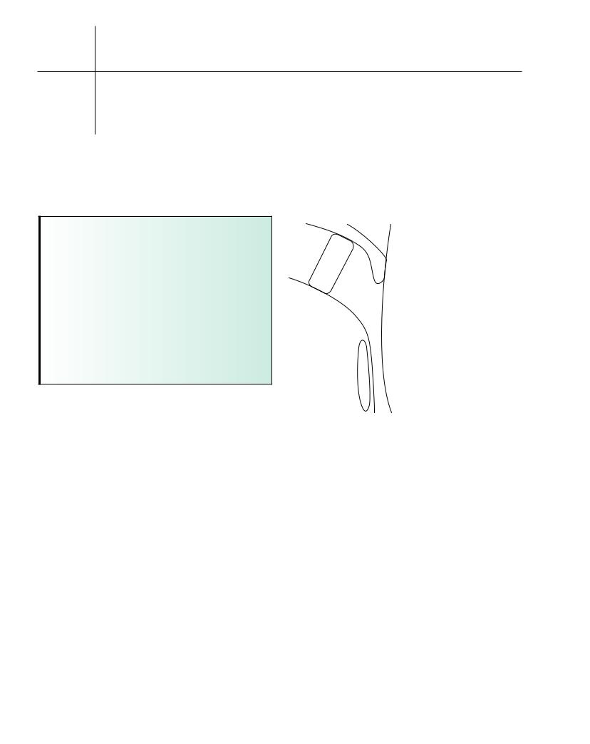
Ideally, a horizontal incision is made 2 cm above the suprasternal notch. Dissection proceeds in the midline to separate the strap muscles and expose the thyroid isthmus, which is ligated and cut. The tracheal rings are exposed and ‘stay’ sutures inserted, especially in children. These help with finding the track should the tube become displaced after operation. A vertical slit tracheostomy is made through the third and fourth rings, and the chosen tracheostomy tube is inserted (Fig. 12.8). The integrity of the tube and the cuff must be checked in advance. The tube is secured in place with sutures and tape as necessary. A tube change is performed after 4 to 7 days, allowing time for the track to mature. An uncuffed tube can be used at this time if there is little concern about significant aspiration. The cricoid cartilage must not be damaged to avoid stenosis.
Tracheostomy tubes
There are many types of tracheostomy tube, made of PVC, silicone or silver. A cuffed tube is usually used in the early days after operation, especially in a ventilated patient. This is changed to an uncuffed tube prior to discharge, unless there are significant problems with aspiration. This is often seen in patients with neurological disabili-
Figure 12.8 Total laryngectomy showing end tracheostoma with speaking valve in place.
Figure 12.9 Tracheostomy tubes.
ties. A fenestrated tube with holes on the shoulder allows phonation when the tube is occluded. Most tracheostomy tubes used in hospital and community practice have an inner tube protruding just beyond the outer tube at its distal tip. The longer end of the inner tube picks up the dried mucus and can be removed for cleaning, while the outer tube is left in place (Fig. 12.9).
Care of a tracheostomy in the community
Patients who have a tracheostomy for chronic airway obstruction or pulmonary toilet may be managed at home. Care in the community needs skilled nursing. A good network of communication needs to be set up before discharge to ensure that the home is equipped with suction apparatus, a humidification system, if required, and a supply of spare tracheostomy tubes. The patient’s family should be taught about tracheostomy care: how to perform competent suction and to replace the tube in the event of a blockage. A community physiotherapist and speech and language therapist may also be needed. Some problems faced in the community, such as narrowing of the tract and persistent granulations with bleeding around the stoma, may need specialist ENT advice.
Further reading
Leung AK, Cho H. (1999) Diagnosis of stridor in children. American Family Physician; 60(8): 2289–96.
Lewarski JS. (2005) Long-term car of the patient with a tracheosotomy. Respiratory Care; 50; (4): 534–7.
Mount J, Uner A, Kaku S. (2004) Pediatric wheezing and stridor. Emergency Medical Services; 33(7): 55–56, 58–60.
Oberwaldner B, Eber E. (2006) Tracheostomy care in the home. Paediatric Respiratory Review 7; (3): 185–90.
Yellon RF, Goldberg H. (2001) Update on gastroesophageal reflux disease in pediatric airway disorders. American Journal of Medicine; 111(Suppl 8A): 78S–84S.
CHAPTER 13
Swallowing Problems
Vinidh Paleri, Patrick J Bradley
OVERVIEW
•Dysphagia is the symptom of swallowing impairment.
•Swallowing can be divided into four stages: oral preparatory, oral, pharyngeal and oesophageal (Box 13.1).
•The majority of the swallowing mechanism is located above the clavicle.
•Weight loss is associated with significant disease or condition, difficult to reverse, and is usually a late sign.
•Aspiration is defined as liquid or solids penetrating below the level of the vocal cord and frequently may not precipitate any symptoms, coughing as a symptom is not reliable.
•Evaluation of the swallowing mechanism should be undertaken by a multidisciplinary team, not only for diagnosis but for treatment and rehabilitation.
Swallowing and its mechanisms
The pharyngeal stage is controlled at brain stem level (Fig. 13.1). The soft palate closes against the nasopharynx to prevent nasal regurgitation and laryngeal closure occurs to prevent aspiration. The epiglottis may play a greater role in directing the bolus into the piriform sinuses than in protecting the airway. Following laryngeal closure by cord adduction, the pharyngeal constrictor muscles sequentially contract to propel the bolus. The suprahyoid muscles raise the larynx, clearing the bolus down as the cricopharyngeal sphincter opens, admitting it to the upper oesophagus. The cricopharyngeus relaxes with laryngeal elevation, which mechanically pulls open the sphincter under bolus pressure. The oesophageal stage then follows for 8–20 seconds.
Symptoms
Swallowing problems present as difficulty in initiating the swallow, choking or coughing upon swallowing (aspiration) or a sensation
Box 13.1 Stages of swallowing
The four stages are:
•oral preparatory;
•oral;
•pharyngeal;
•oesophageal.
T
P
D
E G
(a) |
(b) |
Figure 13.1 (a) Pharyngeal phase of swallowing – early phase. (b) Pharyngeal phase of swallowing – late phase. D, depression; E, elevation; G, gravity; T, tongue; +, positive pressure; – negative pressure.
of obstruction in the neck or behind the sternum. Dysphagia may involve liquids, solids or both. Progressive difficulty with weight loss suggests malignant disease. Slow progression over years occurs in achalasia of the cardia and pharyngeal pouches (Zenker’s diverticulum), associated with regurgitation of undigested food. Dry mouth (xerostomia), caused by autoimmune diseases (Sjogren’s syndrome) and radiation therapy, also causes dysphagia (Box 13.2).
Box 13.2 Investigations for dysphagia
Primary care:
•full blood count;
•contrast swallow. Secondary care:
•flexible and rigid endoscopy;
•videofluoroscopy;
•flexible endoscopic evaluation of swallowing;
•oesophageal manometry.
60