

Учебники / Hair Cell Regeneration, Repair, and Protection Salvi 2008
.pdf2. Morphology of Inner Ear Repair |
41 |
A
B
Figure 2.2. Loss of hair cell number and function in the human cochlea with increasing age. (A) Density of outer hair cells in the cochlea decreases with age, expressed as a percentage of the fetal density (100%). (B) Prevalence of hearing impairment of 25 dB or greater in the better hearing ear among individuals of different ages. The increasing prevalence of hearing loss at increased age correlates strongly with the loss of cochlear hair cells. (A) Modified from Bredberg (1968). (B) Modified from Davis (1989).
2. Ongoing Production of Hair Cells
in Nonmammalian Vertebrates
In many nonmammalian vertebrates, the production of hair cells is not limited to embryonic development (Corwin 1981, 1983, 1985; Popper and Hoxter 1984; Lanford et al. 1996). For example, the ears of sharks and rays add hundreds of thousands of hair cells in the ear and increase in sensitivity as juveniles grow into
42 J.R. Meyers and J.T. Corwin
A MACULA
Non-sensory
epithelium
B
CRISTA
NEUROMAST
D
Striola
Otoconia
Sensory |
Non-sensory |
epithelium |
epithelium |
C
Cupula COCHLEA
|
|
Outer |
|
Tectorial |
||
|
hair cells |
Inner membrane |
||||
Henson’s |
|
|
hair cell |
|||
|
|
|
|
|||
Cells |
|
|
|
|
||
|
|
|
Phalangeal |
|||
Basilar |
Deiter’s |
|
|
Pillar |
||
membrane |
Cells |
|
|
Cells |
Cells |
|
|
|
|
||||
|
|
|
|
|
|
|
E BASILAR PAPILLA
Tectorial membrane
Basilar membrane
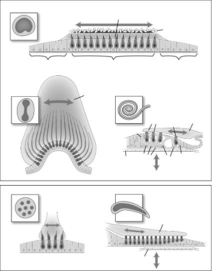
Figure 2.3. Schematic diagrams showing general structural organization of sensory epithelia. (A) Organization of otolith macular organs. The sensory patch, indicated by the darkened color in the inset, is comprised of hair cells and supporting cells in a mosaic pattern. The hair cells insert their hair bundles into a gelatinous matrix covered by otoconial crystals that provide inertial weight to acceleration of the head (arrow). The sensory epithelium is surrounded by a nonsensory epithelium, made up of supporting-like cells. (B) Organization of the semicircular canal cristae. The barbell-shaped sensory patch is organized very similar to that of the otolith macular organs. The hair bundles, however, are inserted into a large cupula, which transduces rotational movement of fluid through the canal to the hair bundles (arrow). (C) Organization of the mammalian cochlea. In contrast to the generally uniform population of supporting cells in vestibular organs,

2. Morphology of Inner Ear Repair |
43 |
adults (Fig. 2.4a; Corwin 1981, 1983). The ears of bony fish and amphibians also add thousands of hair cells during postembryonic growth (Lewis and Li 1973; Li and Lewis 1979; Popper and Hoxter 1984; Corwin 1985; Lombarte and Popper 1994; Lanford et al. 1996).
2.1 Postembryonic Hair Cell Production
These new hair cells are the product of postnatal proliferation, as [3H]thymidine labels both newly produced hair cells and supporting cells (Corwin 1981). The distribution of proliferating cells and newly produced hair cells in elasmobranchs and amphibians is strongly biased toward appositional growth, with cells added at the outer edge of the sensory epithelium (Fig. 2.4b,c), but some interstitial proliferation and addition of new hair cells occurs in the central regions of the maculae as well (Lewis and Li 1973; Li and Lewis 1979; Corwin 1983, 1985).
Appositional expansion is not the only pattern for ongoing growth in the ear. In teleost fish, interstitial addition outweighs appositional growth at the outer margin (Popper and Hoxter 1990; Lombarte and Popper 1994; Lanford et al. 1996). There is also ongoing proliferation and production of hair cells within the vestibular maculae of chickens occurring throughout the epithelium (Jørgensen and Mathiesen 1988; Roberson et al. 1992; Kil et al. 1997).
Thus, the ears of many nonmammalian vertebrate classes show signs of normal continued production of hair cells throughout life. The permanent loss of hair cells that occurs in mammalian ears appears to be unique, as hair cell epithelia retain progenitors in nonmammalian vertebrates that can produce hair cells.
3. Generation and Regeneration of Lateral Line Hair Cells
Hair cells are also continually generated in the sensory organs of the lateral line. Lateral line neuromasts are small collections of hair cells and supporting cells on the heads and bodies of fish and aquatic amphibians. The hair cells in the lateral line are morphologically similar to hair cells from the inner ear. They show expression of the same molecular markers as inner ear hair cells,
Figure 2.3. the cochlear supporting cells (e.g., Deiters’, pillar, and phalangeal cells) show discrete morphological specializations to form the unique structure of the organ of Corti. (D) The lateral line neuromasts found along the body of fish and amphibians is organized similar to vestibular organs, though there are many fewer hair cells per organ. (E) The basilar papilla of birds shows some similarity to the vertebrate cochlea, though the supporting cells do not form morphologically discrete subtypes or show high levels of specialization.

44 J.R. Meyers and J.T. Corwin
Figure 2.4.

2. Morphology of Inner Ear Repair |
45 |
including homologs of atonal (Itoh and Chitnis 2001) and myosin VIIa (Ernest et al. 2000), and are thought to be homologous. During the life of the animal, the lateral line organs bud off accessory neuromasts that lie dorsal or ventral to the primary organ and gradually develop into collections of up to 30 neuromasts (Stone 1937; Ledent 2002). The accessory organs begin as a few supporting cells around a single hair cell, but grow to include several hair cells, indicating that the cells of the neuromasts continue to produce new sensory structures and hair cells (Ledent 2002).
3.1 Regeneration of Hair Cell Epithelia After Amputation
In amphibians, neuromasts also can be replaced during regeneration after amputation of the tail. Cells in the last neuromast on the tail stump divide to give rise to a regenerative placode that is morphologically similar to the embryonic placode that first produces the lateral line organs in embryos and larvae. The regenerative placode migrates along the regenerating tail producing replacement neuromasts similar in number to those lost through amputation (Stone 1933, 1937; Speidel 1947; Wright 1947). These replacement organs develop hair cells with normal morphology and become reinnervated (Stone 1933, 1937; Speidel 1947; Wright 1947; Jørgensen and Flock 1976). Normally, the regenerating placode forms from the posterior edge of the last neuromast, but grafting experiments that reversed the orientation of the neuromasts demonstrated that production of a regenerating placode and neuromasts could develop from the anterior edge as well (Stone 1937). This suggests that the marginal cells closest to the wound are able to respond to some signal from the wound to initiate the regenerative process.
Figure 2.4. Ongoing production of hair cells continues throughout life in the inner ear of many vertebrate classes. (A) A plot of the number of hair cells within the macula neglecta of a ray versus age of the animal. The slope indicates that approximately three hair cells are added per day in the first years of life and approximately one hair cell is added per day beyond 6 years of age. (B) A micrograph of the edge of the sensory epithelium of a toad’s saccule. A [3H]thymidine labeled newborn hair cell is indicated at the edge of the epithelium, and three labeled supporting cells lie just beyond the last hair cell. The transition from the sensory epithelium to the nonsensory epithelium is the primary location for addition of new hair cells in cartilaginous fish and amphibians. (C) Superimposed outlines of the sensory epithelium from the macula neglecta of rays at different ages showing the increase in size of the sensory patch. The arrows indicate the orientation of the hair bundles pointing away from a central point of symmetry. The shaded portion indicates the region of hair cells with immature hair bundles at the outer edge of the epithelium. (A, C) From Corwin (1983); (B) from Corwin (1985).
46 J.R. Meyers and J.T. Corwin
3.2 Identifying the Progenitors of Replacement Hair
Cell Epithelia
It was suggested decades ago that for both neuromast budding and regenerating placodes, the likely source of the cells was the supporting cells of the neuromast (Stone 1933, 1937). Time-lapse microscopy confirmed the identity of progenitor cells of the regenerating placode as mantle-type supporting cells that reside at the edge of the neuromast, adjacent to the internal supporting cells and hair cells (Jones and Corwin 1993). The location of the regenerating cells at the border between the sensory epithelium and surrounding epidermis is similar to the location of the proliferative cells that produce new hair cells throughout the lives of elasmobranches and amphibians (see Section 2.2). Neuromasts formed from a regenerative placode have the ability to form a new regenerating placode in response to reamputation of the tail (Speidel 1947). The progenitor cells, therefore, can make all of the differentiated cell types within the neuromast, and can reproduce latent precursors satisfying two defining characteristics of stem cells: self-renewal and multipotency.
3.3 Regeneration of Lateral Line Hair Cells In Situ
The lateral line can also generate replacement hair cells within individual neuromasts after selective destruction of individual hair cells (Balak et al. 1990; Song et al. 1995). In response to loss of hair cells within a neuromast, the surrounding supporting cells become proliferative and can generate both replacement hair cells and supporting cells (Jones and Corwin 1996). Thus, within the lateral line sensory organs, regeneration can replace lost hair cells or develop multiple new neuromasts, with all of their sensory and nonsensory cells, during tail regeneration.
4. Hair Cell Regeneration in the Inner Ear
Similar to the findings of ongoing addition of hair cells in anamniotes and the regenerative capacity of the lateral line, nonmammalian vertebrates such as fish, amphibians, reptiles, and birds are capable of significant regeneration of hair cells after damage to the inner ear.
4.1 Morphological Recovery in the Avian Hearing Organ
The full complement of hair cells in the chicken basilar papilla sensory epithelium is produced embryonically and the cells all become quiescent after embryogenesis (Tilney et al. 1986; Katayama and Corwin 1989). In response to tonal acoustic overstimulation, hair cells within the region of the basilar papilla tuned to the stimulating tone are lost or suffer significant damage, while hair cells in regions tuned to other frequencies are spared (Fig. 2.5a; Cotanche 1987).
2. Morphology of Inner Ear Repair |
47 |
A B
C D
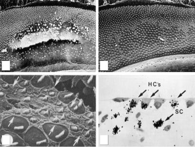
Figure 2.5. Hair cell regeneration in the chicken basilar papilla. (A) After 48 h of acoustic overstimulation at 1.5 kHz, hair cells within the region of the cochlea tuned to 1.5 kHz are lost and can be seen extruding from the epithelium. (B) After a 10-day recovery after acoustic overstimulation, the epithelium has been repaired. Hair cells can be seen throughout the formerly damaged regions. (C) Close-up micrograph of hair cells in the damaged region of a chick cochlea 6 days after damage shows a number of new hair cells with small bundles and small apical surfaces. (D) Injection of the birds with tritiated thymidine during the 10-day recovery period reveals labeling of both hair cells (HC) and supporting cells (SC), indicating that the new hair cells are progeny of dividing supporting cells. (A–D) From Corwin and Cotanche (1988).
This is followed by proliferation at the lesion site, and over 6–10 days there is a dramatic recovery in the number of hair cells (Fig. 2.5b; Cotanche 1987; Corwin and Cotanche 1988). Many of the hair cells within the damaged region exhibit small hair bundles reminiscent of immature hair bundles on developing hair cells (Fig. 2.5c). The ability to stimulate proliferation and regeneration of new hair cells was similar in young chickens and adult quail (Corwin and Cotanche 1988; Ryals and Rubel 1988; Ryals and Westbrook 1990). Thus, cells within the avian cochlea retain the capacity to reenter the cell cycle and generate new hair cells throughout life, even though they normally become mitotically quiescent after embryogenesis. The newly differentiating cochlear hair cells take on the distinct positional morphologies of the cells they replace (Cotanche 1987), suggesting that positional identity such as tonotopy can be passed on to the regenerating cells. Regeneration of hair cells also occurs in birds in response to aminoglycoside toxicity in both the auditory and vestibular systems (Lippe
48 J.R. Meyers and J.T. Corwin
et al. 1991; Weisleder and Rubel 1993). After regeneration of hair cells in birds, the replacement cells become innervated and there is significant recovery of hearing and vestibular function (see discussion by Dooling, Dent, Lauer, and Ryals, Chapter 4).
4.2 Ongoing Proliferation in the Avian Vestibular System
Although the cells in the avian basilar papilla are normally mitotically quiescent until damage induces cell cycle reentry, in the avian vestibular system there is ongoing proliferation throughout life. However, there is also a low level of ongoing cell death, approximately 90% of which are hair cells within the vestibular system (Kil et al. 1997). This suggests that the continual production of new cells in the vestibular system may be in response to the dying cells and serves to replace lost hair cells rather than promote continued growth of the epithelium. Consistent with that hypothesis, inhibition of cell death in the utricle limits ongoing proliferation (Matsui et al. 2002). The mechanism and reason behind the continued loss and regeneration of hair cells in the avian vestibular system remain unclear.
4.3 Regeneration in Fish and Amphibians
The sensory epithelia from fish and amphibians also show substantial damageinduced proliferation and regeneration of hair cells. New hair cells are produced after aminoglycoside treatment in fish (Lombarte et al. 1993) and in bullfrogs (Baird et al. 1993), and after destruction of hair cells in the lateral line (Balak et al. 1990; Song et al. 1995). This generation of hair cells is accompanied by increased proliferation (Balak et al. 1990; Presson and Popper 1990; Baird et al. 1993; Presson et al. 1996; Avallone et al. 2003), similar to that seen in the avian organs after damage. In summary, fish, amphibians, and birds are all capable of significant regeneration of hair cells after damage to the inner ear. The replacement hair cells in these species occurs via cell divisions that occur at the sites of damage.
4.4 Regenerative Responses in Mammalian Ears
The hair cells and supporting cells in the vestibular system of mammals are similar to their counterparts in birds, fish, and amphibians (Fig. 2.3). In contrast, hair cells and supporting cells in mammalian cochleae are highly specialized, without direct counterparts in the auditory organs of other vertebrates. This high degree of differentiation and specialization suggests that regeneration may be more limited in the mammalian cochlea. However„ the similarity between mammalian and nonmammalian vestibular cells paired with the robust regeneration in nonmammalian ears suggests that hair cell regeneration may be more likely in mammalian vestibular epithelia.
2. Morphology of Inner Ear Repair |
49 |
4.4.1 Morphological Recovery in the Mammalian Vestibular Organs
After aminoglycoside-induced hair cell loss, guinea pigs allowed to recover for 4 weeks developed small immature hair bundles in the damaged regions of the utricular epithelium (Fig. 2.6; Forge et al. 1993, 1998). These cells were reminiscent of the appearance of regenerating hair cells in chickens (Cotanche 1987). Utricles from adult guinea pigs maintained in vitro after aminoglycoside treatment had small numbers of [3H]thymidine-labeled cells throughout the sensory epithelium, showing that hair cell damage can trigger proliferation in vestibular organs of mammals (Warchol et al. 1993). Profiles of putative [3H]thymidine-labeled hair cells were also found in those epithelia. When utricles obtained during otologic surgery on adult human patients were treated similarly, that resulted in at least 100 [3H]thymidine-labeled cells in each utricle (Warchol et al. 1993). Thus even supporting cells from the sensory epithelium of adult humans can proliferate after damage.
However, the tens to hundreds of dividing cells in mammalian utricles after damage are considerably less than the thousands that would be seen in avian
B
A
C D
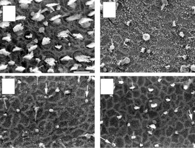
Figure 2.6. Regeneration in the mammalian vestibular epithelium. (A) Scanning electron micrograph of the striola region of a control utricle from a mature guinea pig. (B–D) Damage and recovery of hair bundles in guinea pig utricles after aminoglycoside toxicity. (B) The striola of a utricle 1 week after the end of aminoglycoside treatment, showing loss of most of the hair bundles. (C) The striola of a utricle 2 weeks after the end of aminoglycoside treatment showing initial recovery of hair bundles (arrows). (D) The striola of a utricle 4 weeks after the end of aminoglycoside treatment showing continued replacement of hair bundles (arrows). (A–D) From Forge et al. (1998).
50 J.R. Meyers and J.T. Corwin
utricles after similar damage (Warchol et al. 1993; Matsui et al. 2000). There was also a notable quantitative mismatch between the large numbers of recovering hair bundles reported by Forge and colleagues (1993, 1998) and the limited number of proliferating cells found by Warchol et al. (1993). There may be a substantial difference in the level of damage to the utricles in vivo versus in vitro, which may make it difficult to compare the response between these studies. The studies showing substantial recovery of hair bundle numbers were done in vivo, wherein aminoglycoside nephrotoxicity limits the concentrations that can be used without severe systemic repercussions, while the investigations of proliferation were performed in vitro, wherein high levels of aminoglycoside ensure the death of most of the hair cells. Thus, whether the difference in the magnitude of proliferation versus hair bundle recovery is due to the level of damage, or whether it reflects a nonproliferative mechanism of regenerating hair cells remains an open question.
4.4.2 Minimal Hair Cell Regeneration in the Mammalian Cochlea
In contrast to the mammalian vestibular system, there is, as yet, little evidence for proliferative regeneration within the mammalian cochlea, where the supporting cells show significant morphological specialization. While there have been reports that the cells of the organ of Corti can be stimulated to proliferate and regenerate hair cells after aminoglycoside treatments (Lefebvre et al. 1993), confirmation of those results has proven difficult (Chardin and Romand 1995). The specialization and differentiation of supporting cells into discrete cell types with distinct morphologies within the organ of Corti may limit their ability to reenter the cell cycle.
In summary, while regeneration in the mammalian inner ear does not appear to occur at the robust level as in nonmammalian vertebrates, there is evidence for a limited regenerative response, at least in the vestibular system. By further studying both the strong regenerative processes in nonmammalian systems and the limited regeneration in mammals, it may be possible to override the limitations in the mammalian regenerative response and promote clinically significant recovery from hair cell loss.
5. The Source of Regenerating Hair Cells
The continual production of new hair cells throughout life and the regeneration that follows damage both appear to depend on proliferative precursor cells. The morphological identity of the cells that produce regenerating hair cells within damaged epithelia has been a key question in establishing a mechanism for the observed recovery of functional hair cells. Proposed sources have included the hyaline cells that lie at the margin of the chick basilar papilla (the same position as the cells that result in appositional growth in elasmobranchs and neuromast regeneration in amphibians), the differentiated supporting cells within the sensory organs, or a specialized reserve pool of distinct stem cells.