

Книги фарма 2 / Bertram G. Katzung-Basic & Clinical Pharmacology(9th Edition)
.pdf
|
Familial hypercholesterolemia |
|
|
|
|
|
|
|
Heterozygous |
|
LDL increased |
Reductase |
|
Two or three of the |
|
|
|
|
|
inhibitor, resin, |
|
individual drugs |
|
|
|
|
|
niacin, ezetimibe |
|
|
|
|
Homozygous |
|
LDL increased |
Niacin, |
|
Niacin plus |
|
|
|
|
|
atorvastatin, |
|
reductase inhibitor |
|
|
|
|
|
ezetimibe, |
|
plus ezetimibe |
|
|
|
|
|
rosuvastatin |
|
|
|
|
Familial ligand-defective apoB |
|
LDL increased |
Niacin, reductase |
|
Niacin plus |
|
|
|
|
|
||||
|
|
|
|
inhibitor, |
|
reductase inhibitor |
|
|
|
|
|
ezetimibe |
|
or ezetimibe |
|
|
Lp(a) hyperlipoproteinemia |
|
Lp(a) increased |
Niacin |
|
|
|
1Single-drug therapy should be evaluated before drug combinations are used.
Phenotypes of abnormal lipoprotein distribution are described in this section. Drugs mentioned for use in these conditions are described in the following section on basic and clinical pharmacology.
The Primary Hypertriglyceridemias
Hypertriglyceridemia is linked epidemiologically with increased risk of coronary disease. VLDL and its remnants have been found in atherosclerotic plaques. In some kindreds, hypertriglyceridemia may be the only evident risk factor. These patients tend to have cholesterol-rich VLDL of small particle diameter. Hypertriglyceridemic patients with coronary disease or a family history of premature coronary disease should be treated aggressively. In others, treatment decisions should be based on the aggregate of risk factors. Because clearance of triglycerides by the LPL system is saturated at about 800 mg/dL of triglycerides, patients with higher levels should be treated to prevent acute pancreatitis.
Primary Chylomicronemia
Chylomicrons are not present in the serum of normal individuals who have fasted 10 hours. The recessive traits of lipoprotein lipase deficiency and cofactor deficiency are usually associated with severe lipemia (2000–2500 mg/dL triglycerides when the patient is consuming a typical American diet). These disorders might not be diagnosed until an attack of acute pancreatitis occurs. Patients may have eruptive xanthomas, hepatosplenomegaly, hypersplenism, and lipid-laden foam cells in bone marrow, liver, and spleen. The lipemia is aggravated by estrogens because they stimulate VLDL production, and pregnancy may cause marked increases in triglycerides despite strict dietary control. Although these patients have a predominant chylomicronemia, they may also have moderately elevated VLDL, presenting with a pattern of mixed lipemia (fasting chylomicronemia and elevated VLDL). LPL deficiency is diagnosed by assay of lipolytic activity after intravenous injection of heparin; cofactor deficiency is diagnosed by isoelectric focusing of the VLDL proteins. A presumptive diagnosis of these disorders is made by demonstrating a pronounced decrease in levels of triglycerides a few days after sharp restriction of oral fat intake. Marked restriction of the total fat content in the diet provides effective long-term treatment. Niacin or a fibrate may be of some benefit.
Familial Hypertriglyceridemia
Severe (Usually Mixed Lipemia)
A pattern of mixed lipemia usually results from impaired removal of triglyceride-rich lipoproteins. Factors that increase VLDL production aggravate the lipemia because VLDL and chylomicrons are competing substrates for LPL. The primary mixed lipemias probably represent a variety of modes of inheritance. Most patients have the centripetal pattern of obesity with insulin resistance. Other factors that lead to an increased rate of secretion of VLDL also worsen the lipemia. Eruptive xanthomas, lipemia retinalis, epigastric pain, and pancreatitis are variably present depending on the severity of the lipemia. Treatment is primarily dietary, with restriction of total fat, avoidance of alcohol and exogenous estrogens, and weight reduction. Some patients may require treatment with a fibrate or niacin.
Moderate (Endogenous Lipemia)
Primary increases of VLDL probably reflect a number of genetic determinants and are worsened by factors that increase the rate of VLDL secretion from liver, ie, obesity, alcohol, diabetes, and estrogens. A major indication for treatment is the presence of atherosclerosis in the patient or the patient's family. Treatment includes weight reduction, restriction of all types of dietary fat, and avoidance of alcohol. Fibrates or niacin usually produce further reduction in triglyceride levels if dietary measures are not sufficient. Marine omega fatty acids may also be of value.
Familial Combined Hyperlipoproteinemia
In kindreds with this disorder, individuals may have elevated levels of VLDL, LDL, or both, and the pattern may change with time. Familial combined hyperlipoproteinemia involves an approximate doubling in VLDL secretion. It seems to be transmitted as a semidominant trait. Triglycerides can be increased by the factors noted above. Elevations of cholesterol and triglycerides are generally moderate, and xanthomas are usually absent. Drug treatment is warranted because the risk of coronary atherosclerosis is increased and diet alone does not normalize lipid levels. A reductase inhibitor or ezetimibe in combination with niacin is usually required to treat these patients.
Familial Dysbetalipoproteinemia
In this disorder, remnants of chylomicrons and VLDL accumulate. Levels of LDL are usually decreased. Because remnants are rich in cholesteryl esters, the level of cholesterol may be as high as that of triglycerides. Diagnosis is confirmed by the absence of the E3 and E4 isoforms of apo E. Patients often develop tuberous or tuberoeruptive xanthomas, or characteristic planar xanthomas of the palmar creases. They tend to be obese, and some have impaired glucose tolerance. These factors, as well as hypothyroidism, can aggravate the lipemia. Coronary and peripheral atherosclerosis occur with increased frequency. Weight loss, together with decreased fat, cholesterol, and alcohol consumption, may be sufficient treatment, but a fibrate or niacin is needed in most cases. These agents can be given together in more resistant cases, or a reductase inhibitor may be added.
The Primary Hypercholesterolemias
Familial Hypercholesterolemia
Familial hypercholesterolemia is an autosomal dominant trait. Although levels of LDL tend to increase throughout childhood, the diagnosis can often be made on the basis of elevated umbilical
cord blood cholesterol. In most heterozygotes, cholesterol levels commonly range from 260 to 500 mg/dL. Triglycerides are usually normal, tendinous xanthomatosis is often present, and arcus corneae and xanthelasma may appear in the third decade. Coronary atherosclerosis tends to occur prematurely. Homozygous familial hypercholesterolemia, which can lead to coronary disease in childhood, is characterized by levels of cholesterol often exceeding 1000 mg/dL and early tuberous and tendinous xanthomatosis. These patients may also develop elevated plaque-like xanthomas of the aortic valve, digital webs, buttocks, and extremities.
Defects of LDL receptors underlie this disorder. Some individuals have combined heterozygosity for alleles producing nonfunctional and kinetically impaired receptors. Levels of LDL in compliant heterozygous patients can be normalized with combined drug regimens (Figure 35–2). Those whose receptors retain even minimal function may partially respond to resins or reductase inhibitors. Niacin and atorvastatin may benefit patients with no receptor function.
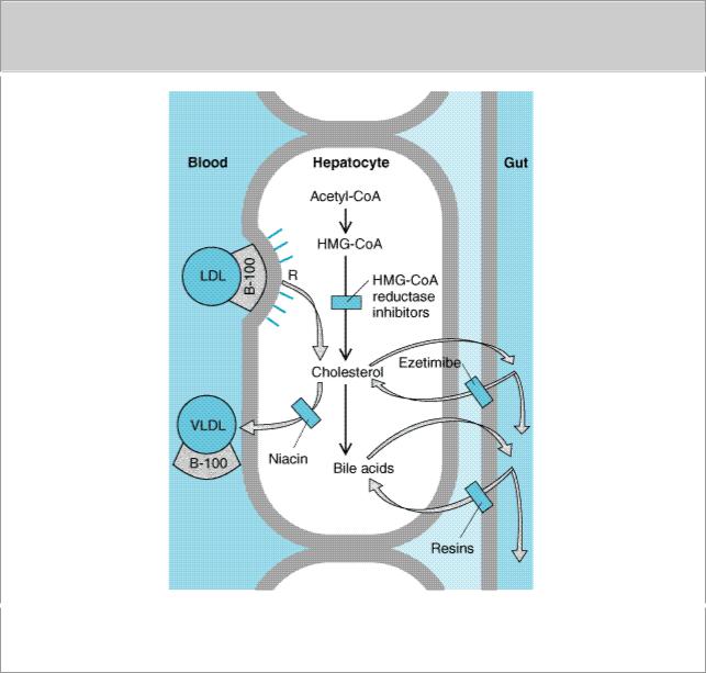
Figure 35–2.
Sites of action of HMG-CoA reductase inhibitors, niacin, ezetimibe, and resins used in treating hyperlipidemias. LDL receptors (R) are increased by treatment with resins and HMG-CoA reductase inhibitors.
Familial Ligand-Defective Apolipoprotein B
Defects in the ligand domain of apo B100 (the region that binds to the LDL receptor) impair the

endocytosis of LDL, leading to hypercholesterolemia of moderate severity. Tendon xanthomas may occur. These disorders are as prevalent as familial hypercholesterolemia. Response to reductase inhibitors is variable. Up-regulation of LDL receptors in liver increases endocytosis of LDL precursors but does not increase uptake of ligand-defective LDL particles. Niacin often has beneficial effects by reducing VLDL production.
Familial Combined Hyperlipoproteinemia (FCH)
As described above, some persons in kindreds with this disorder have only an elevation in LDL. Serum cholesterol is usually less than 350 mg/dL. Premature coronary disease is common. Dietary and drug treatment, usually with niacin or a reductase inhibitor, is indicated. It is frequently necessary to add niacin to normalize LDL or to reduce triglycerides if treatment is initiated with a resin.
Lp(a) Hyperlipoproteinemia
This familial disorder, which is associated with increased atherogenesis, is determined chiefly by alleles that dictate increased production of the Lp(a) lipoprotein. Niacin reduces levels of Lp(a) in many patients.
Other Disorders
Deficiency of cholesterol 7
 -hydroxylase can cause elevated LDL in the heterozygous state. Homozygotes can also have elevated triglycerides, resistance to reductase inhibitors, and premature gallstones. Autosomal recessive hypercholesterolemia is due to mutations in a protein that assists in endocytosis of LDL. Niacin and ezetimibe may be useful in these disorders.
HDL Deficiency
Certain rare genetic disorders are associated with extremely low levels of HDL in serum. These include Tangier disease and disorders of LCAT. Familial hypoalphalipoproteinemia is a more common disorder in which levels of HDL cholesterol are usually below 35 mg/dL, with apparent codominant transmission. These patients tend to have premature atherosclerosis, and the low HDL may be the only identified risk factor. Treatment should include special attention to avoidance of other risk factors. Niacin increases HDL cholesterol in many of these patients. Reductase inhibitors and fibric acid derivatives exert lesser effects.
In the presence of hypertriglyceridemia, HDL cholesterol is low because of exchange of cholesteryl esters from HDL into triglyceride-rich lipoproteins. This may contribute to the atherogenic effect of hypertriglyceridemia. In most, treatment of the hypertriglyceridemia will result in normalization of HDL.
Secondary Hyperlipoproteinemia
Before primary disorders can be diagnosed, secondary causes of the phenotype must be considered. The more common conditions are summarized in Table 35–3. The lipoprotein abnormality usually resolves if the underlying disorder can be treated successfully.
Table 35–3. Secondary Causes of Hyperlipoproteinemia.

Hypertriglyceridemia |
Hypercholesterolemia |
|
|
Diabetes mellitus |
Hypothyroidism |
|
|
Alcohol ingestion |
Early nephrosis |
|
|
Severe nephrosis |
Resolving lipemia |
|
|
Estrogens |
Immunoglobulin-lipoprotein complex disorders |
|
|
Uremia |
Anorexia nervosa |
|
|
Corticosteroid excess |
Cholestasis |
|
|
Myxedema |
Hypopituitarism |
|
|
Glycogen storage disease |
Corticosteroid excess |
|
|
Hypopituitarism |
|
|
|
Acromegaly |
|
|
|
Immunoglobulin-lipoprotein complex disorders |
|
|
|
Lipodystrophy |
|
|
|
Isotretinoin |
|
|
|
Protease inhibitors |
|
|
|
Katzung PHARMACOLOGY, 9e > Section VI. Drugs Used to Treat Disease of the Blood, Inflammation, & Gout > Chapter 35. Agents Used in Hyperlipidemia >
Dietary Management of Hyperlipoproteinemia
Dietary measures are always initiated first and may obviate the need for drugs. Exceptions are patients with familial hypercholesterolemia or familial combined hyperlipidemia in whom diet and drug therapy should be started simultaneously. Cholesterol, saturated fats, and trans fats are the principal factors that influence LDL levels, whereas total fat and calorie restriction is important in management of triglycerides.
Lipoprotein levels are also affected—but to a lesser extent—by other dietary factors. Increases in VLDL occur when carbohydrate intake is increased but tend to normalize after several months. Sucrose and other simple sugars raise VLDL levels in hypertriglyceridemic patients. Some forms of dietary fiber reduce LDL modestly. Alcohol can cause significant hypertriglyceridemia by increasing hepatic secretion of VLDL. Synthesis and secretion of VLDL are increased by excess calories. Caloric restriction, especially in obese subjects, reduces VLDL and often LDL as well. During weight loss, LDL and VLDL levels may be much lower than can be maintained during neutral caloric balance. The conclusion that diet suffices for management can only be arrived at after weight has stabilized for at least 1 month.
Most patients with elevated LDL can be managed with a diet restricted in cholesterol and saturated fat but with sufficient calories to achieve and maintain ideal body weight. Total fat calories should be 20–25%, with saturated fats less than 8% and cholesterol less than 200 mg/d. Reductions in serum cholesterol range from 10% to 20% on this regimen. Use of complex carbohydrates and fiber is recommended, and monounsaturated fats should predominate within the fat allowance. Weight reduction and caloric restriction are especially important for patients with elevated VLDL and IDL. Those with hypertriglyceridemia should avoid alcohol.

The effect of dietary fats on hypertriglyceridemia is dependent upon the disposition of double bonds in the fatty acid. Omega-3 fatty acids found in fish oils—but not those from plant sources—can induce profound lowering of triglycerides in some patients with endogenous or mixed lipemia. In contrast, the omega-6 fatty acids present in vegetable oils may cause triglycerides to increase.
Patients with primary chylomicronemia and some with mixed lipemia must consume a diet severely restricted in total fat—10–15 g/d, of which 5 g should be vegetable oils rich in essential fatty acids. Supplementation of fat-soluble vitamins should be given.
Supplementation with the antioxidant vitamins ascorbic acid (250 mg) and mixed natural tocopherols (50 IU on alternate days) may be beneficial. Higher doses may vitiate the impact of lipid lowering therapy. Other naturally occurring antioxidants such as resveratrol,  -catechin, selenium, and various carotenoids found in a variety of fruits and vegetables may provide additional antioxidant defense. Homocysteine, which initiates proatherogenic changes in endothelium, can be reduced in many patients by restriction of total protein intake to the amount required for amino acid replacement. Daily supplementation with up to 2 mg of folic acid plus other B vitamins is also recommended.
Katzung PHARMACOLOGY, 9e > Section VI. Drugs Used to Treat Disease of the Blood, Inflammation, & Gout > Chapter 35. Agents Used in Hyperlipidemia >
Basic & Clinical Pharmacology of Drugs Used in Hyperlipidemia
The decision to use drug therapy is based on the specific metabolic defect and its potential for causing atherosclerosis or pancreatitis. Suggested regimens for the principal lipoprotein disorders are presented in Table 35–2. Diet should be continued for achievement of the full potential of the drug regimen. Drugs should be avoided in pregnant and lactating women and those likely to become pregnant. All drugs that alter plasma lipoprotein concentrations may require adjustment of doses of warfarin and indandione anticoagulants. Children with heterozygous familial hypercholesterolemia may be treated with a resin or reductase inhibitor, usually after 7 or 8 years of age, when myelination of the central nervous system is essentially complete. The decision to treat a child should be based on the level of LDL, other risk factors, the family history, and the child's age. Drugs are rarely indicated before age 18.
Competitive Inhibitors of HMG-COA Reductase (Reductase Inhibitors; "Statins")
These compounds are structural analogs of HMG-CoA (3-hydroxy-3-methylglutaryl-coenzyme A). (Figure 35–3). Lovastatin, atorvastatin, fluvastatin, pravastatin,simvastatin, and rosuvastatin belong to this class. They are most effective in reducing LDL. Other effects include decreased oxidative stress and vascular inflammation with increased stability of atherosclerotic lesions. It has become standard practice to initiate reductase inhibitor therapy immediately after myocardial infarction, irrespective of lipid levels.
Figure 35–3.
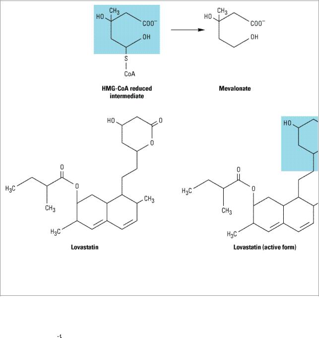
Inhibition of HMG-CoA reductase. Top: The HMG-CoA intermediate that is the immediate precursor of mevalonate, a critical compound in the synthesis of cholesterol. Bottom: The structure of lovastatin and its active form, showing the similarity to the normal HMG-CoA intermediate (shaded areas).
Chemistry & Pharmacokinetics
Lovastatin and simvastatin are inactive lactone prodrugs that are hydrolyzed in the gastrointestinal tract to the active 
 -hydroxyl derivatives, whereas pravastatin has an open, active lactone ring. Atorvastatin, fluvastatin, and rosuvastatin are fluorine-containing congeners that are active as given. Absorption of the ingested doses of the reductase inhibitors varies from 40% to 75% with the exception of fluvastatin, which is almost completely absorbed. All have high first-pass extraction by the liver. Most of the absorbed dose is excreted in the bile; about 5–20% is excreted in the urine. Plasma half-lives of these drugs range from 1 hour to 3 hours except for atorvastatin, which has a half-life of 14 hours, and rosuvastatin, 19 hours.
Mechanism of Action
HMG-CoA reductase mediates the first committed step in sterol biosynthesis. The active forms of the reductase inhibitors are structural analogs of the HMG-CoA intermediate (Figure 35–3) that is formed by HMG-CoA reductase in the synthesis of mevalonate. These analogs cause partial inhibition of the enzyme and thus may impair the synthesis of isoprenoids such as ubiquinone and dolichol and the prenylation of proteins. It is not known whether this has biologic significance.
However, the reductase inhibitors clearly induce an increase in high-affinity LDL receptors. This effect increases both the fractional catabolic rate of LDL and the liver's extraction of LDL precursors (VLDL remnants), thus reducing plasma LDL (Figure 35–2). Because of marked firstpass hepatic extraction, the major effect is on liver. Preferential activity in liver of some congeners appears to be attributable to tissue-specific differences in uptake. Limited reduction of LDL levels in patients who lack functional LDL receptors indicates that decreases in de novo cholesterologenesis also contribute to cholesterol reduction. Modest decreases in plasma triglycerides and small increases in HDL also occur.
Therapeutic Uses & Dosage
Reductase inhibitors are useful alone or with resins, niacin, or ezetimibe in reducing levels of LDL. Women who are pregnant, lactating, or likely to become pregnant should not be given these agents. Use in children is restricted to those with homozygous familial hypercholesterolemia and selected patients with heterozygous familial hypercholesterolemia.
Because cholesterol biosynthesis occurs predominantly at night, reductase inhibitors—except atorvastatin and rosuvastatin—should be given in the evening if a single daily dose is used. Absorption generally (with the exception of pravastatin) is enhanced by taking the dose with food. Daily doses of lovastatin vary from 10 mg to 80 mg. Pravastatin is nearly as potent on a mass basis as lovastatin up to the maximum recommended daily dose, 80 mg. Simvastatin is twice as potent and is given in doses of 5–80 mg daily. Fluvastatin appears to be about half as potent as lovastatin on a mass basis and is given in doses of 10–80 mg daily. Atorvastatin is given in doses of 10–80 mg/d, and rosuvastatin, the most efficacious agent for severe hypercholesterolemia, at 10–40 mg/d. The dose-response curves of pravastatin and especially of fluvastatin tend to level off in the upper part of the dosage range in patients with moderate to severe hypercholesterolemia. Those of lovastatin, simvastatin, and atorvastatin are more nearly linear.
Toxicity
Elevations of serum aminotransferase activity (up to three times the normal level) occur in some patients. These increases are often intermittent and usually not associated with other evidence of hepatic toxicity. Therapy may be continued in such patients in the absence of symptoms if aminotransferase levels are measured frequently. In about 2% of patients, some of whom have underlying liver disease or a history of alcohol abuse, aminotransferase levels may exceed three times the normal limit. This effect, which can occur at any time, portends more severe hepatic toxicity. These patients may present with malaise, anorexia, and precipitous decreases in LDL. Medication should be discontinued immediately in these patients and in asymptomatic patients whose aminotransferase activity is persistently elevated to more than three times the upper limit of normal. These agents should be used with caution and in reduced dosage in patients with hepatic parenchymal disease. In general, aminotransferase activity should be measured at baseline, at 1–2 months, and then every 6 months (if stable).
Minor increases in creatine kinase activity in plasma are observed in some patients receiving reductase inhibitors, frequently associated with heavy physical activity. Rarely, patients may have marked elevations in kinase activity, often accompanied by generalized pain or weakness in skeletal muscles. If the drug is not discontinued, rhabdomyolysis may cause myoglobinuria, which may lead to renal shutdown. Myopathy may occur with monotherapy, but there is an increased incidence in patients receiving a reductase inhibitor concurrently with certain other drugs. The catabolism of lovastatin, simvastatin, and atorvastatin proceeds chiefly through cytochrome P450 3A4, whereas that of fluvastatin and rosuvastatin is mediated by CYP2C9. Pravastatin is catabolized through other
pathways, including sulfation. The 3A4-dependent reductase inhibitors tend to accumulate in plasma in the presence of drugs that inhibit or compete for the 3A4 cytochrome. These include the macrolide antibiotics, cyclosporine, ketoconazole and its congeners, HIV protease inhibitors, tacrolimus, nefazodone, fibrates, and others (see Chapter 4: Drug Biotransformation). Concomitant use of reductase inhibitors with amiodarone or verapamil also causes an increased risk of myopathy. Conversely, drugs such as phenytoin, griseofulvin, barbiturates, rifampin, and thiazolidinediones increase expression of CYP3A4 and can reduce the plasma concentrations of the 3A4-dependent reductase inhibitors. Inhibitors of CYP2C9 such as ketoconazole and its congeners, metronidazole, sulfinpyrazone, amiodarone, and cimetidine may increase plasma levels of fluvastatin and rosuvastatin. Pravastatin appears to be the drug of choice for use with verapamil, the ketoconazole group of antifungal agents, macrolides, and cyclosporine. Plasma levels of lovastatin, simvastatin, and atorvastatin may be elevated in patients ingesting more than 1 liter of grapefruit juice daily.
Creatine kinase activity should be measured frequently in patients receiving potentially interacting drug combinations. In all patients, creatine kinase levels should be measured before treatment and then every 6–12 months (depending on the dose). If significant muscle pain, tenderness, or weakness appears, creatine kinase activity should be measured immediately and the drug discontinued if activity is elevated over baseline. The myopathy usually reverses promptly upon cessation of therapy. If the association is unclear, the patient can be rechallenged under close surveillance. Myopathy in the absence of elevated creatine kinase activity has been reported. Rarely, hypersensitivity syndromes have been reported that include a lupus-like disorder and peripheral neuropathy.
These drugs should be temporarily discontinued in the event of serious illness, trauma, or major surgery.
Niacin (Nicotinic Acid)
Niacin (but not niacinamide) decreases VLDL and LDL levels—and Lp(a) in most patients. It often increases HDL levels significantly.
Chemistry & Pharmacokinetics
Niacin (vitamin B3) is converted in the body to the amide, which is incorporated into niacinamide adenine dinucleotide (NAD). It is excreted in the urine unmodified and as several metabolites.
Mechanism of Action
Niacin inhibits VLDL secretion, in turn decreasing production of LDL (Figure 35–2). Increased clearance of VLDL via the LPL pathway contributes to triglyceride reduction. Niacin has no effect on bile acid production. Excretion of neutral sterols in the stool is increased acutely as cholesterol is mobilized from tissue pools and a new steady state is reached. The catabolic rate for HDL is decreased. Fibrinogen levels are reduced, and levels of tissue plasminogen activator appear to increase. Niacin inhibits the intracellular lipase of adipose tissue via receptor-mediated signaling, possibly reducing VLDL production by decreasing the flux of free fatty acids to liver. Sustained inhibition of lipolysis has not been established, however.
Therapeutic Uses & Dosage
In combination with a resin or reductase inhibitor, niacin normalizes LDL in most patients with heterozygous familial hypercholesterolemia and other forms of hypercholesterolemia. These
combinations are also indicated in some cases of nephrosis. In severe mixed lipemia that is incompletely responsive to diet, niacin often produces marked reduction of triglycerides, an effect enhanced by marine omega-3 fatty acids. It is useful in patients with combined hyperlipoproteinemia and in those with familial dysbetalipoproteinemia. Niacin reduces levels of Lp(a) in many subjects. It is clearly the most effective agent for increasing levels of HDL.
For treatment of heterozygous familial hypercholesterolemia, most patients require 2–6 g of niacin daily; more than this should not be given. For other types of hypercholesterolemia and for hypertriglyceridemia, 1.5–3.5 g daily is often sufficient. The drug should be given in divided doses with meals, starting with 100 mg two or three times daily and increasing gradually.
Toxicity
Most persons experience a harmless cutaneous vasodilation and sensation of warmth after each dose when the drug is started or the dose is increased. Taking 0.3 g of aspirin one half hour beforehand blunts this prostaglandin-mediated effect. Ibuprofen, once daily, also mitigates the flush. Tachyphylaxis to flushing usually occurs within a few days at doses above 1.5–3 g daily. Care providers should warn patients to expect the flush and explain that it is a harmless side effect. Pruritus, rashes, dry skin or mucous membranes, and acanthosis nigricans have been reported. The latter contraindicates use of niacin because of its association with insulin resistance. Some patients experience nausea and abdominal discomfort. Many can continue the drug at reduced dosage, with inhibitors of gastric acid secretion, or use of antacids not containing aluminum. Niacin should be avoided in patients with severe peptic disease.
Reversible elevations in aminotransferases up to twice normal may occur, usually not associated with liver toxicity. However, liver function should be monitored regularly. Rarely, true hepatotoxicity may occur and is an indication for discontinuing the drug. The association of severe hepatic dysfunction, including acute necrosis, with the use of sustained-release preparations of niacin has been reported. Experience to date with one of these, given at bedtime in doses of 2 g or less, suggests that acute liver failure may be avoided. Carbohydrate tolerance may be moderately impaired, but this is also reversible. In some patients with latent diabetes, however, this effect may be incompletely reversible. Niacin may be given to diabetics who are receiving insulin and to some receiving oral agents if insulin resistance is not increased. Hyperuricemia occurs in some patients and occasionally precipitates gout. Allopurinol can be given with niacin if needed. Rarely, niacin is associated with arrhythmias, mostly atrial, and a reversible toxic amblyopia. Patients should be instructed to report blurring of distance vision. Niacin may potentiate the action of antihypertensive agents, requiring adjustment of their dosages. Dryness of mucous membranes is occasionally reported.
Fibric Acid Derivatives
Gemfibrozil and fenofibrate decrease levels of VLDL and, in some patients, LDL as well. Another fibrate, bezafibrate, is not yet available in the USA.
Chemistry & Pharmacokinetics
Gemfibrozil is absorbed quantitatively from the intestine and is tightly bound to plasma proteins. It undergoes enterohepatic circulation and readily passes the placenta. The plasma half-life is 1.5 hours. Seventy percent is eliminated through the kidneys, mostly unmodified. The liver modifies some of the drug to hydroxymethyl, carboxyl, or quinol derivatives. Fenofibrate is a methylethyl ester that is hydrolyzed completely in the intestine. Its plasma half-life is 20 hours. Sixty percent is