

Universal definition of myocardial infarction
.pdfESC/ACCF/AHA/WHF Expert Consensus Document
Third Universal Definition of Myocardial Infarction
Kristian Thygesen, Joseph S. Alpert, Allan S. Jaffe, Maarten L. Simoons, Bernard R. Chaitman, and Harvey D. White: the Writing Group on behalf of the Joint ESC/ACCF/AHA/WHF Task Force for the Universal Definition of Myocardial Infarction
AUTHORS/TASK FORCE MEMBERS CHAIRPERSONS
Kristian Thygesen (Denmark),* Joseph S. Alpert (USA),* Harvey D. White (New Zealand),* Biomarker Subcommittee: Allan S. Jaffe (USA), Hugo A. Katus (Germany), Fred S. Apple (USA), Bertil Lindahl (Sweden), David A. Morrow (USA), ECG Subcommittee: Bernard R. Chaitman (USA), Peter M. Clemmensen (Denmark), Per Johanson (Sweden),
Hanoch Hod (Israel), Imaging Subcommittee: Richard Underwood (UK), Jeroen J. Bax (The Netherlands), Robert O. Bonow (USA), Fausto Pinto (Portugal), Raymond J. Gibbons (USA), Classification Subcommittee: Keith A. Fox (UK), Dan Atar (Norway), L. Kristin Newby (USA), Marcello Galvani (Italy),
Christian W. Hamm (Germany), Intervention Subcommittee: Barry F. Uretsky (USA), Ph. Gabriel Steg (France), William Wijns (Belgium), Jean-Pierre Bassand (France), Phillippe Menasché (France), Jan Ravkilde (Denmark), Trials & Registries Subcommittee: E. Magnus Ohman (USA), Elliott M. Antman (USA), Lars C. Wallentin (Sweden), Paul W. Armstrong (Canada), Maarten L. Simoons (The Netherlands), Heart Failure Subcommittee:
James L. Januzzi (USA), Markku S. Nieminen (Finland), Mihai Gheorghiade (USA), Gerasimos Filippatos (Greece), Epidemiology Subcommittee: Russell V. Luepker (USA), Stephen P. Fortmann (USA), Wayne D. Rosamond (USA), Dan Levy (USA), David Wood (UK), Global Perspective Subcommittee: Sidney C. Smith (USA), Dayi Hu (China), José-Luis Lopez-Sendon (Spain), Rose Marie Robertson (USA), Douglas Weaver (USA), Michal Tendera (Poland), Alfred A. Bove (USA), Alexander N. Parkhomenko (Ukraine), Elena J. Vasilieva (Russia), Shanti Mendis (Switzerland)
ESC COMMITTEE FOR PRACTICE GUIDELINES (CPG)
Jeroen J. Bax (CPG Chairperson) (Netherlands), Helmut Baumgartner (Germany), Claudio Ceconi (Italy), Veronica Dean (France), Christi Deaton (UK), Robert Fagard (Belgium), Christian Funck-Brentano (France), David Hasdai (Israel), Arno Hoes (Netherlands), Paulus Kirchhof (Germany/UK), Juhani Knuuti (Finland), Philippe Kolh (Belgium), Theresa McDonagh (UK), Cyril Moulin (France), Bogdan A. Popescu (Romania), Željko Reiner (Croatia), Udo Sechtem (Germany), Per Anton Sirnes (Norway), Michal Tendera (Poland), Adam Torbicki (Poland), Alec Vahanian (France), Stephan Windecker (Switzerland)
*Corresponding authors/co-chairpersons: Professor Kristian Thygesen, Department of Cardiology, Aarhus University Hospital, Tage-Hansens Gade 2, DK-8000 Aarhus C, Denmark. Tel: 45 7846-7614; fax: 45 7846-7619: E-mail: kristhyg@rm.dk. Professor Joseph S. Alpert, Department of Medicine, Univ. of Arizona College of Medicine, 1501 N. Campbell Ave., P.O. Box 245037, Tucson AZ 85724, USA, Tel: 1 520 626 2763, Fax: 1 520 626 0967, E-mail: jalpert@email.arizona.edu. Professor Harvey D. White, Green Lane Cardiovascular Service, Auckland City Hospital, Private Bag 92024, 1030 Auckland, New Zealand. Tel: 64 9 630 9992, Fax: 64 9 630 9915, E-mail: harveyw@adhb.govt.nz.
The European Society of Cardiology, American College of Cardiology Foundation, American Heart Association, and the World Heart Federation make every effort to avoid any actual or potential conflicts of interest that may arise as a result of an outside relationship or a personal, professional, or business interest of a member of the writing panel. Specifically, all members of the writing group are required to complete and submit a Disclosure Questionnaire showing all such relationships that might be perceived as real or potential conflicts of interest.
This document was approved by the European Society of Cardiology, American College of Cardiology Foundation, American Heart Association, and World Heart Federation in July 2012.
The American Heart Association requests that this document be cited as follows: Thygesen K, Alpert JS, Jaffe AS, Simoons ML, Chaitman BR, White HD; the Writing Group on behalf of the Joint ESC/ACCF/AHA/WHF Task Force for the Universal Definition of Myocardial Infarction. Third universal definition of myocardial infarction. Circulation. 2012;126:2020 –2035.
This article has been copublished in the European Heart Journal, Journal of the American College of Cardiology, and Global Heart.
Copies: This document is available on the World Wide Web sites of the European Society of Cardiology (www.escardio.org.), American College of Cardiology (www.cardiosource.org), American Heart Association (my.americanheart.org), ), and the World Heart Federation (www.world-heart-federation. org). A copy of the document is available at http://my.americanheart.org/statements by selecting either the “By Topic” link or the “By Publication Date” link. To purchase additional reprints, call 843-216-2533 or e-mail kelle.ramsay@wolterskluwer.com.
The disclosure forms of the authors and reviewers are available as a Data Supplement on the Circulation Web site.
The online-only Data Supplement is available with this article at http://circ.ahajournals.org/lookup/suppl/doi:10.1161/CIR.0b013e31826e1058/-/DC1.
Expert peer review of AHA Scientific Statements is conducted by the AHA Office of Science Operations. For more on AHA statements and guidelines development, visit http://my.americanheart.org/statements and select the “Policies and Development” link.
Permissions: Multiple copies, modification, alteration, enhancement, and/or distribution of this document are not permitted without the express permission of the American Heart Association. Instructions for obtaining permission are located at http://www.heart.org/HEARTORG/General/Copyright- Permission-Guidelines_UCM_300404_Article.jsp. A link to the “Copyright Permissions Request Form” appears on the right side of the page.
(Circulation. 2012;126:2020-2035.)
© 2012 by The European Society of Cardiology, American College of Cardiology Foundation, American Heart Association, Inc., and the World Heart Federation.
Circulation is available at http://circ.ahajournals.org DOI: 10.1161/CIR.0b013e31826e1058
Downloaded from http://circ.ahajournals2020 .org/ by guest on April 13, 2015

Thygesen et al |
ESC/ACCF/AHA/WHF Third Universal Definition of MI |
2021 |
DOCUMENT REVIEWERS
Joao Morais (CPG Review Co-ordinator) (Portugal), Carlos Aguiar (Portugal),
Wael Almahmeed (United Arab Emirates), David O. Arnar (Iceland), Fabio Barili (Italy), Kenneth D. Bloch (USA), Ann F. Bolger (USA), Hans Erik Bøtker (Denmark), Biykem Bozkurt (USA), Raffaele Bugiardini (Italy), Christopher Cannon (USA), James de Lemos (USA), Franz R. Eberli (Switzerland), Edgardo Escobar (Chile), Mark Hlatky (USA), Stefan James (Sweden), Karl B. Kern (USA), David J. Moliterno (USA),
Christian Mueller (Switzerland), Aleksandar N. Neskovic (Serbia), Burkert Mathias Pieske (Austria), Steven P. Schulman (USA), Robert F. Storey (UK), Kathryn A. Taubert (Switzerland), Pascal Vranckx (Belgium),
Daniel R. Wagner (Luxembourg)
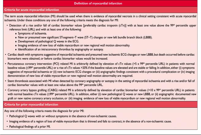
Table of Contents
Abbreviations and Acronyms . . . . . . . . . . . . . . . . . . . .2021 Definition of Myocardial Infarction . . . . . . . . . . . . . . .2022 Criteria for Acute Myocardial Infarction. . . . . . . . . . . .2022 Criteria for Prior Myocardial Infarction . . . . . . . . . . . .2022 Introduction. . . . . . . . . . . . . . . . . . . . . . . . . . . . . . . . . .2022 Pathological Characteristics of Myocardial Ischaemia
and Infarction . . . . . . . . . . . . . . . . . . . . . . . . . . . . . . . .2023 Biomarker Detection of Myocardial Injury With Necrosis. . .2023 Clinical Features of Myocardial Ischaemia and Infarction . . .2024 Clinical Classification of Myocardial Infarction . . . . . .2024 Spontaneous Myocardial Infarction (MI Type 1). . . .2024
Myocardial Infarction Secondary to an Ischaemic Imbalance (MI Type 2) . . . . . . . . . . . . . . . . . . . . . . . .2024 Cardiac Death Due to Myocardial Infarction (MI Type 3) . .2025 Myocardial Infarction Associated With Revascularization Procedures (MI Types 4 and 5) . . . . . . . . . . . . . . . . . . .2026
Electrocardiographic Detection of Myocardial Infarction. .2026 Prior Myocardial Infarction. . . . . . . . . . . . . . . . . . . . . .2027 Silent Myocardial Infarction . . . . . . . . . . . . . . . . . . . . .2027 Conditions that Confound the ECG Diagnosis of Myocardial Infarction . . . . . . . . . . . . . . . . . . . . . . . . . .2027 Imaging Techniques . . . . . . . . . . . . . . . . . . . . . . . . . . .2027
Echocardiography . . . . . . . . . . . . . . . . . . . . . . . . . . .2028 Radionuclide Imaging . . . . . . . . . . . . . . . . . . . . . . . .2028 Magnetic Resonance Imaging . . . . . . . . . . . . . . . . . .2028 Computed Tomography . . . . . . . . . . . . . . . . . . . . . . .2028 Applying Imaging in Acute Myocardial Infarction . .2028 Applying Imaging in Late Presentation of
Myocardial Infarction . . . . . . . . . . . . . . . . . . . . . . .2029 Diagnostic Criteria for Myocardial Infarction With
PCI (MI Type 4) . . . . . . . . . . . . . . . . . . . . . . . . . . . . . .2029 Diagnostic Criteria for Myocardial Infarction With
CABG (MI Type 5) . . . . . . . . . . . . . . . . . . . . . . . . . . .2029 Assessment of MI in Patients Undergoing Other
Cardiac Procedures . . . . . . . . . . . . . . . . . . . . . . . . . . . .2030 Myocardial Infarction Associated With
Non-Cardiac Procedures . . . . . . . . . . . . . . . . . . . . . . . .2030 Myocardial Infarction in the Intensive Care Unit . . . . .2030 Recurrent Myocardial Infarction . . . . . . . . . . . . . . . . . .2030 Reinfarction. . . . . . . . . . . . . . . . . . . . . . . . . . . . . . . . . .2030 Myocardial Injury or Infarction Associated With Heart
Failure . . . . . . . . . . . . . . . . . . . . . . . . . . . . . . . . . . . . . .2031 Application of MI in Clinical Trials and Quality
Assurance Programmes . . . . . . . . . . . . . . . . . . . . . . . . .2031 Public Policy Implications of the Adjustment of
the MI Definition. . . . . . . . . . . . . . . . . . . . . . . . . . . . .2031 Global Perspectives of the Definition of Myocardial
Infarction . . . . . . . . . . . . . . . . . . . . . . . . . . . . . . . . . . . .2032 Conflicts of Interest. . . . . . . . . . . . . . . . . . . . . . . . . . . .2032 Acknowledgments . . . . . . . . . . . . . . . . . . . . . . . . . . . . .2033 References. . . . . . . . . . . . . . . . . . . . . . . . . . . . . . . . . . .2033
Abbreviations and Acronyms
ACCF |
American College of Cardiology Foundation |
ACS |
acute coronary syndrome |
AHA |
American Heart Association |
CAD |
coronary artery disease |
CABG |
coronary artery bypass grafting |
CKMB |
creatine kinase MB isoform |
cTn |
cardiac troponin |
CT |
computed tomography |
CV |
coefficient of variation |
ECG |
electrocardiogram |
ESC |
European Society of Cardiology |
FDG |
fluorodeoxyglucose |
h |
hour(s) |
HF |
heart failure |
LBBB |
left bundle branch block |
LV |
left ventricle |
LVH |
left ventricular hypertrophy |
MI |
myocardial infarction |
mlBG |
meta-iodo-benzylguanidine |
min |
minute(s) |
MONICA |
Multinational MONItoring of trends and determinants in |
|
CArdiovascular disease |
MPS |
myocardial perfusion scintigraphy |
MRI |
magnetic resonance imaging |
mV |
millivolt(s) |
ng/L |
nanogram(s) per litre |
Non-Q Ml |
non-Q wave myocardial infarction |
NSTEMI |
non-ST-elevation myocardial infarction |
PET |
positron emission tomography |
pg/mL |
pictogram(s) per millilitre |
PCI |
percutaneous coronary intervention |
Q wave Ml |
Q wave myocardial infarction |
RBBB |
right bundle branch block |
sec |
second(s) |
SPECT |
single photon emission computed tomography |
STEMI |
ST elevation myocardial infarction |
ST-T |
ST-segment-T wave |
URL |
upper reference limit |
WHF |
World Heart Federation |
WHO |
World Health Organization |
|
|
Downloaded from http://circ.ahajournals.org/ by guest on April 13, 2015
2022 |
Circulation |
October 16, 2012 |
Introduction
Myocardial infarction (MI) can be recognised by clinical features, including electrocardiographic (ECG) findings, elevated values of biochemical markers (biomarkers) of myocardial necrosis, and by imaging, or may be defined by pathology. It is a major cause of death and disability worldwide. MI may be the first manifestation of coronary artery disease (CAD) or it may occur, repeatedly, in patients with established disease. Information on MI rates can provide useful information regarding the burden of CAD within and across populations, especially if standardized data are collected in a manner that distinguishes between incident and recurrent events. From the epidemiological point of view, the incidence of MI in a population can be used as a proxy for the prevalence of CAD in that population. The term ‘myocardial infarction’ may have major psychological and legal implications for the individual and society. It is an indicator of one of the leading health problems in the world and it is an outcome measure in clinical trials, observational studies and quality assurance programmes. These studies and programmes require a precise and consistent definition of MI.
In the past, a general consensus existed for the clinical syndrome designated as MI. In studies of disease prevalence, the World Health Organization (WHO) defined MI from symptoms, ECG abnormalities and cardiac enzymes. However, the development of ever more sensitive and myocardial tissue-specific cardiac biomarkers and more sensitive imaging techniques now allows for detection of very small
amounts of myocardial injury or necrosis. Additionally, the management of patients with MI has significantly improved, resulting in less myocardial injury and necrosis, in spite of a similar clinical presentation. Moreover, it appears necessary to distinguish the various conditions which may cause MI, such as ‘spontaneous’ and ‘procedure-related’ MI. Accordingly, physicians, other healthcare providers and patients require an up-to-date definition of MI.
In 2000, the First Global MI Task Force presented a new definition of MI, which implied that any necrosis in the setting of myocardial ischaemia should be labelled as MI.1 These principles were further refined by the Second Global MI Task Force, leading to the Universal Definition of Myocardial Infarction Consensus Document in 2007, which emphasized the different conditions which might lead to an MI.2 This document, endorsed by the European Society of Cardiology (ESC), the American College of Cardiology Foundation (ACCF), the American Heart Association (AHA), and the World Heart Federation (WHF), has been well accepted by the medical community and adopted by the WHO.3 However, the development of even more sensitive assays for markers of myocardial necrosis mandates further revision, particularly when such necrosis occurs in the setting of the critically ill, after percutaneous coronary procedures or after cardiac surgery. The Third Global MI Task Force has continued the Joint ESC/ACCF/AHA/WHF efforts by integrating these insights and new data into the current document, which now recognizes that very small amounts of myocardial
Downloaded from http://circ.ahajournals.org/ by guest on April 13, 2015
Thygesen et al |
ESC/ACCF/AHA/WHF Third Universal Definition of MI |
2023 |
injury or necrosis can be detected by biochemical markers and/or imaging.
Pathological Characteristics of Myocardial
Ischaemia and Infarction
MI is defined in pathology as myocardial cell death due to prolonged ischaemia. After the onset of myocardial ischaemia, histological cell death is not immediate, but takes a finite period of time to develop—as little as 20 min, or less in some animal models.4 It takes several hours before myocardial necrosis can be identified by macroscopic or microscopic post-mortem examination. Complete necrosis of myocardial cells at risk requires at least 2– 4 h, or longer, depending on the presence of collateral circulation to the ischaemic zone, persistent or intermittent coronary arterial occlusion, the sensitivity of the myocytes to ischaemia, pre-conditioning, and individual demand for oxygen and nutrients.2 The entire process leading to a healed infarction usually takes at least 5– 6 weeks. Reperfusion may alter the macroscopic and microscopic appearance.
Biomarker Detection of Myocardial Injury
With Necrosis
Myocardial injury is detected when blood levels of sensitive and specific biomarkers such as cTn or the MB fraction of creatine kinase (CKMB) are increased.2 Cardiac troponin I and T are components of the contractile apparatus of myocardial cells and are expressed almost exclusively in the heart. Although elevations of these biomarkers in the blood reflect injury leading to necrosis of myocardial cells, they do not indicate the underlying mechanism.5 Various possibilities have been suggested for release of structural proteins from the myocardium, including normal turnover of myocardial cells, apoptosis, cellular release of troponin degradation products, increased cellular wall permeability, formation and release of membranous blebs, and myocyte necrosis.6 Regardless of the pathobiology, myocardial necrosis due to myocardial ischaemia is designated as MI.
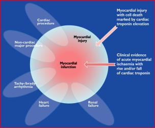
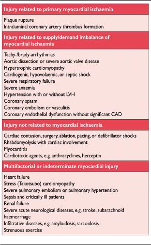
Also, histological evidence of myocardial injury with necrosis may be detectable in clinical conditions associated with predominantly non-ischaemic myocardial injury. Small amounts of myocardial injury with necrosis may be detected, which are associated with heart failure (HF), renal failure, myocarditis, arrhythmias, pulmonary embolism or otherwise uneventful percutaneous or surgical coronary procedures. These should not be labelled as MI or a complication of the procedures, but rather as myocardial injury, as illustrated in Figure 1. It is recognized that the complexity of clinical circumstances may sometimes render it difficult to determine where individual cases may lie within the ovals of Figure 1. In this setting, it is important to distinguish acute causes of cTn elevation, which require a rise and/or fall of cTn values, from chronic elevations that tend not to change acutely. A list of such clinical circumstances associated with elevated values of cTn is presented in Table 1. The multifactorial contributions resulting in the myocardial injury should be described in the patient record.
The preferred biomarker— overall and for each specific category of MI—is cTn (I or T), which has high myocardial
Figure 1. This illustration shows various clinical entities: for example, renal failure, heart failure, tachyor bradyarrhythmia, cardiac or non-cardiac procedures that can be associated with myocardial injury with cell death marked by cardiac troponin elevation. However, these entities can also be associated with myocardial infarction in case of clinical evidence of acute myocardial ischaemia with rise and/or fall of cardiac troponin.
tissue specificity as well as high clinical sensitivity. Detection of a rise and/or fall of the measurements is essential to the diagnosis of acute MI.7 An increased cTn concentration is defined as a value exceeding the 99th percentile of a normal reference population [upper reference limit (URL)]. This discriminatory 99th percentile is designated as the decision level for the diagnosis of MI and must be determined for each specific assay with appropriate quality control in each laboratory.8,9 The values for the 99th percentile URL defined by manufacturers, including those for many of the highsensitivity assays in development, can be found in the package inserts for the assays or in recent publications.10,11,12
Values should be presented as nanograms per litre (ng/L) or picograms per millilitre (pg/mL) to make whole numbers. Criteria for the rise of cTn values are assay-dependent but can be defined from the precision profile of each individual assay, including high-sensitivity assays.10,11 Optimal precision, as described by coefficient of variation (CV) at the 99th percentile URL for each assay, should be defined as 10%. Better precision (CV 10%) allows for more sensitive assays and facilitates the detection of changing values.13 The use of assays that do not have optimal precision (CV 10% at the 99th percentile URL) makes determination of a significant change more difficult but does not cause false positive results. Assays with CV 20% at the 99th percentile URL should not be used.13 It is acknowledged that pre-analytic and analytic problems can induce elevated and reduced values of cTn.10,11
Blood samples for the measurement of cTn should be drawn on first assessment and repeated 3– 6 h later. Later samples are required if further ischaemic episodes occur, or when the timing of the initial symptoms is unclear.14 To establish the diagnosis of MI, a rise and/or fall in values with at least one value above the decision level is required, coupled with a strong pre-test likelihood. The demonstration of a rising and/or falling pattern is needed to distinguish
Downloaded from http://circ.ahajournals.org/ by guest on April 13, 2015
2024 |
Circulation |
October 16, 2012 |
Table 1. Elevations of Cardiac Troponin Values Because of Myocardial Injury
acute-from chronic elevations in cTn concentrations that are associated with structural heart disease.10,11,15–19 For example, patients with renal failure or HF can have significant chronic elevations in cTn. These elevations can be marked, as seen in many patients with MI, but do not change acutely.7 However, a rising or falling pattern is not absolutely necessary to make the diagnosis of MI if a patient with a high pre-test risk of MI presents late after symptom onset; for example, near the peak of the cTn time-concentration curve or on the slow-declining portion of that curve, when detecting a changing pattern can be problematic. Values may remain elevated for 2 weeks or more following the onset of myocyte necrosis.10
Sex-dependent values may be recommended for highsensitivity troponin assays.20,21 An elevated cTn value ( 99th percentile URL), with or without a dynamic pattern of values or in the absence of clinical evidence of ischaemia, should prompt a search for other diagnoses associated with myocardial injury, such as myocarditis, aortic dissection, pulmonary embolism, or HF. Renal failure and other more non-ischaemic chronic disease states, that can be associated with elevated cTn levels, are listed in Table 1.10,11
If a cTn assay is not available, the best alternative is CKMB (measured by mass assay). As with troponin, an
increased CKMB value is defined as a measurement above the 99th percentile URL, which is designated as the decision level for the diagnosis of MI.22 Sex-specific values should be employed.22
Clinical Features of Myocardial Ischaemia
and Infarction
Onset of myocardial ischaemia is the initial step in the development of MI and results from an imbalance between oxygen supply and demand. Myocardial ischaemia in a clinical setting can usually be identified from the patient’s history and from the ECG. Possible ischaemic symptoms include various combinations of chest, upper extremity, mandibular or epigastric discomfort (with exertion or at rest) or an ischaemic equivalent such as dyspnoea or fatigue. The discomfort associated with acute MI usually lasts 20 min. Often, the discomfort is diffuse—not localized, nor positional, nor affected by movement of the region—and it may be accompanied by diaphoresis, nausea or syncope. However, these symptoms are not specific for myocardial ischaemia. Accordingly, they may be misdiagnosed and attributed to gastrointestinal, neurological, pulmonary or musculoskeletal disorders. MI may occur with atypical symptoms—such as palpitations or cardiac arrest— or even without symptoms; for example in women, the elderly, diabetics, or post-operative and critically ill patients.2 Careful evaluation of these patients is advised, especially when there is a rising and/or falling pattern of cardiac biomarkers.
Clinical Classification of
Myocardial Infarction
For the sake of immediate treatment strategies, such as reperfusion therapy, it is usual practice to designate MI in patients with chest discomfort, or other ischaemic symptoms that develop ST elevation in two contiguous leads (see ECG section), as an ‘ST elevation MI’ (STEMI). In contrast, patients without ST elevation at presentation are usually designated as having a ‘non-ST elevation MI’ (NSTEMI). Many patients with MI develop Q waves (Q wave MI), but others do not (non-Q MI). Patients without elevated biomarker values can be diagnosed as having unstable angina. In addition to these categories, MI is classified into various types, based on pathological, clinical and prognostic differences, along with different treatment strategies (Table 2).
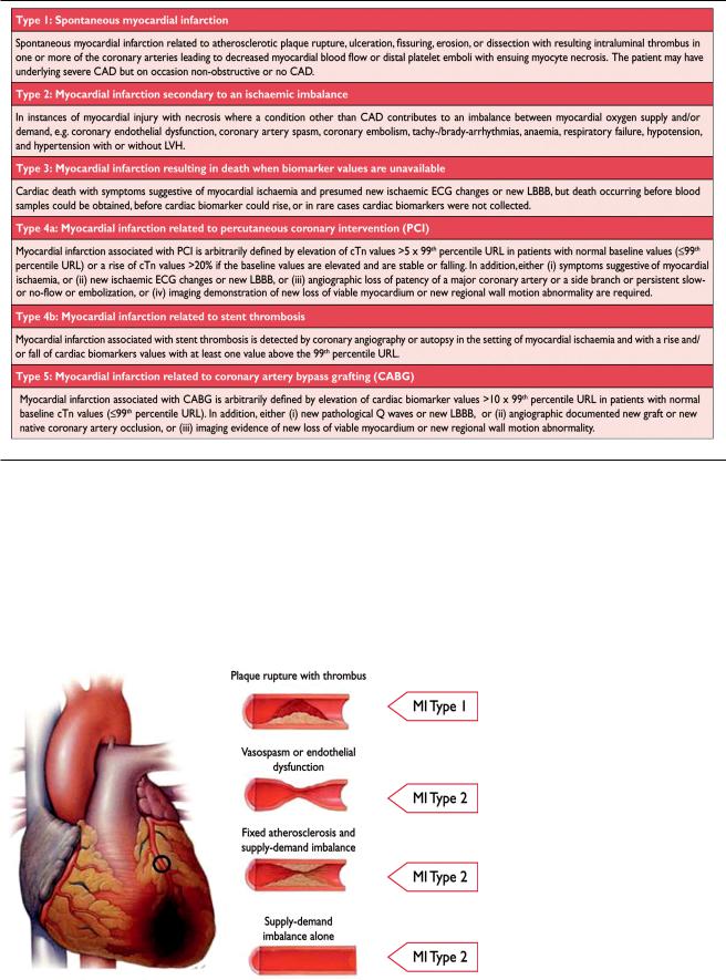
Spontaneous Myocardial Infarction (MI Type 1)
This is an event related to atherosclerotic plaque rupture, ulceration, fissuring, erosion, or dissection with resulting intraluminal thrombus in one or more of the coronary arteries, leading to decreased myocardial blood flow or distal platelet emboli with ensuing myocyte necrosis. The patient may have underlying severe CAD but, on occasion (5 to 20%), nonobstructive or no CAD may be found at angiography, particularly in women.23–25
Myocardial Infarction Secondary to an Ischaemic Imbalance (MI Type 2)
In instances of myocardial injury with necrosis, where a condition other than CAD contributes to an imbalance be-
Downloaded from http://circ.ahajournals.org/ by guest on April 13, 2015
Thygesen et al |
ESC/ACCF/AHA/WHF Third Universal Definition of MI |
2025 |
Table 2. Universal Classification of Myocardial Infarction
tween myocardial oxygen supply and/or demand, the term ‘MI type 2’ is employed (Figure 2). In critically ill patients, or in patients undergoing major (non-cardiac) surgery, elevated values of cardiac biomarkers may appear, due to the direct toxic effects of endogenous or exogenous high circulating catecholamine levels. Also coronary vasospasm and/or endothelial dysfunction have the potential to cause MI.26 –28
Cardiac Death Due to Myocardial Infarction (MI Type 3)
Patients who suffer cardiac death, with symptoms suggestive of myocardial ischaemia accompanied by presumed new ischaemic ECG changes or new LBBB— but without available biomarker values—represent a challenging diagnostic group. These individuals may die before blood samples for
Figure 2. Differentiation between myocardial infarction (MI) types 1 and 2 according to the condition of the coronary arteries.
Downloaded from http://circ.ahajournals.org/ by guest on April 13, 2015
2026 |
Circulation |
October 16, 2012 |
biomarkers can be obtained, or before elevated cardiac biomarkers can be identified. If patients present with clinical features of myocardial ischaemia, or with presumed newischaemic ECG changes, they should be classified as having had a fatal MI, even if cardiac biomarker evidence of MI is lacking.
Myocardial Infarction Associated With Revascularization Procedures (MI Types 4 and 5)
Periprocedural myocardial injury or infarction may occur at some stages in the instrumentation of the heart that is required during mechanical revascularization procedures, either by PCI or by coronary artery bypass grafting (CABG). Elevated cTn values may be detected following these procedures, since various insults may occur that can lead to myocardial injury with necrosis.29 –32 It is likely that limitation of such injury is beneficial to the patient: however, a threshold for a worsening prognosis, related to an asymptomatic increase of cardiac biomarker values in the absence of procedural complications, is not well defined.33–35 Subcategories of PCI-related MI are connected to stent thrombosis and restenosis that may happen after the primary procedure.
Electrocardiographic Detection of
Myocardial Infarction
The ECG is an integral part of the diagnostic work-up of patients with suspected MI and should be acquired and interpreted promptly (i.e. target within 10 min) after clinical presentation.2 Dynamic changes in the ECG waveforms during acute myocardial ischaemic episodes often require acquisition of multiple ECGs, particularly if the ECG at initial presentation is non-di- agnostic. Serial recordings in symptomatic patients with an initial non-diagnostic ECG should be performed at 15–30 min intervals or, if available, continuous computer-assisted 12-lead ECG recording. Recurrence of symptoms after an asymptomatic interval are an indication for a repeat tracing and, in patients with evolving ECG abnormalities, a pre-discharge ECG should be acquired as a baseline for future comparison. Acute or evolving changes in the ST-T waveforms and Q waves, when present, potentially allow the clinician to time the event, to identify the infarct-related artery, to estimate the amount of myocardium at risk as well as prognosis, and to determine therapeutic strategy. More profound ST-segment shift or T wave inversion involving multiple leads/territories is associated with a greater degree of myocardial ischaemia and a worse prognosis. Other ECG signs associated with acute myocardial ischaemia include cardiac arrhythmias, intraventricular and atrioventricular conduction delays, and loss of pre-cordial R wave amplitude. Coronary artery size and distribution of arterial segments, collateral vessels, location, extent and severity of coronary stenosis, and prior myocardial necrosis can all impact ECG manifestations of myocardial ischae-mia.36 Therefore the ECG at presentation should always be compared to prior ECG tracings, when available. The ECG by itself is often insufficient to diagnose acute myocardial ischaemia or infarction, since ST deviation may be observed in other conditions, such as acute pericarditis, left ventricular hypertrophy (LVH), left bundle branch block (LBBB), Brugada syndrome, stress cardiomyopathy, and early repolarization patterns.37 Prolonged new ST-segment elevation (e.g. 20 min), particularly when associated with reciprocal
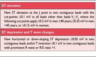
Table 3. ECG Manifestations of Acute Myocardial Ischaemia
ST-segment depression, usually reflects acute coronary occlusion and results in myocardial injury with necrosis. As in cardiomyopathy, Q waves may also occur due to myocardial fibrosis in the absence of CAD.
ECG abnormalities of myocardial ischaemia or infarction may be inscribed in the PR segment, the QRS complex, the ST-segment or the T wave. The earliest manifestations of myocardial ischaemia are typically T wave and ST-segment changes. Increased hyperacute T wave amplitude, with prominent symmetrical T waves in at least two contiguous leads, is an early sign that may precede the elevation of the ST-segment. Transient Q waves may be observed during an episode of acute ischaemia or (rarely) during acute MI with successful reperfusion. Table 3 lists ST-T wave criteria for the diagnosis of acute myocardial ischaemia that may or may not lead to MI. The J point is used to determine the magnitude of the ST-segment shift. New, or presumed new, J point elevation 0.1 mV is required in all leads other than V2 and V3. In healthy men under age 40, J-point elevation can be as much as 0.25 mV in leads V2 or V3, but it decreases with increasing age. Sex differences require different cut-points for women, since J point elevation in healthy women in leads V2 and V3 is less than in men.38 ‘Contiguous leads’ refers to lead groups such as anterior leads (V1–V6), inferior leads (II, III, aVF) or lateral/apical leads (I, aVL). Supplemental leads such as V3R and V4R reflect the free wall of the right ventricle and V7–V9 the infero-basal wall.
The criteria in Table 3 require that the ST shift be present in two or more contiguous leads. For example, 0.2 mV of ST elevation in lead V2, and 0.1 mV in lead V1, would meet the criteria of two abnormal contiguous leads in a man 40 years old. However, 0.1 mV and 0.2 mV of ST elevation, seen only in leads V2–V3 in men (or 0.15 mV in women), may represent a normal finding. It should be noted that, occasionally, acute myocardial ischaemia may create sufficient ST-segment shift to meet the criteria in one lead but have slightly less than the required ST shift in a contiguous lead. Lesser degrees of ST displacement or T wave inversion do not exclude acute myocardial ischaemia or evolving MI, since a single static recording may miss the more dynamic ECG changes that might be detected with serial recordings. ST elevation or diagnostic Q waves in contiguous lead groups are more specific than ST depression in localizing the site of myocardial ischaemia or necrosis.39,40 Supplemental leads, as well as serial ECG recordings, should always be considered in patients that present with
Downloaded from http://circ.ahajournals.org/ by guest on April 13, 2015
Thygesen et al |
ESC/ACCF/AHA/WHF Third Universal Definition of MI |
2027 |
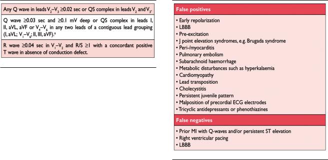
Table 4. ECG Changes Associated With Prior Myocardial Infarction (in Absence of LVH and LBBB)
Table 5. Common ECG Pitfalls in Diagnosing Myocardial
Infarction
aThe same criteria are used for supplemental leads V7–V9.
ischaemic chest pain and a non-diagnostic initial ECG.41,42 Electrocardiographic evidence of myocardial ischaemia in the distribution of a left circumflex artery is often overlooked and is best captured using posterior leads at the fifth intercostal space (V7 at the left posterior axillary line, V8 at the left mid-scapular line, and V9 at the left paraspinal border). Recording of these leads is strongly recommended in patients with high clinical suspicion for acute circumflex occlusion (for example, initial ECG non-diagnostic, or ST-segment depression in leads V1–3).41 A cut-point of 0.05 mV ST elevation is recommended in leads V7–V9; specificity is increased at a cut-point 0.1 mV ST elevation and this cut-point should be used in men 40 years old. ST depression in leads V1–V3 may be suggestive of infero-basal myocardial ischaemia (posterior infarction), especially when the terminal T wave is positive (ST elevation equivalent), however this is non-specific.41– 43 In patients with inferior and suspected right ventricular infarction, right precordial leads V3R and V4R should be recorded, since ST elevation 0.05 mV ( 0.1 mV in men 30 years old) provides supportive criteria for the diagnosis.42
During an episode of acute chest discomfort, pseudonormalization of previously inverted T waves may indicate acute myocardial ischaemia. Pulmonary embolism, intracranial processes, electrolyte abnormalities, hypothermia, or peri-/myocarditis may also result in ST-T abnormalities and should be considered in the differential diagnosis. The diagnosis of MI is more difficult in the presence of LBBB.44,45 However, concordant ST-segment elevation or a previous ECG may be helpful to determine the presence of acute MI in this setting. In patients with right bundle branch block (RBBB), ST-T abnormalities in leads V1–V3 are common, making it difficult to assess the presence of ischaemia in these leads: however, when new ST elevation or Q waves are found, myocardial ischaemia or infarction should be considered.
Prior Myocardial Infarction
As shown in Table 4, Q waves or QS complexes in the absence of QRS confounders are pathognomonic of a prior MI in patients with ischaemic heart disease, regardless of symptoms.46,47 The specificity of the ECG diagnosis for MI is greatest when Q waves occur in several leads or lead groupings. When the Q waves are associated with ST deviations or T wave changes in the same leads, the likelihood of MI is increased; for example, minor Q waves 0.02 sec and 0.03 sec that are 0.1 mV deep are suggestive of prior MI if accompanied by inverted T waves in the same lead group. Other validated MI coding algorithms,
such as the Minnesota Code and WHO MONICA, have been used in epidemiological studies and clinical trials.3
Silent Myocardial Infarction
Asymptomatic patients who develop new pathologic Q wave criteria for MI detected during routine ECG follow-up, or reveal evidence of MI by cardiac imaging, that cannot be directly attributed to a coronary revascularization procedure, should be termed ‘si lent MI.’48 –51 In studies, silent Q wave MI accounted for 9 –37% of all non-fatal MI events and were associated with a significantly increased mortality risk.48,49 Improper lead placement or QRS confounders may result in what appear to be new Q waves or QS complexes, as compared to a prior tracing. Thus, the diagnosis of a new silent Q wave MI should be confirmed by a repeat ECG with correct lead placement, or by an imaging study, and by focussed questioning about potential interim ischaemic symptoms.
Conditions That Confound the ECG Diagnosis
of Myocardial Infarction
A QS complex in lead V1 is normal. A Q wave 0.03 sec and25% of the R wave amplitude in lead III is normal if the frontal QRS axis is between 30° and 0°. A Q wave may also be normal in aVL if the frontal QRS axis is between 60° and 90°. Septal Q waves are small, non-pathological Q waves 0.03 sec and 25% of the R-wave amplitude in leads I, aVL, aVF, and V4–V6. Pre-excitation, obstructive, dilated or stress cardiomyopathy, cardiac amyloidosis, LBBB, left anterior hemiblock, LVH, right ventricular hypertrophy, myocarditis, acute cor pulmonale, or hyperkalaemia may be associated with Q waves or QS complexes in the absence of MI. ECG abnormalities that mimic myocardial ischaemia or MI are presented in Table 5.
Imaging Techniques
Non-invasive imaging plays many roles in patients with known or suspected MI, but this section concerns only its role
Downloaded from http://circ.ahajournals.org/ by guest on April 13, 2015
2028 |
Circulation |
October 16, 2012 |
in the diagnosis and characterisation of MI. The underlying rationale is that regional myocardial hypoperfusion and ischaemia lead to a cascade of events, including myocardial dysfunction, cell death and healing by fibrosis. Important imaging parameters are therefore perfusion, myocyte viability, myocardial thickness, thickening and motion, and the effects of fibrosis on the kinetics of paramagnetic or radioopaque contrast agents.
Commonly used imaging techniques in acute and chronic infarction are echocardiography, radionuclide ventriculography, myocardial perfusion scintigraphy (MPS) using single photon emission computed tomography (SPECT), and magnetic resonance imaging (MRI). Positron emission tomography (PET) and X-ray computed tomography (CT) are less common.52 There is considerable overlap in their capabilities and each of the techniques can, to a greater or lesser extent, assess myocardial viability, perfusion, and function. Only the radionuclide techniques provide a direct assessment of myocyte viability, because of the inherent properties of the tracers used. Other techniques provide indirect assessments of myocardial viability, such as contractile response to dobutamine by echocardiography or myocardial fibrosis by MR.
Echocardiography
The strength of echocardiography is the assessment of cardiac structure and function, in particular myocardial thickness, thickening and motion. Echocardiographic contrast agents can improve visualisation of the endocardial border and can be used to assess myocardial perfusion and microvascular obstruction. Tissue Doppler and strain imaging permit quantification of global and regional function.53 Intravascular echocardiographic contrast agents have been developed that target specific molecular processes, but these techniques have not yet been applied in the setting of MI.54
Radionuclide Imaging
Several radionuclide tracers allow viable myocytes to be imaged directly, including the SPECT tracers thallium-201, technetium-99m MIBI and tetrofosmin, and the PET tracers F-2-fluorodeoxyglucose (FDG) and rubidium-82.18,52 The strength of the SPECT techniques is that these are the only commonly available direct methods of assessing viability, although the relatively low resolution of the images leaves them at a disadvantage for detecting small areas of MI. The common SPECT radiopharmaceuticals are also tracers of myocardial perfusion and the techniques thereby readily detect areas of MI and inducible perfusion abnormalities. ECG-gated imaging provides a reliable assessment of myocardial motion, thickening and global function. Evolving radionuclide techniques that are relevant to the assessment of MI include imaging of sympathetic innervation using iodine-123-labelled meta-iodo-benzylguanidine (mIBG),55 imaging of matrix metalloproteinase activation in ventricular remodelling,56,57 and refined assessment of myocardial metabolism.58
Magnetic Resonance Imaging
The high tissue contrast of cardiovascular MRI provides an accurate assessment of myocardial function and it has similar
capability to echocardiography in suspected acute MI. Paramagnetic contrast agents can be used to assess myocardial perfusion and the increase in extracellular space that is associated with the fibrosis of prior MI. These techniques have been used in the setting of acute MI,59,60 and imaging of myocardial fibrosis by delayed contrast enhancement is able to detect even small areas of subendocardial MI. It is also of value in detecting myocardial disease states that can mimic MI, such as myocarditis.61
Computed Tomography
Infarcted myocardium is initially visible as a focal area of decreased left ventricle (LV) enhancement, but later imaging shows hyperenhancement, as with late gadolinium imaging by MRI.62 This finding is clinically relevant because contrastenhanced CT may be performed for suspected pulmonary embolism and aortic dissection— conditions with clinical features that overlap with those of acute MI— but the technique is not used routinely. Similarly, CT assessment of myocardial perfusion is technically feasible but not yet fully validated.
Applying Imaging in Acute Myocardial Infarction
Imaging techniques can be useful in the diagnosis of acute MI because of their ability to detect wall motion abnormalities or loss of viable myocardium in the presence of elevated cardiac biomarker values. If, for some reason, biomarkers have not been measured or may have normalized, demonstration of new loss of myocardial viability in the absence of nonischaemic causes meets the criteria for MI. Normal function and viability have a very high negative predictive value and practically exclude acute MI.63 Thus, imaging techniques are useful for early triage and discharge of patients with suspected MI. However, if biomarkers have been measured at appropriate times and are normal, this excludes an acute MI and takes precedence over the imaging criteria.
Abnormal regional myocardial motion and thickening may be caused by acute MI or by one or more of several other conditions, including prior MI, acute ischaemia, stunning or hibernation. Non-ischaemic conditions, such as cardiomyopathy and inflammatory or infiltrative diseases, can also lead to regional loss of viable myocardium or functional abnormality. Therefore, the positive predictive value of imaging for acute MI is not high unless these conditions can be excluded, and unless a new abnormality is detected or can be presumed to have arisen in the setting of other features of acute MI.
Echocardiography provides an assessment of many nonischaemic causes of acute chest pain, such as peri-myocarditis, valvular heart disease, cardiomyopathy, pulmonary embolism or aortic dissection.53 It is the imaging technique of choice for detecting complications of acute MI, including myocardial free wall rupture, acute ventricular septal defect, and mitral regurgitation secondary to papillary muscle rupture or ischaemia.
Radionuclide imaging can be used to assess the amount of myocardium that is salvaged by acute revascularization.64 Tracer is injected at the time of presentation, with imaging deferred until after revascularization, providing a measure of myocardium at risk. Before discharge, a second resting
Downloaded from http://circ.ahajournals.org/ by guest on April 13, 2015
Thygesen et al |
ESC/ACCF/AHA/WHF Third Universal Definition of MI |
2029 |
injection provides a measure of final infarct size, and the difference between the two corresponds to the myocardium that has been salvaged.
Applying Imaging in Late Presentation of Myocardial Infarction
In case of late presentation after suspected MI, the presence of regional wall motion abnormality, thinning or scar in the absence of nonischaemic causes, provides evidence of past MI. The high resolution and specificity of late gadolinium enhancement MRI for the detection of myocardial fibrosis has made this a very valuable technique. In particular, the ability to distinguish between subendocardial and other patterns of fibrosis provides a differentiation between ischaemic heart disease and other myocardial abnormalities. Imaging techniques are also useful for risk stratification after a definitive diagnosis of MI. The detection of residual or remote ischaemia and/or ventricular dysfunction provides powerful indicators of later outcome.
Diagnostic Criteria for Myocardial Infarction With PCI (MI Type 4)
Balloon inflation during PCI often causes transient ischaemia, whether or not it is accompanied by chest pain or ST-T changes. Myocardial injury with necrosis may result from recognizable peri-procedural events—alone or in combina- tion—such as coronary dissection, occlusion of a major coronary artery or a side-branch, disruption of collateral flow, slow flow or no-reflow, distal embolization, and microvascular plugging. Embolization of intracoronary thrombus or atherosclerotic particulate debris may not be preventable, despite current anticoagulant and antiplatelet adjunctive therapy, aspiration or protection devices. Such events induce inflammation of the myocardium surrounding islets of myocardial necrosis.65 New areas of myocardial necrosis have been demonstrated by MRI following PCI.66
The occurrence of procedure-related myocardial cell injury with necrosis can be detected by measurement of cardiac biomarkers before the procedure, repeated 3–6 h later and, optionally, further re-measurement 12 h thereafter. Increasing levels can only be interpreted as procedure-related myocardial injury if the pre-procedural cTn value is normal ( 99th percentile URL) or if levels are stable or falling.67,68 In patients with normal pre-procedural values, elevation of cardiac biomarker values above the 99th percentile URL following PCI are indicative of procedure-related myocardial injury. In earlier studies, increased values of post-procedural cardiac biomarkers, especially CKMB, were associated with impaired outcome.69,70 However, when cTn concentrations are normal before PCI and become abnormal after the procedure, the threshold above the 99th percentile URL—whereby an adverse prognosis is evi- dent—is not well defined71 and it is debatable whether such a threshold even exists.72 If a single baseline cTn value is elevated, it is impossible to determine whether further increases are due to the procedure or to the initial process causing the elevation. In this situation, it appears that the prognosis is largely determined by the pre-procedural cTn level.71 These relationships will probably become even more complex for the new highsensitivity troponin assays.70
In patients undergoing PCI with normal ( 99th percentile URL) baseline cTn concentrations, elevations of cTn 5 99th percentile URL occurring within 48 h of the procedure— plus either (i) evidence of prolonged ischaemia ( 20 min) as demonstrated by prolonged chest pain, or (ii) ischaemic ST changes or new pathological Q waves, or (iii) angiographic evidence of a flow limiting complication, such as of loss of patency of a side branch, persistent slow-flow or no-reflow, embolization, or (iv) imaging evidence of new loss of viable myocardium or new regional wall motion abnormality—is defined as PCI-related MI (type 4a). This threshold of cTn values 5 99th percentile URL is arbitrarily chosen, based on clinical judgement and societal implications of the label of peri-procedural MI. When a cTn value is 5 99th percentile URL after PCI and the cTn value was normal before the PCI—or when the cTn value is 5 99th percentile URL in the absence of ischaemic, angiographic or imaging findings—the term ‘myocardial injury’ should be used.
If the baseline cTn values are elevated and are stable or falling, then a rise of 20% is required for the diagnosis of a type 4a MI, as with reinfarction. Recent data suggest that, when PCI is delayed after MI until biomarker concentrations are falling or have normalized, and elevation of cardiac biomarker values then reoccurs, this may have some longterm significance. However, additional data are needed to confirm this finding.73
A subcategory of PCI-related MI is stent thrombosis, as documented by angiography and/or at autopsy and a rise and/or fall of cTn values 99th percentile URL (identified as MI type 4b). In order to stratify the occurrence of stent thrombosis in relation to the timing of the PCI procedure, the Academic Research Consortium recommends temporal categories of ‘early’ (0 to 30 days), ‘late’ (31 days to 1 year), and ‘very late’ ( 1 year) to distinguish likely differences in the contribution of the various pathophysiological processes during each of these intervals.74 Occasionally, MI occurs in the clinical setting of what appears to be a stent thrombosis: however, at angiography, restenosis is observed without evidence of thrombus (see section on clinical trials).
Diagnostic Criteria for Myocardial Infarction With CABG (MI Type 5)
During CABG, numerous factors can lead to periprocedural myocardial injury with necrosis. These include direct myocardial trauma from (i) suture placement or manipulation of the heart, (ii) coronary dissection, (iii) global or regional ischaemia related to inadequate intra-operative cardiac protection, (iv) microvascular events related to reperfusion, (v) myocardial injury induced by oxygen free radical generation, or (vi) failure to reperfuse areas of the myocardium that are not subtended by graftable vessels.75–77 MRI studies suggest that most necrosis in this setting is not focal but diffuse and localized in the subendocardium.78
In patients with normal values before surgery, any increase of cardiac biomarker values after CABG indicates myocardial necrosis, implying that an increasing magnitude of biomarker concentrations is likely to be related to an impaired outcome. This has been demonstrated in clinical studies employing CKMB, where elevations 5, 10 and 20 times the URL after
Downloaded from http://circ.ahajournals.org/ by guest on April 13, 2015