

Hart C. Anthony, Shears Paul. Color Atlas of Medical Microbiology.pdf
.pdfThe Physiology of Metabolism and Growth in Bacteria 165
Growth and Cell Death
Bacteria reproduce asexually by means of simple transverse binary fission. Their numbers (n) increase logarithmically (n = 2G). The time required for a reproduction cycle (G) is called the generation time (g) and can vary greatly from species to species. Fast-growing bacteria cultivated in vitro have a generation time of 15–30 minutes. The same bacteria may take hours to reproduce in vivo. Obligate anaerobes grow much more slowly than aerobes; this is
true in vitro as well. Tuberculosis bacteria have an in-vitro generation time of 3 12–24 hours. Of course the generation time also depends on the nutrient con-
tent of the medium.
The so-called normal growth curve for bacteria is obtained by inoculating a nutrient broth with bacteria the metabolism of which is initially quiescent, counting them at intervals and entering the results in a semilog coordinate system (Fig. 3.16). The lag phase (A) is characterized by an increase in bacterial mass per unit of volume, but no increase in cell count. During this phase, the metabolism of the bacteria adapts to the conditions of the nutrient medium. In the following log (or exponential) phase (C), the cell count increases logarithmically up to about 109/ml. This is followed by growth deceleration and transition to the stationary phase (E) due to exhaustion of the nutrients and the increasing concentration of toxic metabolites. Finally, death phase (F) processes begin. The generation time can only be determined during phase C, either graphically or by determining the cell count (n) at two different times and applying the formula:
g |
t2 |
" t1 |
: |
|
¼ log2 n2 |
||||
|
" log2 n1 |
|
Normal Growth Curve of a Bacterial Culture
Number of surviving cells (log)
D E
C F
A B
(Hours) Time (Days)
Fig. 3.16 A = lag phase, B = acceleration phase,
C = log (exponential) phase, D = deceleration phase,
E = stationary phase, F = death phase.

166 3 General Bacteriology
Bacterial Cell Count and Bacterial Mass
The colony counting method. The number of living cells in a given culture or material can be determined by means of the colony counting method. The samples are diluted logarithmically by a dilution factor of 10. Using the pour plate technique, each dilution is mixed with 1 ml of liquid agar and poured out in a plate. In the surface inoculation method, 0.1 ml of each dilution is plated out on a nutrient agar surface. The plates are incubated, resulting in colony growth. The number of colonies counted, multiplied by the dilution factor, results in the original number
3of viable bacterial cells (CFU = colony forming units).
Bacterial mass. The bacterial mass can be established by weighing (dry or wet weight). The simplest way to determine the mass is by means of photometric adsorption measurement. The increases in mass and cell count run parallel during phase C on the growth curve.
The Molecular Basis of Bacterial Genetics
& Bacteria possess two genetic structures: the chromosome and the plasmid. Both of these structures consist of a single circular DNA double helix twisted counterclockwise about its helical axis. Replication of this DNA molecule always starts at a certain point (the origin of replication) and is “semiconservative,” that is, one strand in each of the two resulting double strands is conserved. Most bacterial genes code for proteins (polypeptides). Noncoding interposed sequences (introns), like those seen in eukaryotes, are the exception. Certain bacterial genes have a mosaic structure. The phases of transcription are promoter recognition, elongation, and termination. Many bacterial mRNAs are polycistronic, meaning they contain the genetic information for several polypeptides. Translation takes place on the 70S ribosomes. Special mRNA codons mark the start and stop of polypeptide synthesis. Many genes that code for functionally related polypeptides are grouped together in chromosome or plasmid segments known as operons. The most important regulatory mechanism is the positive or negative control of transcription initiation. This control function may be exercised by individual localized genes, the genes of an operon or genes in a regulon. &

The Molecular Basis of Bacterial Genetics 167
The Structure of Bacterial DNA
A bacterium’s genetic information is stored in its chromosome and plasmids. Each of these structures is made of a single DNA double helix twisted to the right, then additionally twisted to the left about its helical axis (supercoiled, see p. 148ff. and Fig. 3.17). Plasmids consisting of linear DNA also occur, although this is rare. This DNA topology solves spatial problems and enables
such functions as replication, transcription, and recombination. Some genes 3 are composed of a mosaic of minicassettes interconnected by conserved DNA sequences between the cassettes (see Fig. 1.2, p. 14).
Chromosome. The chromosome corresponds to the nucleoid (p. 148ff.). The E. coli chromosome is composed of 4.63 ! 106 base pairs (bp). It codes for 4288 proteins. The gene sequence is colinear with the expressed genetic products. The noncoding interposed sequences (introns) normally seen in eukaryotic genes are very rare. The chromosomes of E. coli and numerous other pathogenic bacteria have now been completely sequenced.
Plasmids. The plasmids are autonomous DNA molecules of varying size (3 ! 103 to 4.5 ! 105 bp) localized in the cytoplasm. Large plasmids are usually present in one to two copies per cell, whereas small ones may be present in 10, 40, or 100 copies. Plasmids are not essential to a cell’s survival.
Resistance Plasmid in Escherichia coli
Fig. 3.17 a Covalently closed circle (CCC), also known as a “supercoil” or “supertwist.”
b Open circle. This open form is an artifact produced by a nick in one strand of the DNA double helix.

168 3 General Bacteriology
Many of them carry genes that code for certain phenotypic characteristics of the host cell. The following plasmid types are medically relevant:
&Virulence plasmids. Carry determinants of bacterial virulence, e.g., enterotoxin genes or hemolysin genes.
&Resistance plasmids. Carry genetic information bearing on resistance to anti-infective agents. R plasmids may carry several R genes at once (see also Fig. 3.23, p. 176). Plasmids have also been described that carry both virulence
3 and resistance genes.
DNA Replication
The identical duplication process of DNA is termed semiconservative because the double strand of DNA is opened up during replication, whereupon each strand serves as the matrix for synthesis of a complementary strand. Thus each of the two new double strands “conserves” one old strand. The doubling of each DNA molecule (replicon) begins at a given starting point, the so-called origin of replication. This process continues throughout the entire fission cycle.
Transcription and Translation
& Transcription. Copying of the sense strand of the DNA into mRNA. The continuous genetic nucleotide sequence is transcribed “colinearly” into mRNA. This principle of colinearity applies with very few exceptions. The transcription process can be broken down into the three phases promoter recognition, elongation, and termination. The promoter region is the site where the RNA polymerase begins reading the DNA sequence. A sigma factor is required for binding to the promoter. Sigma factors are proteins that associate temporarily with the RNA polymerase (core enzyme) to form a holoenzyme, then dissociate themselves once the transcription process has begun, making them available to associate once again. Specific sigma factors recognize the standard promoters of most genes. Additional sigma factors, the expression of which depends on the physiological status of the cell, facilitate the transcription of special determinants. Genes that code for functionally related proteins, for example proteins that act together to catalyze a certain metabolic step, are often arranged sequentially at specific locations on the chromosome or plasmid. Such DNA sequences are known as operons (Fig. 3.18). The mRNA synthesized by the transcription of an operon is polycistronic, i.e., it contains the information sequences of several genes. The information sequences are
|
|
|
The Molecular Basis of Bacterial Genetics |
169 |
||||||
Bacterial Operon and Regulator Gene |
|
|
|
|
|
|||||
5' |
P Regulator gene T |
P |
O |
Gene A |
|
Gene B |
T DNA |
3' |
||
3' |
|
5' |
||||||||
|
|
|
|
Transcription |
|
Transcription |
|
|
||
|
|
|
|
Start |
Stop |
Start |
Stop |
mRNA |
||
5' |
|
|
|
|
CR |
IR |
|
CR |
|
3' |
|
|
|
|
|
|
|
|
|||
|
|
|
|
Translation |
|
Translation |
|
3 |
||
|
Regulator protein |
|
|
Protein A |
|
Protein B |
|
|||
|
|
|
|
|
|
|||||
|
|
Effector |
|
|
|
|
CR = Cistron region |
|
||
|
|
|
|
|
|
|
|
|||
P |
Promoter |
T Terminator |
O |
Operator |
|
|
(coding region) |
|
||
|
IR = Intercistron region |
|||||||||
|
|
|
|
|
|
|
||||
Fig. 3.18 An operon is a DNA sequence that usually includes several structural genes. These genes code for proteins that are functionally related. Transcription of an operon is often activated or repressed by the product of a regulator gene located elsewhere on the chromosome.
separated by intercistronic regions. Each cistron has its own start and stop codon in the mRNA.
& Translation. Transformation of the nucleotide sequence carried by the mRNA into the polypeptide amino acid sequence at the 70S ribosomes. In principle, bacterial and eukaryotic translation is the same. The enzymes and other factors involved do, however, differ structurally and can therefore be selectively blocked by antibiotics (p. 198ff.).
Regulation of Gene Expression
Bacteria demonstrate a truly impressive capacity for adapting to their environment. A number of regulatory bacterial mechanisms are known, for example posttranslational regulation, translational regulation, transcription termination, and quorum sensing (see Fig. 1.5, p. 20). The details of all these mechanisms would exceed the scope of this book. The most important is regulation of the initiation of transcription by means of activation or repression, a process not observed in this form in eukaryotes: a single gene, or several genes in an operon at one DNA location, may be affected (see Fig. 3.18). The mechanism that has been investigated most thoroughly is transcriptional regulation of catabolic and anabolic operons by a repressor or activator.

170 3 General Bacteriology
Transcriptional Regulation of an Operon:
&Catabolic operons have genes that code for enzymes of catabolic metabolism. Anabolic operons code for enzymes of anabolic metabolism.
&Regulators. Code for proteins that can repress or activate transcription by binding to the operator or promoter of an operon.
&Effectors. Low-molecular-weight signal molecules from the immediate environment of the bacterial cell. Can activate (= corepressor) or inactivate (= inducer) the
3repressor by means of an allosteric effect.
&Induction of a catabolic operon. The effector molecule is a nutrient substrate that is broken down by the products of the operon genes (e.g., lactose). Lactose inactivates the repressor, initializing transcription of the genes for b-galactosidase and b-galactoside permease in the lactose operon. These genes are normally not read off because the repressor is bound to the operator. The cell is not induced to produce the necessary catabolic enzymes until the nutrient substrate is present.
&Repression of an anabolic operon. The signal molecule is the final product of an anabolic process, for instance an amino acid. If this acid is present in the medium, it can be obtained from there and the cell need not synthesize the anabolic enzymes it would require to produce it. In such a case, binding to the effector is what turns the regulator protein into an active repressor.
A single regulator protein can also activate or repress several genes not integrated in an operon, i.e., at various locations on the DNA. Such functional gene groups are called regulons. Alternative sigma factors (see p. 168) may be involved in the transcriptional activation of special genes with special promoters. Physiological cell status determines whether or not these alternative factors are produced.
The Genetic Variability of Bacteria
& Changes in bacterial DNA are the result of spontaneous mutations in individual genes as well as recombination processes resulting in new genes or genetic combinations. Based on the molecular mechanisms involved, bacterial recombinations are classified as homologous, site-specific, and transpositional. The latter two in particular reflect the high level of mobility of many genes and have made essential contributions to the evolution of bacteria.
Although sexual heredity is unknown in bacteria, they do make use of the mechanisms of intercellular transfer of genomic material known as parasexual processes. Transformation designates transfer of DNA that is essentially
|
The Genetic Variability of Bacteria |
171 |
|
|
|
|
|
||
chemically pure from a donor into a receptor cell. In transduction, bacte- |
|
|
||
riophages serve as the vehicles for DNA transport. Conjugation is the transfer |
|
|
||
of DNA by means of cell-to-cell contact. This process, made possible by con- |
|
|
||
jugative plasmids and transposons, can be a high-frequency one and may |
|
|
||
even occur between partners of different species, genera, or families. The |
|
|
||
transfer primarily involves the conjugative elements themselves. Conjugative |
|
|
||
structures carrying resistance or virulence genes are of considerable medical |
|
|
||
significance. |
|
|
|
|
|
|
3 |
||
The processes of restriction and modification are important factors |
|
|||
limiting genetic exchange among different taxa. Restriction is based on |
|
|
||
the effects of restriction endonucleases capable of specific excision of foreign |
|
|
||
|
|
|||
DNA sequences. These enzymes have become invaluable tools in the field of |
|
|
||
genetic engineering. |
& |
|
|
|
Molecular Mechanisms of Genetic Variability
Spontaneous Mutation
In the year 1943, Luria and Delbru¨ck used the so-called fluctuation test to demonstrate that changes in the characteristics of bacterial populations were the results of rare, random mutations in the genes of individual cells, which then were selected. Such mutations may involve substitution of a single nucleotide, frame-shifts, deletions, inversions, or insertions. The frequency of mutations is expressed as the mutation rate, which is defined as the probability of mutation per gene per cell division. The rate varies depending on the gene involved and is approximately 10–6 to 10–10. Mutation rates may increase drastically due to mutagenic factors such as radioactivity, UV radiation, alkylating chemicals, etc.
Recombination
The term recombination designates processes that lead to the restructuring of DNA, formation of new genes or genetic combinations.
Homologous (generalized) recombination. A precise exchange of DNA between corresponding sequences. Several enzymes contribute to the complex breakage and reunion process involved, the most important being the RecA enzyme and another the RecBC nuclease. Fig. 1.2 (p. 14) shows an example of homologous recombination resulting in the exchange of minicassettes between two genes.
172 3 General Bacteriology
Site-specific recombination. Integration or excision of a sequence in or from target DNA. Only a single sequence of a few nucleotides of the integrated DNA needs to be homologous with the recombination site on the target DNA. The integration of bacteriophage genomes is an example of what this process facilitates (p. 184f.) Integration of several determinants of antibiotic resistance in one integron can also utilize this process (Fig. 3.19). Resistance integrons may be integrated in transposable DNA.
3Transposition. The transposition process does not require the donor and target DNA to be homologous. DNA sequences can either be transposed to a different locus on the same molecule or to a different replicon. Just as in sitespecific recombination, transposition has always played a major role in the evolution of multi-resistance plasmids (see Fig. 3.23, p. 176).
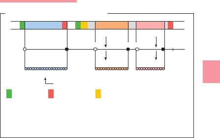
Site-Specific Recombination (Integron)
Integron
|
|
Pint |
Pant |
|
|
|
|
||||
|
|
|
|
|
|
|
|
+ |
|
||
|
|
|
|
|
|
|
|
|
|
||
|
intI |
att |
I |
sulI |
|
||||||
|
|
|
Integrase |
Integration |
|
|
Excision |
||||
|
|
|
|
|
|||||||
|
|
|
|
|
|
|
|||||
|
|
Pint Pant |
|
|
|
|
|
|
|||
|
|
|
|
|
|
|
|
||||
|
|
|
|
|
|
|
|
||||
|
|
|
|
|
|
|
|||||
|
|
|
|
|
|
||||||
|
intI |
attI |
|
|
|
|
|
|
|||
|
|
|
|
|
|
|
|
|
|
|
|
a
Mobile gene cassette
Resistance gene (without promoter)
59 bp-element
sulI
|
Pint Pant |
|
|
|
|
|
|
|
|
|
|
|||
|
|
|
Resistance genes |
|
|
|||||||||
|
|
|
|
|
|
|
|
|
||||||
|
|
|
|
|
|
|
|
|
|
|
|
|
|
|
|
intI |
attI |
|
aacC1 |
|
orfE |
|
aadA2 |
|
cmlA |
|
sul |
I |
|
|
|
|
|
|
|
|
|
|
|
|
|
|
|
|
Fig. 3.19 a Integration or excision of a circular gene cassette into or out of an integron. An integron is a genetic structure containing the determinants of a sitespecific recombination system. This structure is capable of capturing or mobilizing mobile gene cassettes. It also provides the promoter for transcription of the cassette genes, which themselves have no promoter. The cassettes occur in both free circularized and integrated forms.
bintI Integrase gene. Codes for site-specific integrase (int = integrase). attI Attachment sequence for the integrase. Site of recombination
(att = attachment).
Promoter for the integrated gene cassette (P = promoter). Promoter for the integrase gene.
sulI Gene for sulfonamide resistance (sul = sulfonamide). 59bp Integrase-specific recombination site in cassette.
b Model of integron 4 (In4), which contains three resistance genes and an open reading frame (orfE), the function of which is unknown. In4 is the result of successive integration of several resistance genes at the att1 site.

The Genetic Variability of Bacteria 173
Transposable DNA Elements |
|
|
|
|
|
|
|
|
|
|
|
|
|
|
|
|
|
|
|
|
|
|
||||||||||||||||
|
|
|
Tn3-like transposons |
|
|
|
|
|
|
|
||||||||||||||||||||||||||||
|
|
|
|
|
|
|
|
|
|
|
|
|
|
|
|
|
|
|
|
|
|
|
|
|
|
|
||||||||||||
|
IS elements |
|
|
|
|
|
|
|
|
Transposon 3 (Tn3), 4.9 kbp, |
|
|
|
|
|
|
|
|||||||||||||||||||||
|
800–1500 bp |
|
|
|
|
|
|
|
|
codes for betalactamase (= bla) |
|
|
||||||||||||||||||||||||||
|
|
|
|
|
|
|
|
|
|
|
|
|
|
|
|
|
|
|
|
|
|
|
|
|
|
|
|
|
|
|
|
|
|
|
|
|
|
|
|
|
|
|
|
|
tnp |
|
|
|
|
|
|
|
|
|
|
|
|
|
|
|
tnp |
tnpR |
res |
|
|
bla |
|
|
|
|
|
|
|||||
a |
|
|
|
|
|
|
|
|
|
|
|
|
|
|
b |
|
|
|
|
|
|
|
|
|
|
|
|
|
|
|
||||||||
|
Trans- |
|
|
|
|
|
|
|
|
|
|
|
|
|
|
|
|
|
|
|
|
|
||||||||||||||||
|
|
|
|
|
|
posase- |
|
|
|
|
|
|
|
|
|
|
|
|
|
|
|
|
|
|
|
|
|
|
|
|
|
|
|
|
|
|
||
|
|
|
|
|
|
gene |
|
|
|
|
|
|
|
Composite transposons |
|
|
|
|
|
|
3 |
|||||||||||||||||
|
|
|
|
|
|
Inverted |
|
|
|
|
|
|
|
|
|
|
|
|
|
|
||||||||||||||||||
|
|
|
|
|
|
|
|
|
|
|
|
|
|
Transposon 10 (Tn10), 9.3 kbp, tetB = determinant |
|
|||||||||||||||||||||||
|
|
|
|
|
|
|
|
|
|
|
|
|
|
|||||||||||||||||||||||||
|
|
|
|
|
|
repeats |
|
|
|
|
|
of resistance to tetracyclines |
|
|
|
|
|
|
|
|||||||||||||||||||
|
|
|
|
|
Direct repeats |
|
|
|
|
|
|
|
|
|
|
|
|
|
|
|
|
|
|
|
|
|
|
|
|
|
|
|
|
|
||||
|
|
|
|
|
|
|
|
|
|
|
|
|
|
|
IS10L |
|
|
tetB |
|
|
|
|
|
IS10R |
|
|
|
|
|
|||||||||
|
|
|
|
|
|
|
|
|
|
|
|
|
|
|
|
|
|
|
|
|
|
|
|
|
|
|||||||||||||
|
|
|
|
|
|
|
|
|
|
|
|
|
|
|
|
|
|
|
|
|
|
|
|
|
|
|
|
|
||||||||||
|
|
|
|
|
|
|
|
|
|
|
|
|
|
c |
|
|
|
|
|
|
|
|
|
|
|
|
|
|
|
|||||||||
|
Conjugative transposons |
|
|
|
|
|
|
|
|
|
|
|
|
|
|
|
|
|
|
|
|
|
|
|
|
|
||||||||||||
|
Transposon 916 (Tn916), 18 kbp, determinant of resistance to tetracyclines (tetM) |
|
|
|||||||||||||||||||||||||||||||||||
|
|
|
|
|
|
|
|
|
|
|
|
|
|
|
|
|
|
|
|
|
|
|
|
|
|
|
|
|
|
|
|
|
|
|
|
|
||
|
|
tn |
|
tra |
tetM |
tn |
tra |
tn |
tra |
tn |
tra |
|
|
tra |
|
|
|
|
|
|
|
|
|
|
||||||||||||||
|
|
|
|
|
|
|
|
|
|
|
|
|
|
|
|
|
|
|
|
|
|
|
|
|
|
|
|
|
|
|
|
|
|
|
|
|
|
|
d
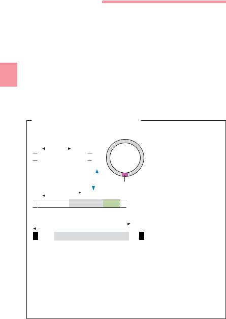
Fig. 3.20 a–d Explanation of the structures and abbreviations used in the text box “Details of Transposable DNA elements” below.
Details of Transposable DNA Elements
&Insertion sequences (IS elements, Fig. 3.20). These are the simplest transposable DNA sequences. They are terminated by identical, but reversed, sequences of 10–40 nucleotides known as inverted repeats (IR). They frame the segment that codes for the enzyme transposase. The target structures for this enzyme are the socalled direct repeats, nucleotide sequences comprising 5–9 bp that are duplicated in the integration process.
&Tn3 transposons (Fig. 3.20b). In addition to the transposase gene tnpA, they contain the regulator sequence tnpR and the res site to which resolvase must bind. Tn3-like transposons are duplicated in the transposition process, so that one copy remains at the original location and the other is integrated at the new location.
&Composite transposons (Fig. 3.20c). They consist of two IS elements framing a sequence of variable size that is not required for transposition, e.g., a resistance gene.
&Conjugative transposons (Fig. 3.20d). These genetic elements code in certain regions for factors that control the transfer (Tra) and transposition (Tn) processes. Conjugative transposons have been discovered mainly in Gram-positive cocci and Gram-negative anaerobes (Bacteroides).
174 3 General Bacteriology
Intercellular Mechanisms of Genetic Variability
Although bacteria have no sexual heredity in the strict sense, they do have mechanisms that allow for intercellular DNA transfer. These mechanisms, which involve a unilateral transfer of genetic information from a donor cell to a receptor cell, are subsumed under the term parasexuality.
3 Transformation
Transfer of “naked” DNA. In 1928, Griffith demonstrated that the ability to produce a certain type of capsule could be transferred between different pneumococci. Then Avery showed in 1944 that the transforming principle at work was DNA. This transformation process has been observed mainly in the genera Streptococcus, Neisseria, Helicobacter and Haemophilus.
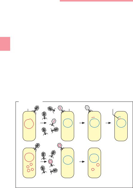
Transduction
Transfer of DNA from a donor to a receptor with the help of transport bacteriophages (Fig. 3.21).
Transduction |
|
|
Donor cell |
Receptor cell |
Recombination |
a |
|
|
b |
|
|
Fig. 3.21 |
Transduction of a chromosomal DNA sequence (a) and a plasmid (b). |
|