

Hart C. Anthony, Shears Paul. Color Atlas of Medical Microbiology.pdf
.pdfImmunological Test Methods 125
Western Blotting
2
Fig. 2.24 Antigen samples separated in a gel are transferred to nitrocellulose. Non-specific binding of the antibodies to the filter is then prevented with serum albumin or irrelevant proteins that do not cross-react with any of the antibodies used. Antibodies specific for the antigens being sought are then added. Once immune complexes have formed, the unbound antibodies are thoroughly washed away and the remaining bound antibodies are labeled using anti-immunoglobulin antibodies. These are in turn rendered visible by the autoradiographic procedure.
cubated in vitro with erythrocytes or antigenic particles. In all cases agglutination is detected using anti-Ig antibodies. Antigens can also be adsorbed to latex.
Complement Fixation Test (CFT)
CFT was formerly used to measure complement consumption by preformed antigen-antibody complexes. The unused complement is then detected by addition of a known amount of antibody-loaded erythrocytes. Should all of the erythrocytes be lysed, this indicates that no complement had been consumed and the CFT is negative. This method is no longer used very frequently, with the newer immunosorbent tests being preferred (RIA, ELISA, RAST, see below).
Direct and Indirect Immunofluorescence
Direct immunofluorescence. Immunofluorescence can be used for in-vivo detection of antibodies, complement, viruses, fungi, bacteria, or other im-

126 2 Basic Principles of Immunology
Hemagglutination
Erythrocyte |
Antigen |
antigen |
artificially fixed |
|
on erythrocyte |
2
|
|
Reciprocal serum dilution |
Control |
|
2 |
4 |
8 |
16 32 64 128 256 |
512 1024 pos. neg. |
Test serum a positive1 32
Test serum b negative
Test serum c positive 1/8  with prozone 1/2
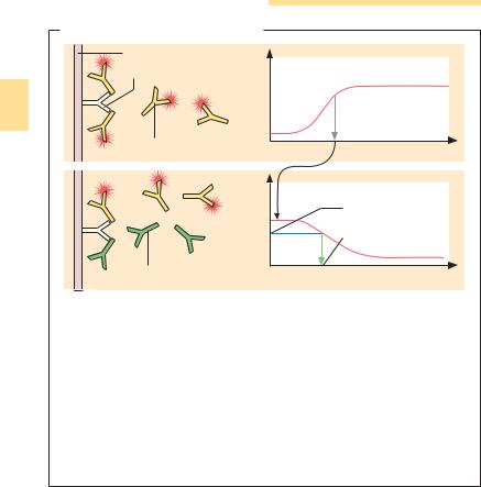
Fig. 2.25 The hemagglutination test is based on the principle that erythrocytes cross-linked by antibodies settle to the bottom of the microtiter plate wells in matlike aggregates (test sera a and c), whereas non-agglutinated erythrocytes collect at the lowest point of the wells to form a single “button” in the middle (test serum b). The test sera are first pipetted into the wells at the indicated dilutions, then the erythrocyte suspension is added. Non-specific agglutination is prevented by addition of an irrelevant protein. The test can be carried out using erythrocyte antigens (above left). Alternatively, other antigens can be fixed to the erythrocyte surface and the agglutination monitored (above right). The so-called “prozone” phenomenon results from non-specific blocking mechanisms present in sera which has not been sufficiently diluted.
mune factors present within patient cells and tissues. For this purpose tissue sections, or cell preparations, are treated with specific antibodies (anti-sera) which have been labeled with a fluorochrome (Fig. 2.26a). Antigen-antibody reactions can thus be detected using a fluorescence microscope. The fluorochrome absorbs light of a certain wavelength (e.g., UV light), and emits the light energy in the form of light at a different (visible) wavelength. The fluorochrome fluorescein isothiocyanate (FITC), which absorbs UV light and emits it as green light, is used most frequently (caution: bleaches out quickly!).

Immunological Test Methods 127
Antigen Detection Methods
2
Fig. 2.26 Immunofluorescence (a, b) is particularly suitable for the detection of antigens, or specific antibodies, fixed on plastic (solid phase) (ELISA) or present within a tissue section (immunohistology). For direct immunofluorescence (a) the specific primary antibody is labeled with a fluorochrome, or an enzyme (ELISA = enzyme-linked immunosorbent assay). The term indirect immunofluorescence is used when it is not the primary antibody being detected, but a secondary antibody which is directed against the unlabeled primary antibody and has also been labeled with a fluorochrome or enzyme (b). In most cases, this method achieves a certain degree of amplification. However, an even higher level of amplification can be achieved using preformed complexes of secondary antibody and enzyme (c). For the peroxidase method the detector enzyme is bound directly to the secondary antibody (peroxidase catalyzes a color reaction). In the biotin-avidin method the detector enzymes are coupled to either biotin or avidin.
Indirect immunofluorescence and enzyme histology. In this technique the specific or “first” antibody can be unlabeled. The antigen–antibody complexes that form are then detected using a labeled or “second” antibody, directed against the first antibody (Fig. 2.26b). Instead of fluorochromes, enzy- me-labeled antibodies are now frequently used for tissue sections. The enzyme catalyzes the formation of a color signal following addition of a previously colorless detector substance. This color precipitate allows the direct observation of signals using a light microscopic, and exhibits little bleaching.
Indirect immunofluorescence can be used for the qualitative and quantitative analysis of antibodies directed against particular microbial antigens, or self-tissue antigens, within a patients serum. In the quantitative test, the antigen is fixed in a well or to a tissue section on a slide. The patient sample is repeatedly diluted by a factor of two and added to the antigen or section then rendered visible with a labeled anti–antibody.
There are two main methods of amplifying the immunohistological color signal:

128 2 Basic Principles of Immunology
&The direct ’primary’ antibody, or the detected ’secondary’ antibody, is labeled with peroxidase. Following the antigen-antibody reaction, large preformed peroxidase-antiperoxidase complexes are added to the tissue section; these complexes can attach to the peroxidase-labeled antibodies, which are
2alreadyspecificallybound,thusamplifying thesignalconsiderably(Fig. 2.26c).
&Similarly, biotinylated antibodies can be used. The vitamin biotin is bound with strong affinity by avidin, a basic glycoprotein. Various colorants or enzymes coupled to avidin thus facilitate the color reactions. Such reactions can be amplified on the tissue section by adding preformed biotin-avidin-perox- idase complexes that bind to those biotin-coupled antibodies which have already been bound.
Radioimmunological and Enzyme Immunological Tests
Radioimmunoassay (RIA) and enzyme immunoassay (EIA), also known as ELISA (enzyme-linked immunosorbent assay) (Fig. 2.28), are now used very frequently to test for antigens and antibodies. All absorbency tests involve the fixation of antigens or antibodies to a plastic surface. The lower detection limit is a few nanograms. This method forms the basis of modern hepatitis serology, HIV tests, and tests for autoantibodies, lymphokines, cytokines, etc. All of these assays can be performed in a direct form (different sandwich combinations of antigen, antibody and anti-antibody, Fig. 2.27)
Fig. 2.27 For solid phase tests both the antigen and antibody are bound to a solid phase (e.g., plastic surface). Various methods are then used to detect any interaction between the antigen and antibody. In the direct test (a) an immobilized, unknown, antigen can be detected using a fluorescent-labeled antibody. If the immobilized antigen is known, this test method can also be used to detect an antibody bound to the antigen. In the sandwich method (b) a known antibody is immobilized. Detection of antibody-antigen binding is then performed using a second, labeled antibody which interacts with the antigen at a different site. The capture method (c) can be used to detect any antigen, for instance IgM antibodies. First, anti-IgM antibodies are immobilized, then serum containing IgM is added to them. The bound IgM can then bind a foreign antigen (e.g., a virus). The detection procedure next makes use of either the labeled foreign antigen or a specific, additionally labeled, antibody which binds to the bound antigen but not to the plastic bound antibody. In the competition or competitive inhibition test (d) antibodies are immobilized, and labeled antigens are then bound to them. An unlabeled (unknown) antigen is added, which competes with the labeled antigen. The level of interaction between the antibody and the unknown antigen is then determined by measuring attenuation of the signal. "

Immunological Test Methods 129
or as competition assays. Fig. 2.28 illustrates the quantitative IgE assay, Fig. 2.29 the procedure for detection of specific IgE in patient sera. Analogous procedures are used to detect specific antibody-binding cells or cytokine-releas- ing T cells (Fig. 2.30).
2
In-Vitro Cellular Immunity Reactions
Isolation of Lymphocytes
The methods used to measure cellular immunity are experimentally complex. The first step is to isolate human lymphocytes from blood, which can be achieved using Ficoll density gradient centrifugation. Certain lymphocyte
|
Basic Solid Phase Test Types |
|
|
|
Conjugate |
|
Conjugate |
|
|
|
Unknown antibody |
|
Unknown antigen |
|
Known antigen |
a |
Direct test |
|
|
|
Unknown antigen |
|
Conjugate |
|
|
(labeled antigen) |
|
|
|
|
|
|
Conjugate |
|
|
|
|
|
Unknown |
|
|
|
IgM antibody |
|
Known antibody |
|
|
|
|
|
Anti-IgM |
b |
Sandwich method |
c |
Capture method |
|
Known antibody |
|
Known antibody |
|
Labeled |
|
Unknown |
|
antigen |
|
|
|
|
antigen |
|
|
|
|
|
|
Strong signal |
|
Signal reduction |
|
|
|
Labeled antigen |
d |
Competitive test |
|
|
130 2 Basic Principles of Immunology
Radioimmunosorbent Test (RIST) |
|
||
|
Plastic surface |
cpm |
Plateau |
|
Anti-IgE |
||
|
|
(max. binding capacity) |
|
2 |
|
|
|
|
|
|
80% |
a |
Labeled IgE* |
|
Amount of labeled IgE* |
|
|
||
|
|
|
|
|
|
cpm |
Standard curve |
|
|
|
|
|
|
|
Radioactivity |
|
|
|
in test tube |
|
|
|
IgE in patient serum |
|
IgE from patient serum |
Amount of unlabeled IgE in IgE* test solution |
|
b |
|
||
|
|
|
|
Fig. 2.28 RIST is a competitive radioimmunoassay (RIA) used for quantitative measurement of antibodies of any given Ig class (in this example total IgE) within patient serum. Anti-IgE antibodies are allowed to adsorb to the solid phase (plastic surface). In the first instance, defined concentrations of radiolabeled IgE (IgE*) are used to determine the maximum binding capacity of these antibodies (a). The actual test (b) is then performed using the IgE* concentration determined to result in 80% saturation of the fixed antibodies: The IgE* test solution is added to the fixed anti-IgE antibodies and the patient serum is then added by pipette. The more IgE the serum contains, the more IgE* will be displaced by the patients antibodies, and the lower the radioactivity level will be in the test tube. The IgE concentration in the patient serum is then calculated based on a standard curve established previously by progressively “diluting” the IgE* test solution with unlabeled IgE.
populations can be coated with magnetic beads, or sheep erythrocytes loaded with specific antibodies, then purified using a magnet or a Ficoll gradient. The fluorescence-activated cell sorter (FACS, Fig. 2.31, p. 133) is now regularly used for this purpose. In this assay, monoclonal antibodies labeled with various fluorochromes directed against cell surface antigens (such as CD4, CD8), or against intracellular cytokines (which involves the use of detergents to increase the permeability of the cell membrane), are incubated with the isolated blood lymphocytes. Alternatively, antigen-specific T lymphocytes can be labeled with MHC class I or II plus peptide tetramers (see below). Following incubation, and several washing steps, the equipment identifies and counts the antibody-loaded lymphocytes, employing magnetic pulse sorting as required.
|
Immunological Test Methods 131 |
|
Radioallergosorbent Test (RAST) |
|
|
Addition |
Cellulose plate |
|
of antigen |
|
|
(solid phase) |
(solid phase) |
|
Wash |
Antigen |
2 |
|
|
|
Patient serum with |
IgE |
|
IgE? |
|
|
Wash |
Anti-IgE |
|
Addition of |
|
|
labeled Anti-IgE |
|
|
Fig. 2.29 This test is a highly sensitive detection method for the presence of specific IgE in patient serum. Antigen is bound covalently to a cellulose plate (solid phase). Any IgE in the serum that binds to the antigen is then detected using radiolabeled anti-IgE antibodies.
Tetramer test for detection of specific T cells (Fig. 2.32, p. 134): recombinant MHC class I antigen coupled to biotin, labeled with avidin, and correctly folded together with peptide and b2 microglobulin, forms tetramers; which are recognized by specific TCRs. Subsequent analysis of tetramer binding using FACS equipment is based on the color indicator of the avidin (fluorescein, phycoerythrin, etc.). Tetramers specific for MHC class II antigens plus
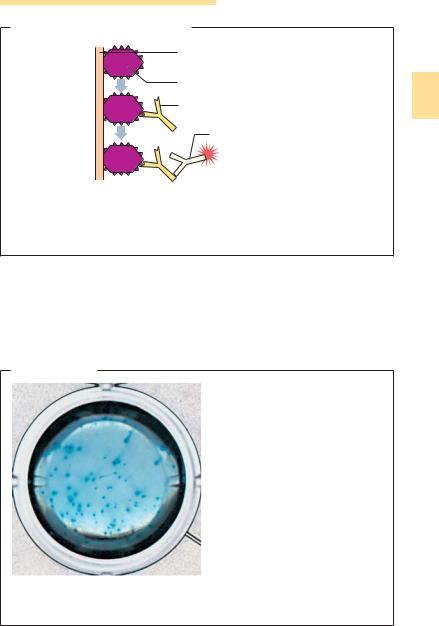
ELISPOT Assay
Fig. 2.30 In the ELISPOT assay antigens, or specific anti-IL antibodies, are applied to the plastic surface. It is then possible to determine the number of immune cells releasing antibodies specific for the applied antigen, or releasing interleukins that are recognized by the applied anti-IL antibodies. Following incubation at 37 8C, the immune complexes which form around these cells can be visualized using a covering agarose layer which includes an enzyme-coupled antibody. These enzymes catalyze a color reaction, resulting in the formation of color spots, each of which will correspond to a single cell producing the specific antibody or interleukin.

132 2 Basic Principles of Immunology
peptide can theoretically be used to assay specific CD4+ T cells, but are still difficult to manufacture. Using the tetramer test, specific T cells can be detected directly from blood or lymphoid organs. Histological applications are feasible, but still difficult.
2
Lymphocyte Function Tests
Certain functions of isolated lymphocyte populations can be determined by a number of methods:
&Determination of the number of cells producing antibodies, e.g., the hemolytic plaque assay in which antibody production is tested by adding antigencoupled erythrocytes. In the vicinity of antibody-secreting cells, the erythrocytes are covered with antibodies and can be lysed by addition of complement. Today, ELISA methods are more often used than erythrocytes (ELISPOT).
&ELISPOT ASSAY: used to measure antibody-producing, or IL-releasing, lymphocytes. The antigen or anti-IL antibody is fixed on a plastic surface. Lymphocytes are then placed over this, within a thin layer of agar medium. When the cells are incubated at 37 8C, they may secrete the antibodies or IL recognized by the corresponding test substances. After a certain period of time, the cell layer is shaken off and the preparation is thoroughly washed. The bound material can then be developed using an overlaid semisolid agar, as for the ELISA method. The enzyme reaction generates spots of color, each of which corresponds to a cell, and which can be counted (Fig. 2.30).
&Measurement of the release capacity of cytokines, or detection of mRNA, is also possible with the ELISPOT assay.
&Lymphocyte stimulation assay: isolated lymphocytes are incubated with antigen in culture medium. Measuring the 3H-thymidine incorporation,
Fig. 2.31 This device analyzes cells by means of fluorescent-labeled antibodies directed against cell surface antigens – or for permeabilized cells, directed against internal cell antigens. In the example shown, peripheral blood lymphocytes (PBL) are incubated with monoclonal antibodies specific for CD4 or CD8, resulting in the distribution of fluorescence intensity as indicated in a. In b the labeling of different cell populations with anti-CD4 or anti-CD8 is shown. By this means, the percentages of the subpopulations in the total population can be determined. The fluor- escence-activated cell sorter shown in (c) makes use of this data. By means of vibration, the cell stream is broken up into fine droplets which, depending on the fluorescence and sorting settings used, are charged just before they are separated and ideally contain one cell each. Certain parameters are measured for each cell with the help of a laser beam, where-upon the droplets are deflected into the intended containers by the + and – plate fields. "

Immunological Test Methods 133
interleukin release, or a pH transition, can determine whether antigen-spe- cific lymphocytes are present or whether polyclonal T-cell responses (concanavalin [ConA], phytohemagglutinin [PHA]) or B-cell responses (lipopolysaccharide [LPS], pokeweed mitogen [PMA]) were induced.
& Mixed lymphocyte reactions are used to measure alloreactivity (prolifera- |
2 |
tion, cytotoxicity), mainly between recipients and donors of organ or bone |
|
Fluorescence-Activated Cell Sorter (FACS) |
|
|
||||
|
Histogram |
|
Two-channel analysis |
|||
|
Anti-CD4 |
|
|
of peripheral blood lymphocytes |
||
Numberofcells |
|
|
|
40– 60% |
<1% |
|
Anti-CD8 |
|
|
|
|||
|
|
|
CD4 |
|
|
|
a |
Fluorescence intensity (log 10) |
Anti- |
|
|
||
|
|
|
|
|
|
|
|
|
|
Cells |
|
15– 25% |
20– 30% |
|
|
|
|
|
||
|
Buffer |
|
|
b |
Anti-CD8 |
|
|
|
|
|
|
||
|
|
|
|
|
|
|
|
solution |
|
|
|
|
|
|
|
|
|
|
|
Red |
|
|
|
|
|
|
fluorescence |
|
Laser |
|
|
Prism |
|
|
|
|
|
|
|
|
Green |
|
|
|
|
|
|
fluorescence |
|
Application |
|
|
|
90° deflection |
|
|
of a charge |
|
|
Forward |
(granulation) |
|
|
|
|
|
|
|
|
|
|
|
|
scattered light |
|
|
|
Polarized field |
– |
+ |
(cell size) |
|
|
|
for droplet |
|
|
|
||
|
deflection |
|
|
|
|
|
c |
|
|
|
|
|
|

134 2 Basic Principles of Immunology
Tetramer Assay
Labeling of avidin |
Avidin with 4 |
|
|
binding sites for |
|
||
with fluorescein, |
|
||
biotin |
T cell |
||
phycoerythrin, etc. |
|||
|
|||
|
|
2
Specific peptide MHC class I β2-microglobulin Biotin
T cell
Fig. 2.32 The tetramer assay is an antigen-specific binding test for living T cells. Complexes comprising biotin-coupled MHC class I heavy chains, b2 microglobulin, and specific peptide are properly folded, washed, then bound to avidin (which contains four binding sites for biotin). The resulting tetrameric complexes are then incubated with a population of T cells. Those T cells expressing the appropriate T-cell receptor will bind to two or three of the exposed MHC class I-peptide complexes present on each tetramer. Labeling of the avidin component with fluorescein, phycoerythrin, or other fluorescent substances then permits FACS analysis of tertramer binding T cells.
marrow transplants. This test is based on the principle that T lymphocytes are stimulated to proliferate by nonself MHC class I or II antigens and to develop into cytotoxic T cells directed against class I.
&Chromium release assay measures cytolytic activity, mainly by CD8+ Tcells, directed against allogeneic, virus-infected, or peptide-loaded target cells. The target cells are incubated with 51Cr which the cells incorporate. They are then cultivated with effector cells for 4–6 hours. When the target cells are lysed chromium is released into the culture medium, following which it can be quantitatively measured.
&Assay of intracellular cytokines. Following a brief stimulatory culture (six hours), the cells are rendered permeable using a mild detergent so that specifically labeled antibodies can diffuse into the cells. Labeled cells
can then be analyzed by FACS equipment (or by a microscope).