

Hart C. Anthony, Shears Paul. Color Atlas of Medical Microbiology.pdf
.pdfTransplantation Immunity 115
basic protein in multiple sclerosis, against collagen determinants in polyarthritis, and against islet cell components in diabetes.
Transplantation Immunity
2
& Transplant rejection within the same species is largely a consequence of MHC-restricted T-cell recognition of foreign MHC antigens. Interspecies rejection is additionally contributed to by antibodies, and intolerance between complement activation mechanisms. Methods for reducing, or preventing, rejection include general immunosuppression, tolerance induction by means of cell chimerism, and sequestering of the transplanted cells or organ. &
The strong transplantation antigens are encoded within the MHC complex (see p. 58ff.), whilst the weak antigens constitute the MHC-presented allelic differences of non MHC-encoded host proteins or peptides. It is possible to differentiate between the host-versus-graft (HVG) reaction of the recipient against a genetically foreign tissue or organ, and the graft-versus-host (GVH) reaction.
The GVH reaction. This type of reaction results when immunologically responsive donor T cells are transferred to an allogeneic recipient who is unable to reject them (e.g., following a bone marrow transplant into an immuno-in- competent or immuno-suppressed recipient). The targets against which the transplanted T cells generate an immune response include the MHC class I and II molecules of the recipient. The recipient’s transplantation antigens also present allelic variants of recipient self-peptides, which can be recognized by donor T cells as weak transplantation antigens when presented by common MHC alleles (it is conceivable that strong recipient transplantation antigens could be accepted and processed by donor APCs, however even if this did occur it would be of limited functional consequence as they would not be presented by the recipient APC in the correct antigen configuration). Weak histocompatibility antigens—for instance those peptide variants recognized as nonself when presented in combination with essentially histocompatible MHC molecules—play a more significant role in bone marrow transplants. The existence, and pathological role, of weak transplantation antigens has only been demonstrated in completely histocompatible siblings or within inbred animal strains with identical MHC. The wide variety of alloreactive T cells can be explained by cross-reactivity, as well as by the enormous number of different combinations of MHC molecules and cellular peptides. It must be emphasized that allogeneic MHC antigens on APCs and lymphocytes (socalled passenger lymphocytes) derived from the donor organ are particularly immunogenic since they express high levels of antigens and can traffic to

116 2 Basic Principles of Immunology
secondary lymphatic organs. Indeed the same foreign transplantation antigens are hardly immunogenic when expressed on fibroblasts or on epithelial or neuroendocrine cells, unless these cells are able to reach local lymphoid tissue.
2 To avoid a GVH reaction in immunoincompetent or suppressed bone marrow recipients, immunocompetent T cells must first be eliminated from the transplanted bone marrow. This can be achieved by using anti-T-cell antibodies, anti-lymphocyte antisera, and complement or magnetic bead cell-se- paration techniques. However, it is noteworthy that complete elimination of mature T cells leads to a reduction in the acceptance rate for bone marrow transplants, and that it may also weaken the anti-tumor effect of the transplant (desirable in leukemia). It seems that the small number of T cells transplanted with the bone marrow can mediate a subclinical GVH reaction, thus preventing rejection of the transplant but retaining the ability to destroy the recipient’s leukemia cells and preventing tumor re-emergence.
Bone Marrow Transplants Today
&Reconstitution of immune defects involving B and T cells
&Reconstitution of other lymphohematopoietic defects
&Gene therapy via insertion of genes into lymphohematopoietic stem cells
&Leukemia therapy with lethal elimination of tumor cells and reconstitution with histocompatible, purified stem cells, either autologous or allogenic.
HVG reactions, that is immune responses of the recipient against transplanted cells or organs, are not generated in autotransplants (for instance transplantation of skin from one part of the body to another on the same individual). This also applies to transplants between monozygotic twins or genetically identical animals (syngeneic transplants). However, transplants between non-related or non-inbred animals of the same species (allogeneic transplants), and transplants between individuals of different species (xenogeneic transplants) are immunologically rejected. Because T cells recognition is subject to MHC restriction, cellular rejection within a species is even more pronounced than between different species, although the latter procedure involves other transplantation complications. These include the occurrence of natural cross-reactive antibodies, and a lack of complement inactivation by anti-complement factors (which are often species-incompatible and therefore absent in xenogeneic transplants), which together often results in hyperacute rejection within minutes, hours, or a few days—that is before any specific immune responses can even be induced.
Three types of transplant rejection have been characterized:
& Hyperacute rejection of vascularized transplants, occurring within minutes to hours and resulting from preformed recipient antibodies reacting

Immune Defects and Immune Response Modulation 117
against antigens present on the donor endothelium, resulting in coagulation, thromboses, and infarctions with extensive necrosis.
& Acute rejection, occurring within days or weeks. This is accompanied by a |
|
perivascular and prominent occurrence of T lymphocyte infiltrates. Acute re- |
2 |
jection can be prevented by immunosuppression. |
& Chronic rejection, occurring within months to years. This is caused by low-level chronic T-cell responses, and can be mediated by cellular and humoral mechanisms. This can include obliterative vascular intima proliferation, vasculitis, toxic, and immune complex glomerulonephritis.
Antigenicity and Immunogenicity of MHC in Organ Transplants
A thyroid gland from donor “a,” freshly transplanted under the renal capsule of an MHC (H-2)-incompatible recipient mouse “b” is acutely rejected (within seven to nine days). If the organ is treated in such a way as to kill the migratory APCs and leukocytes before it is transplanted, then transplant “a” will be accepted by recipient “b” (often permanently). However, should fresh spleen cells (APCs) from donor “a” be transferred by infusion 100 days later into the recipient “b,” the previously accepted transplant “a,” can sometimes be acutely rejected (i.e., within 10 days).
This experiment demonstrates that it is not the MHC antigens per se that are potently immunogenic, but rather that they only show this immunogenicity when they are located on cells capable of migration to local lymph nodes. Methods of implanting foreign tissue cells or small organs strictly extralymphatically, without inducing immune responses, are currently undergoing clinical trials (i.e., with islet cells in diabetes and neuronal cells in parkinsons disease).
Methods of measurement. The main methods used for follow-up analysis of HVG and GVH reactions are biopsies and histological evaluation, evaluation of blood cells and in-vitro mixed lymphocyte reactions (p. 132).
Immune Defects and Immune Response
Modulation
& Immune defects are frequently acquired by therapy or viral infections, or as a consequence of advanced age. In rare cases immune defects can also result from congenital defects, these include severe combined immunodeficiency’s (SCID) or transient partial immune defects (mainly involving IgA responses). Immunomodulation can be attempted using interleukins or monoclonal antibodies directed against lymphocyte surface molecules or antigenic peptides. Immunostimulation is achieved using adjuvants or
|
|
118 2 Basic Principles of Immunology |
|
|
|
|
|
|
|
|
|
|
|
the genetically engineered insertion of costimulatory molecules into tumor |
|
|
|
cells. Immunosuppression can be induced globally using drugs, or specifi- |
|
|
|
cally using antibodies, interleukins or soluble interleukin receptors; this can |
|
|
|
also be achieved by means of tolerance induction with proteins, peptides, or |
|
2 |
|
cell chimerism. |
& |
|
|
|
|
Immune Defects
The most important and frequent immune defects are acquired, e.g., iatrogenic (cytostatics, cortisone, irradiation, etc.), age-induced, or the result of viral infections (above all HIV). Congenital defects are rare; examples include Bruton’s X-chromosome-linked B-cell defect, thymic hypoplasia (DiGeorge), and combined T- and B-cell deficiency resulting from MHC defects (bare lymphocyte syndrome) or from enzyme defects (adenosine deaminase [ADA] deficiency or purine nucleoside phosphorylase [PNP] deficiency). These defects can also be repaired by reconstitution (thymic transplants), or in some cases through the use of stem cells (gene therapy; one of the very first successful gene therapies was the treatment of ADA deficiency). More frequent congenital defects involve selective deficiencies, for example a relative-to-absolute IgA deficiency, normally being more prominent in infants than later in life. Children with such deficiencies are more susceptible to infection with Haemophilus influenzae, pneumococci, and meningococci.
General consequences of immune defects include recurring and unusual infections, eczemas, and diarrhea.
Immunoregulation
This area of immunology is difficult to define and remains elusive. Antigens represent the most important positive regulator of immunity; since there is simply no immune stimulation when antigens have been eliminated or are absent. Other important regulators include interferon gamma (IFNc) for TH1 responses, and IL-4 for TH2 responses. Further IL-dependent regulatory functions are in the process of being defined. The existence of specific CD8+ T suppressor cells, capable of downregulating immune responses, has been postulated and their role was assumed to be that of counteracting the inflammatory CD4+ T cell response. However, to date there has been no convincing proof of their existence. The term CD8+ T suppressor cells, which is used frequently, is therefore misleading and inaccurate. In relatively rare cases, cyto-

Immune Defects and Immune Response Modulation 119
toxic CD8+ T cells do exercise a regulatory effect by lysing infected APCs or B cells (see also p. 106). It is unclear whether CD4+ T cells could have similar effects. Regulation via idiotypic/anti-idiotypic antibody networks (i.e., antibodies directed against the ABS of other antibodies), or anti-TCR networks,
have also been postulated—but remain hypothetical. Although attractive 2 hypothesis, for most cases such regulatory pathways have only proved disappointing theoretical concepts, and as such should no longer be employed in
the explanation of immunoregulation. In isolated cases, anti-idiotypic, or anti-TCR peptide-specific feedback, mechanisms can be modeled under forced experimental conditions. However such conditions probably fail to model normal situations, therefore they cannot accurately indicate whether these feedback mechanisms have a role in regulating the immune system as a whole.
Immunostimulation
The aim of immunological treatment of infections and tumors is to enhance immune responsiveness via the use of thymic hormones (thymopoietin, pentapeptides), leukocyte extracts, or interferons. Derivatives or synthetic analogs of microorganisms such as BCG, components of Corynebacterium parvum and peptidoglycans (e.g., muramyl peptide), or oligonucleic acids (CpG), are used as adjuvants. Components of streptococci and Streptomyces, eluates and fractions of bacterial mixtures, and the related synthetic substance levamisole are also used. The role of Toll-like receptors in these adjuvant effects is becoming increasingly understood, with a major role of these molecules being to link non-specific innate resistance to specific immunity. .
Recently developed immune therapy strategies aim to improve antigen presentation. For instance interleukins, or costimulatory molecules such as B7 or CD40, have been inserted into tumor cells by means of transfection. Hybrid antibodies have been constructed in an attempt to improve antigen recognition and phagocytosis (one such example is the coupling of an antiCD3 antibody with tumor antigen-specific antibodies). Other ideas tested successfully in model experiments include systemic treatment with interleukins (this presents with frequent toxicity problems) or targeted insertion of GM-CSF, TNF, or IL-2. Alternatively, the production of IFNc or IFNb by cells, or the use of molecules capable of polyclonal T- and B-cell stimulation has been employed. This concept utilizes local chronic or acute infections with the aim of achieving inflammation surrounding, or direct infection of, tumor cells resulting in their cytolytic destruction. Such concepts have also been used to force phagocytosis and uptake of antigens by APCs with the aim of inducing or enhancing tumor immunity (e.g., BCG infections in bladder carcinoma treatment).

120 2 Basic Principles of Immunology
Immunosuppression
Various methods are employed to inhibit, or suppress, the immune response:
& Generalized immunosuppression; glucocorticoids (inhibition of inflam-
2matory cells), cytostatic drugs (endoxan, DNA alkylating agents, methotrexate, antimetabolites), and more specific immunosuppressants, e.g., cyclosporine A, FK506, rapamycin (inhibition of signal transduction in T cells, see Fig. 2.11, p. 73).
&Immunosuppression by antibodies, soluble cytokine receptors, deletion of T cells or T-cell sub-populations (anti-CD4, anti-CD8, anti-CD3, anti-Thy1, etc.). Administration of monoclonal antibodies directed against adhesion molecules and accessory molecules or cytokines and cytokine receptors. Administration of soluble cytokine receptors, or soluble CTLA4, in order to block B7- 1 and B7-2 (important costimulators, see p. 71ff.).
&Specific tolerance induction or “negative immunization.” Massive and depletive T-cell activation brought about by systemic administration of large amounts of peptides, proteins (risk of immunopathology), or cells (chimerism).
&Complete neutralization and elimination of the antigen with the purpose of preventing induction of an antibody response. Example; rhesus prophylaxis with hyperimmune serum.
Adaptive Immunotherapy
This involves in-vitro antigen stimulation, and consequent proliferation, of patient T-cell effector clones or populations (CD8+ T cells or less specific lymphokine-activated killer cells, LAK cells), followed by transfusion of these cells back into the patient. This method is sometimes used as a means of limiting cytomegaly or Epstein-Barr virus infection of bone marrow recipients. The LAK cells also include less specific NK-like cells, which can be expanded with IL-2 in the absence of antigen stimulation.
Toxic antibodies are monoclonal antibodies to which toxins have been coupled. These are used as specific toxin transporters, administered directly, or with liposomes bearing anchored antibodies and containing a toxin or cytostatic drug.

Immunological Test Methods 121
Immunological Test Methods
Antigen and Antibody Assays
2
Immunoprecipitation in Liquids and Gels
Immunoprecipitate. Maximum precipitation results when both reaction partners are present in an approximately equivalent ratio (Fig. 2.20). In antibody excess, or antigen excess, the amount of precipitate is considerably reduced.
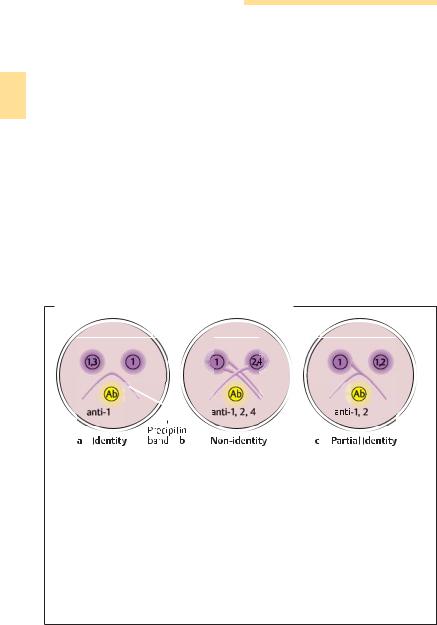
Double diffusion according to Ouchterlony. This technique allows for a qualitative evaluation of whether certain antibodies or antigens are present or not, plus determination of the degree of relationship between antibodies and antigens. It also provides information on whether different antigenic de-
Immunoprecipitation
Fig. 2.20 To identify the
unknown antigen, a known
specific antibody is added to an antigen mixture which is radio-, or other-
wise-, labeled. The immune
complexes are precipitated
with the help of co-precipi-
tating reagents (e.g., anti-
immunoglobulin antibo-
dies). The precipitate is
thoroughly washed to re-
move unbound antigen, then dispersed into solution once again (e.g., in
SDS), after which the components are separated using SDS polyacrylamide gel electrophoresis (SDS-
PAGE). The labeled antigen
is then rendered visible by
means of autoradiography.
122 2 Basic Principles of Immunology
terminants are localized on the same, or on different, antigens; or whether different antibodies can bind to the same antigen (Fig. 2.21).
Radial immunodiffusion according to Mancini. This is a quantitative antigen assay based on a predetermined standard curve (Fig. 2.22).
2Nephelometry. This method measures the amount of light scatter as a quantification of precipitation turbidity.
Immunoprecipitation Combined with Electrophoresis. Antigens are separated in an agarose gel by applying an electric current. The antibodies react by migrating in the gel, either without an electric field, or simultaneously within the electric field; and either in the same dimension as the antigens or in a second vertical step (“rocket” electrophoresis).
Immunoelectrophoresis according to Grabar and Williams. In the first instance serum proteins are electrophoretically separated within a thin agarose gel layer. A trough is then cut into the agar, next to the separated sample and parallel to the direction of migration along the entire migration distance, and anti-serum is applied to the trough. The antibodies diffuse into the gel, and precipitation lines are formed wherever they encounter their antigens. The
Double Diffusion According to Ouchterlony
Fig. 2.21 This technique facilitates assignment of antigens (violet) to a certain test antibody (yellow), or vice versa. The antigens and antibodies are pipetted into troughs within the gel and diffuse through this medium (the numbers designate the epitopes present). Where they meet lines of precipitation (known as precipitin bands) develop, indicating immune complex formation. a The antibodies precipitate identical epitopes (epitope 1) of both antigens, resulting in formation of precipitin bands which flow together to form an arch, mutually inhibiting their migration. In b, three independent precipitin bands form, indicating that the antibodies differentiate three different epitopes on three different antigens. c Epitope 1 of both antigen samples forms precipitin bands which flow together. Anti-2 migrates beyond the line of confluence into the area in which it precipitates with free antigen 1, 2 and forms a spur.

Immunological Test Methods 123
Radial Immunodiffusion According to Mancini
Gel containing Ab
Ag |
Ag |
Ag |
Ag |
2 |
|
|
|
|
|
|
Precipitin ring |
|
|
|
Standard curve |
|
|
|
|
2
(Ring diameter)
0 |
10 |
25 |
50 |
100 |
|
|
Antigen concentration |
|
|
Fig. 2.22 Quantitative assay of an antigen using a monospecific anti-serum which is mixed with agar and poured into a plate. The antigen is then diluted to different concentrations, and pipetted into wells that have been previously punched into the plate. Antigen-antibody complexes precipitate in the form of a ring around the well, the diameter of which is proportional to the antigen concentration. The result is a standard curve from which unknown test antigens can be quantified. Analogously, antibodies can also be quantified by mixing antigens into the gel.
precipitate can then be stained and evaluated. This older method is still used to identify paraproteins, monoclonal immunoglobulins, etc. (Fig. 2.23).
Electrophoresis plus antibody reaction: Western blotting. This method involves electrophoresis of proteins in a gel, coupled with detection by specific antibodies. The separated proteins are transferred to nitrocellulose, where they are identified with the help of specific antibodies (Fig. 2.24). Polyclonal sera is normally used for this purpose as monoclonal antibodies only rarely bind to denaturated and separated proteins.
Agglutination Reaction
Antibodies can agglutinate antigen-loaded particles (Fig. 2.25), whilst antigens can agglutinate antibody-loaded particles. Application: agglutination of bacteria or erythrocytes (e.g., blood group tests).

124 2 Basic Principles of Immunology
Immunoelectrophoresis According to Grabar and Williams
Undiluted serum
2 |
|
|
|
|
|
_ |
|
+ |
|
|
|
||
|
|
Albumin |
|
Antihuman serum |
|
|
|
|
|
|
|
||
|
|
|
|
|
|
|
1 : 6 |
α- |
β- |
γ-globulins |
|
||
|
|
|
|
|
|
|
|
|
|
|
|
|
|
|
|
Undiluted |
|
|
|
|
|
|
|
|
IgM |
IgA |
IgG |
Anti-IgG, anti-IgA, anti-IgM
IgG
1 : 6
Fig. 2.23 Serum is separated within agarose by an electric field, and rendered visible with anti-serum directed against human serum (above), or with selected specific antibodies (below).
&Indirect hemagglutination. An antigen is fixed on the surface of erythrocytes and the antigen-loaded erythrocytes are then agglutinated using specific antibodies.
&Hemagglutination inhibition test. The ability of a sample containing antigen to inhibit hemagglutination between antigen-loaded erythrocytes and antiserum is measured. This test is frequently used to quantify antibodies againsthemagglutinatingviruses(mainlyinfluenzaandparainfluenzaviruses).
&Antiglobulin tests according to Coombs. The direct Coombs test determines antibody binding directly to erythrocytes (e.g., anti-Rh antibodies agglutinate Rh+ erythrocytes of neonates). The indirect Coombs test is suitable for detection of antibodies that have already bound to the Rh+ erythrocytes of newborns (second pregnancy or sensitized mother), or which have been in-