

Иностранный язык / Єрьомкіна Г.Г. Англійська мова _ навч. посібник _ Г.Г. Єрьомкіна, А.Н. Некрасова, Є.Г. Кривда та ін. - О._ ОДМУ, 2009. - 164 с
.pdfThe uterus is a hollow, pear-shaped organ situated between the bladder and rectum. It is supported by heavy ligaments to keep it in place. It is a muscular and elastic organ which expands greatly during pregnancy and contracts to expel the baby at birth. It nourishes and protects the fetus during pregnancy.
The vagina is an elastic canal located behind the urethra. Its lower end opens to the exterior of the body. Its upper end extends to the cervix, the lower part of the uterus. The vagina serves as an outlet for secretions of the uterus and for the baby to pass through at birth.
There are a few additional terms which you must know:
Ovulation is the process of casting off an ovum from the ovary. This occurs midway between menstrual periods. The ovum is picked up by the end of one of the fallopian tubes and is carried to the uterus.
Menstruation is the process of casting off the unfertilized ovum from the uterus along with a bloody excretion.
Pregnancy is a normal condition in which a woman has a fertilized ovum within her.
Menopause is the time when the menstrual cycle stops.
Corpus luteum is a yellow mass in the ovary which secretes the female hormone, progesterone, and induces the final premenstrual changes in the uterus.
Estrogen is the hormone produced by the Graafian follicle as it matures.
MALE ORGANS
The organs of reproduction in the male are especially adapted to transport the male reproductive cells to the female.
The testes, two glands which lie in a sac (scrotum) behind the penis and below the groin, correspond in function to the female ovaries. The testes manufacture the male reproductive cells, spermatozoa. These spermatozoa are discharged by the testes into a fluid (seminal fluid) and are carried through the urethra. The urethra serves two purposes: as a channel through which urine is voided from the bladder, and as a passageway for the spermatozoa. The urethra extends from the bladder inside the body through the penis outside the body. The male hormones are called androgens. One of these androgens, testosterone, is responsible for the male characteristics.
It is through the union of the spermatozoa of the male and the ovum of the female (zygote) that a new life begins.
DISORDERS OF THE
REPRODUCTIVE SYSTEM
Objective:
• To become familiar with some of the disorders of the reproductive system
FEMALE REPRODUCTIVE SYSTEM
Carcinoma is a cancer which may occur in the breasts or uterus.
Dysmenorrhea is a term used for painful menstruation.
Endocervicitis is inflammation in the canal of the cervix.
Fibroid tumors are fibrous tumors in the uterus. They are usually benign.
Hysterectomy is the surgical removal of the uterus.
Mastectomy is the surgical removal of the breast.
Retroversion is the backward displacement of the uterus.
Salpingitis is inflammation of the fallopian tubes.
Sterility is the inability to reproduce. It may occur in either sex.
MALE REPRODUCTIVE SYSTEM
Epididymitis is the painful swelling in groin and scrotum due to infection.
Prostatitis is an inflammation of the prostate gland. By pressing on the bladder, the prostate gland causes frequent, painful urination or, if pressure on the urethra is severe, may cause urine to be retained.
Prostatectomy is the surgical removal of all or part of the prostate gland.
Orchitis is the inflammation of a testis which may be a complication of mumps, influenza, etc. Symptoms are the swelling of scrotum, accompanied by high temperature.
CHECK YOURSELF
1.Lactation refers to that time in the life of a woman when ... .
a) fertilization takes place b) milk is secreted
c) menstruation stops d) menstruation begins e) during menstruation
2.Carcinoma is ... .
a)a malignant tumor
b)a benign tumor
c)an inflammation
d)a cyst
e)an abscess
29
3.The menopause refers to the time when ...
a) pregnancy begins b) menstruation stops
c) secretion of milk stops d) birth takes place
e) menses begin
4.Genetics is ... .
a)the science of reproduction
b)a condition of pregnancy
c)the science of heredity
d)the study of fertilization
e)inheritance
5.The unborn baby in the uterus is called the ...
a) embryo b) fetus c) gene d) egg
e) newborn
6.The union of the ovum and sperm cell is ...
a) reproduction b) delivery
c) pregnancy d) fertilization e) birth
7.… are running from the ovaries to the uterus through which a female reproductive cell travels.
a) ureter b) urethra
c) fallopian tubes d) windpipe
e) cervix
8.Surgical removal of the uterus:
a)mastectomy
b)hysterectomy
c)appendectomy
d)endometritis
e)hysteroscopy
9.Surgical removal of the breast: a) mastectomy
b) hysterectomy c) appendectomy d) endometritis e) hysteroscopy
10.Inflammation of lining of the uterus: a) leucorrhea
b) salpingitis
c) dysmenorrhea d) endometritis e) endometriosis
11.Conception:
a)pregnancy
b)reproduction
c)inflammation
d)fertilization
e)delivery
12.Female sex cell: a) egg
b) sperm c) ovum d) embryo e) fetus
13.Male sex cell: a) egg
b) sperm c) ovum d) embryo e) fetus
14.What disease is not related to females? a) mastitis
b) orchitis c) salpingitis
d) endocervicitis e) endometritis
15.What disease is not related to males? a) prostatitis
b) orchitis c) salpingitis
d) epididymitis e) impotence
16.The process of casting off an ovum from the ovary is called ... .
a) fertilization b) ovulation
c) menstruation d) pregnancy e) coupling
17.The vagina is located ... .
a)over the anus
b)in front of the uterus
c)behind the urethra
d)in the uterus
e)behind the anus
18.The male hormones are called ... .
a) estrogens b) androgens
c) progesterone d) thyroxin
e) gene
19.The female hormones are called ... .
a) estrogens b) androgens
c) progesterone d) thyroxin
e) gene
20.Fertilization takes place in the ... .
a) uterus b) ovary c) vagina
d) fallopian tube e) cervix
30

Lesson 7
WHY ARE WE WHAT WE ARE?
INTRODUCTION |
PITUITARY GLAND |
Objective:
• To learn which glands make up the endocrine system and how they affect the body activities
The endocrine glands are organized groups of cells which draw upon materials from the blood or lymph to make new compounds called hormones. These secretions are picked up by the blood as the circulation passes through the gland. The secretions are then transported to all areas of the body where they have a special influence on cells, tissues, organs and systems. Endocrine glands are also called ductless glands and glands of internal secretion because their secretions go directly into the bloodstream.
A few of the glands manufacture two secretions: an internal secretion and an external secretion. Such a gland is the pancreas whose internal secretion, insulin, is discharged directly into the bloodstream and whose external secretion is discharged through the pancreatic duct into the small intestine where it aids the digestive process. Pancreatic juice, therefore, is an external secretion because it discharges its secretion through a duct into the small intestines, instead of directly into the bloodstream (Fig. 13).
There are these six important endocrine glands or groups of glands in the body:
—The pituitary gland in the skull
—The thyroid gland in the neck
—The parathyroid glands near the thyroid
gland
—The pancreas in back of the stomach
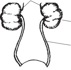
—The two adrenal glands, one over each kidney
—The gonads, or sex glands
Objective:
• To learn the location and functions of the pituitary gland
Each endocrine gland has specific functions to perform. Any disturbance in the functioning of these glands may cause changes in the appearance or functioning of the body. Sometimes both conditions arise.
The pituitary gland is located at the base of the brain. It is called the master gland because it secretes several different hormones into the bloodstream which affect other endocrine glands. For instance, the activity of the thyroid glands and the gonads is affected by the amount of hormone secretion discharged into the bloodstream by the pituitary gland.
pineal pituitary
parathyroids thyroid
thymus
adrenal
islands of Langerhans
gonads
Fig. 13. Location of the endocrine glands
31
The pituitary gland is responsible for the growth of the long bones; therefore, it controls the height of the individual. Circus giants are frequently the result of overgrowth of the long bones due to oversecretion of the pituitary gland. The gland influences the organs of reproduction. Its secretions are essential to pregnancy and lactation. It is responsible for maintaining the water balance of the body.
It affects the bodily use of starches and sugars because of its control over the formation of insulin in the pancreas.
It secretes adenocorticotropic hormone (ACTH), one of the hormones used in the treatment of arthritis.
The pituitary gland, therefore, is responsible for many of the secretions which help to keep us well-balanced individuals.
THYROID AND PARATHYROID GLANDS
Objective:
• To learn the important functions of the thyroid and parathyroid glands
The thyroid gland is located in the front middle portion of the neck on either side of the windpipe. It has two connected lobes. The chief function of the thyroid gland is to regulate the body metabolism. Metabolism is the rate at which the body functions take place. The gland helps to regulate rate of physical growth, mental development, sexual maturity, and the distribution and exchange of water and salts in the body. It can speed up or slow down the body’s activities as needed. Iodine is stored in the thyroid gland because it is essential for the manufacture of its hormone, thyroxin.
The parathyroid glands, usually four in number, are tiny glands with the size of a grain of rice attached to the posterior surface of the thyroid lobes. Only one function of these glands is known. They control the use of calcium and phosphorus in the body. The thymus gland is located under the breastbone or the sternum. It is fairly large during childhood but begins to disappear at puberty. Very little is known about the gland, but it is thought to be associated with growth and maturity.
FUNCTIONS OF ADRENAL GLANDS AND GONADS
Objective:
• To learn the location and functions of the adrenals and gonads
The adrenal glands lie on top of each kidney. The secretions of the adrenal glands per-
form several functions. Two of these functions are: (1) to release extra energy to help the body meet physical emergencies, and (2) to control the salt and water usage by the body. The cortex or outer part of the gland secretes a compound from which cortisone, used in the treatment of rheumatoid arthritis, is prepared. The medulla, or inner portion, secretes adrenalin, a powerful heart stimulant (Fig. 14).
The gonads, or sex organs, include the ovaries in the female and the testes in the male. The gonads are organs of both internal and external secretion. The ovaries are located in the pelvic cavity, one on either side of the uterus. The testes are located in the scrotum.
The secretion of the ovaries, estrogen and progesterone, are necessary for the normal female appearance, ovulation and characteristics. The secretion of the testes, testosterone, is essential to the development of the male sex characteristics. The gonads are responsible for fertility and reproduction of both sexes.
THE PANCREAS
Objective:
• To learn the location and endocrine functions of the pancreas
The pancreas is an organ which is located behind the stomach. The gland cells of the pancreas are concerned with the production of pancreatic juice, a digestive juice. The islet cells secrete the hormone, insulin. Thus the pancreas is a gland of both external secretion and internal secretion.
The islet cells are groups of pale cells distributed throughout the pancreas. These cells have been named the islands of Langerhans for the doctor who discovered them. Their function is
adrenal
kidney
ureter
Fig. 14. The adrenal glands
32
the production of insulin which controls the use of carbohydrates and, consequently, fat metabolism by the body. The lack of insulin secretion by the islet cells causes diabetes mellitus.
Insulin was prepared for human use in 1921 by Doctors Banting and Best. This discovery has saved millions of lives throughout the world.
SOME ENDOCRINE DISEASES
Objective:
• To become familiar with the disorders of the endocrine glands which interfere with proper functioning of the bodily organs
Endocrine gland disturbances may be caused by several factors such as:
—Disease of the gland itself
—Infections in other parts of the body
—Dietary deficiencies
Most endocrine disturbances may be the result of hyperactivity (oversecretion) of the gland or hypoactivity (undersecretion) of the gland.
THYROID DISTURBANCES
Hyperthyroidism or overactivity of the thyroid gland is usually shown by enlargement of the gland. This condition of enlargement is frequently noted in marked prominence of the eyeballs, rapid heartbeat, hand tremors and irritability.
Simple goiter is an enlargement of the thyroid gland due to a deficiency of iodine in the diet.
Hypothyroidism is a condition in which the underactivity of the thyroid gland slows down all body processes. Hypothyroidism may lead to:
Cretinism — a condition of infancy and early childhood in which mental and physical development are retarded.
The treatment for thyroid deficiency usually consists of giving the person enough thyroid extract to bring the metabolism up to normal. The basal metabolism test is an aid in diagnosing thyroid functioning. It measures the amount of oxygen the person uses in his many activities. This test is being rapidly displaced by the Pro- tein-Bound Iodine (P.B.I.) test. This is a blood test which determines the concentration of thyroxin in the bloodstream. This test is performed on an empty stomach. The iodine intake is eliminated for the week previous to the test.
The Radioactive Iodine Uptake test determines the degree of activity of the thyroid gland. Dilute radioactive iodine is given by mouth.
PARATHYROID DISTURBANCES
Disturbances in functioning of the parathyroid gland cause a disturbance in the use of calcium by the body.
Hypofunctioning of the glands may cause tetany. This is a disease marked by convulsive twitchings. Vitamin D and calcium are given to restore normal balance.
Hyperfunctioning of the glands may cause the calcium in the blood to increase, thereby causing a tendency to crystallize in the kidneys as kidney stones.
PITUITARY DISTURBANCES
Disturbances in functioning of the pituitary gland may produce many body changes, chiefly in the growth function.
Hypofunctioning of the gland may cause: dwarfism, diabetes insipidus, menstrual disturbances.
Hyperfunctioning of the gland may cause overgrowth of the long bones leading to gigantism.
ADRENAL DISTURBANCES
Disturbances in adrenal gland functioning are usually due to new growths of the glands or to tuberculosis of the glands (Addison’s disease).
GONADAL DISTURBANCES
Disturbances in the ovaries may consist of cysts and tumors, disturbances in menstruation, and changes at the menopause due to stopping or cessation of the endocrine activity of the ovaries.
PANCREAS DISTURBANCES
Diabetes mellitus is a condition caused by lack of secretion of insulin from the islet cells of the pancreas and in which carbohydrate and, therefore, fat metabolism are disturbed.
CHECK YOURSELF
1.Endocrine glands are called ... glands. a) colorless
b) tasteless c) ductless d) invisible e) visible
2.Secretions of the endocrine glands go directly into the ... .
a) brain
b) bloodstream c) uterus
d) spinal column e) lymph flow
33
3.What organ does not concern the endocrine glands?
a) thyroid b) pituitary
c) duodenum d) gonads
e) parathyroid
4.The secretion of the endocrine gland is called ... .
a) hormone b) juice
c) urine d) liquor e) lymph
5.The internal secretion of the pancreas is ...
a) thyroxin b) insulin c) pepsin d) rennin
e) gastric acid
6.What gland is called a master gland?
a)thyroid
b)gonads
c)pituitary
d)adrenals
e)parathyroid
7.The pituitary gland controls … of the individual.
a) complexion b) stature
c) colour of the eyes d) height
e) colour of the hair
8.ACTH is secreted by the ... .
a)thyroid
b)pancreas
c)adrenals
d)pituitary gland
e)hypothalamus
9.The pituitary gland is located ... .
a) in the middle of the neck b) at the base of the brain c) on the back
d) in the groin e) on the neck
10.The thyroid gland is located ... .
a) in the middle of the neck b) at the base of the brain c) on the back
d) in the groin e) in the chest
11.The parathyroid glands are usually … in number.
a) two b) three c) four d) five e) six
12. The chief function of the thyroid gland is ...
a)to control height of the individual
b)to maintain water balance
c)to regulate the body metabolism
d)to control lactation
e)to control growth
13. ... is stored in the thyroid gland.
a)glycogen
b)adrenalin
c)calcium
d)iodine
e)potassium
14. Iodine is essential for the manufacture of ...
a)insulin
b)thyroxin
c)adrenalin
d)estrogen
e)lactic acid
15. The parathyroid glands control the use of … in the body.
a)sodium
b)potassium
c)phosphorus
d)calcium
e)magnesium
16. The gonads in the male include ... .
a)testes
b)ovaries
c)uterus
d)prostate
e)cervix
17. The adrenal glands are located ... .
a)on the spine
b)in the groin
c)on the top of kidneys
d)on the liver
e)in the abdomen
18. The pancreas is located ... .
a)on the spine
b)behind the stomach
c)on the top of the kidneys
d)in front of the stomach
e)in the spleen
19. The islet cells of the pancreas secrete the hormone ... .
a)thyroxin
b)insulin
c)androgen
d)estrogen
e)iodine
20. When was insulin prepared for human use?
a)in 1918
b)in 1921
c)in 1930
d)in 1935
e)in 1945
34

Lesson 8
HOW ARE BODY FUNCTIONS COORDINATED?
INTRODUCTION
Objective:
• To study the overall structure of the nervous system
The nervous system is the communication system of the body. It helps sending messages throughout the body, and it also controls many of our bodily actions. Without this network of tiny nerve tissues, our body systems would be unable to work together as one.
The basis of the nervous system, as with other systems, is the cell. The nerve cell or neuron is specially constructed to carry out efficiently its function of communication. In addition to the nucleus, cytoplasm and cell membrane, the neuron has extensions from the cell body. These extensions or processes are called dendrites and axons and there may be several or only one, depending upon the function of the particular nerve cell. These dendrites and axons form the paths along which impulses travel.
All cells possess the characteristic of being able to respond when excited and being able to pass along from cell to cell the reaction to a stimulus. We call these characteristics, irritability and conductivity.
Irritability is the ability to react when stimulated.
Conductivity is the ability to transmit a disturbance to distant points.
The nervous system consists of three main parts:
—The columnar central nervous system — brain and spinal cord
—The peripheral nervous system — cranial nerves (12 pairs), spinal nerves (31 pairs)
—The autonomic nervous system is ganglia on either side of the spinal cord
Where decision is called for and we must think about an action, the central and peripher-
al nervous systems carry the message to the brain and carry back to the organs or muscles whatever command the brain gives. The autonomic nervous system supplies the heart muscle, smooth muscles of the blood vessels, digestive organs and other organs and glands with nervous impulses as needed.
THE BRAIN AND SPINAL CORD
Objective:
• To study the structure of the brain and spinal cord
The brain is a soft mass of the nervous tissue. Its under side is flattened and rests on the floor of the skull. Its upper surface is curved and lies underneath the roof of the skull. The brain is covered by three membranes called meninges. These are the dura mater (outer one), the pia mater (over the brain tissue and innermost one) and the arachnoid which separates the innerand outermost ones. The space between the arachnoid and pia mater is filled with spinal fluid which is made in the ventricles of the brain. Between the three layers of meninges is the cer- ebro-spinal fluid. Inflammation of the brain membranes is called meningitis.
The brain itself is divided into three parts: the cerebrum, the cerebellum, and the brain stem. The brain stem is further divided into several parts, one of which is the medulla (Fig. 15).
The surface of the cerebrum is covered with wrinkles causing the brain to have the appearance of little mounds separated by grooves. These mounds are called convolutions and they serve to increase the surface area of the brain.
The outer surface of the brain is grayish and the center, white. The outside folded portion called the cortex is the highest center of the brain and is made up of so-called gray matter. The
35

|
parietal lobe |
|
|
ventricle |
|
|
dura mater |
|
frontal lobe |
arachnoid meninges |
|
pia mater |
||
|
cerebrum
occipital lobe vision
speech center
convolution
pons
medulla |
cerebellum |
|
spinal cord
Fig. 15. Cross section of the brain
gray matter really consists of millions of nerve cells and naked nerve fibers; the white matter contains millions of nerve cell fibers with their myelin sheaths, thus accounting for the difference in appearance.
The cerebrum is divided into the right and left hemispheres. Cells in the right hemisphere control the voluntary movements which occur in the left side of the body. The left hemisphere controls voluntary movements of the right side of the body. The cerebrum contains the centers of reasoning, memory, thought, speaking and sensation. It is the largest part of the brain.
The cerebellum is also divided into two hemispheres, contains both gray and white matter, but is much smaller than the cerebrum. The function of the cerebellum is to coordinate the muscular movements of the body and thus govern the steadiness of our movements.
Beneath the cerebral hemispheres are the thalamus which relays messages, and the hypothalamus which helps regulate the body temperature.
The medulla controls the involuntary movements of the vital organs, such as the heart, blood vessels, lungs, stomach, and intestines.
The spinal cord continues down from the medulla. It is white and soft and lies within the vertebrae of the spinal column. Nerves enter and leave the spinal cord to carry messages between the parts of the body and the central nervous
system. The spinal cord is surrounded by the three meninges which also surround the brain. The gray matter is located in the center of the cord, the white matter on the outside. Some of the nerve fibers carry impulses from the sense organs to the brain; these are the sensory or afferent fibers. Others carry impulses from the brain to the muscles and glands of the body; these are the motor or efferent fibers.
THE REFLEX ACT AND
AUTONOMIC NERVOUS
SYSTEM
Objective:
• To understand how a simple reflex act is carried out by the nervous system
The simplest type of nervous response is the reflex act. This is an unconscious, automatic, involuntary act. The blinking of the eye when a particle of dust touches it, the removing of your finger from a hot object, the secretion of saliva when we see or smell food, the movements of the heart, stomach and intestines, are all examples of reflex actions. Every reflex act is preceded by a stimulus. Anything in the environment which causes activity is called a stimulus. Ex-
36
amples of stimuli are sound waves, light waves, and odours. The special organs which pick up these stimuli are called receptors. For example, the retina located in the eye is the receptor for light; special cells in the inner ear are receptors for sound waves. The reaction to the stimulus is called the response. The response may be in the form of movement, in which case the muscles are the effectors or responding organs; or, the response may be in the form of a secretion, in which case glands are the effectors.
The autonomic nervous system, as the name suggests, plays a part in these involuntary, reflex actions. It consists of a series of ganglia located on either side of the spinal cord. Let us analyze a typical reflex action, such as the removal of a finger from a burning object. The receptors located in the skin pick up the stimulus; the impulse travels through a sensory nerve to the ganglia outside the spinal cord and then enters the spinal cord. Here the impulse travels to a motor neuron directly, or, in some cases, there may be one or more intermediate neurons for connections. The impulse then leaves the spinal cord via a motor neuron which goes to the muscles in the finger, causing them to jerk the finger away from the hot object.
The center for some reflexes, such as the blinking of the eye, heartbeat, breathing movements and stomach movements, is located in the medulla. Other reflexes, such as the knee jerk and the removal of a finger from a hot stove, have their centers in the spinal cord.
SPECIAL SENSE ORGANS — THE EYE AND THE EAR
Objective:
• To become familiar with the general nature of some sense organs, especially the eye and the ear
Sense organs or receptors are special structures which are stimulated by changes in the environment. General sense organs, such as touch, pain, temperature and pressure receptors, are found all over the body, located either in the skin or connective tissues. Special sense organs include the taste buds of the tongue, special cells in the nose, the retina of the eye and the special cells in the inner ear which make up the organ of Corti. When special sense organs are stimulated, the impulse travels along nerve pathways to the brain, where it is registered in certain areas. Sensation actually takes place in the brain but we refer the sensation back to the sense organ mentally. This is called projection of the sensation.
THE EYE
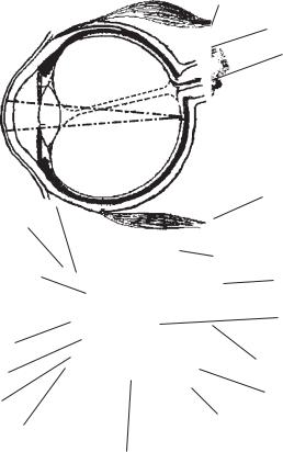
The eye is the sense organ which is stimulated by light rays. The wall of the eye is made up of three layers or coats: the outer, the middle and the inner. The outer layer is called the sclerotic coat. It is tough in order to protect the delicate structure within. The rear portion of this coat is the so-called “white of the eye”. The front, in the very center of the sclerotic coat, is a circular, clear area which is called the cornea. This is the so-called “window of the eye.” It is transparent to permit light rays to pass through it. The middle layer is the choroid coat; it is pigmented and has blood vessels to nourish the eye. In front, the choroid coat has a hole in it called the pupil.
The pupil is in reality a hole which lies behind the cornea. The circular band of the choroid coat which surrounds the pupil is the familiar iris of the eye. It may be brown, blue, black, hazel or whatever colour your eye happens to be. The iris, by contraction of its muscles, regulates the size of the pupil and thus determines the amount of light which may enter the eye. The pupil gets smaller when we enter the bright sunshine so that the eye will not be flooded by too much light; it gets bigger when we enter a darkened room or theater, in order to permit as much light as possible to enter. In this way, the eye may be compared to a camera; the iris corresponds to the shutter or diaphragm.
The retina of the eye is the innermost or third coat of the eye. It is upon this sensitive layer that the light rays from an image are focused. It corresponds to the film or plate in a camera. The retina contains pigment and specialized cells known as rods and cones which are sensitive to light. The portion of the retina where rods and cones are missing is the pathway of the optic nerve from the retina to the brain.
Vision is impossible when the rods and cones are missing. Thus, it is often called the “blind spot.” The lens is a crystal structure behind the iris. Light rays travel through it and are bent or refracted so that they may focus on the retina.
The aqueous humor is a watery fluid which fills the compartment in front of the lens and which fills the compartment in back of it is called the vitreous body. It is jelly-like. Both aid in refraction of light. If rays of light do not focus correctly on the retina, we correct this by wearing glasses with properly fitted lenses which will bend the rays of light accurately.
The eyeball is moved by muscles. The eye is protected by the bones surrounding it and by the eyebrows, eyelids, and eyelashes.
37
eyelach |
upper eyelid |
|
white of eye |
pupil |
|
|
iris |
|
lower eyelid |
|
|
|
a |
|
ciliary muscle |
muscles of eyeball |
|
|
||
cornea |
|
|
suspensory |
sclerotic coat |
|
optic |
||
ligament |
||
|
nerve |
|
|
rays |
|
pupil |
of light |
|
|
||
lens |
focus point |
|
|
||
iris |
retina |
|
choroid coat |
||
|
||
aqueus humor |
|
|
|
vitreous body humor |
|
|
b |
Fig. 16. The eye: a) external view; b) internal view
THE EAR
The ear is a special sense organ which is especially adapted to pick up sound waves and send these impulses to the auditory center of the brain, which is located in the temporal area just above the ears. The inner ear contains three semicircular canals which regulate the sense of equilibrium. These have nothing to do with the sense of hearing. The receptor for hearing is the delicate organ of Corti which is located within the cochlea of the inner ear (Fig. 17).
The ear has three parts: the outer ear, the middle ear and the inner ear. The outer ear consists of the visible portion and a canal which leads to the ear drum.
The middle ear is really a cavity in the temporal bone. It connects with the pharynx by means of a tube called the Eustachian tube. This tube serves to equalize the air pressure in the middle ear with that of the outside atmosphere. A chain of three tiny bones are found in the middle ear. These are called the hammer, the anvil, and the stirrup. These three bones carry the sound waves across the middle ear, from the ear drum to the inner ear.
The inner ear consists of several membranelined channels, which lie deep within the temporal bone. The special organ of hearing is a spiralshaped passage known as the cochlea which contains a membranous tube called the cochlear duct.
This duct is filled with a fluid, which vibrates when the sound waves from the stirrup bone hit it. Located in the cochlear duct are the delicate cells which make up the organ of Corti. These hairlike cells pick up the vibrations of the fluid caused by the sound waves and transmit them through the auditory nerve to the hearing center in the brain.
Three semicircular canals also lie within the inner ear. They contain a liquid and delicate cells. These hair-like cells bend when the liquid in the canals is set in motion by head and body movements. These impulses are sent to the brain and proper body balance is maintained.
DISEASES OF THE NERVOUS SYSTEM
Objective:
• To become familiar with some diseases of the nervous system
You will become familiar with many disorders of the nervous system. Among these are the following:
NERVOUS SYSTEM DISORDERS
Chorea or St. Vitus’ Dance is characterized by involuntary twitching of the muscles of the legs, arms, and face. The disease may last from three to six months. It usually occurs in children. Treatment consists of rest, nourishing food, and protection of the child from fright and excitement.
Shingles or Herpes Zoster results in eruptions on the skin, accompanied by pain along the nerves in some parts of the body. The involved area must be treated by protecting it from air and from the irritation of clothing.
Neuralgia is a pain along a nerve. It is usually a symptom of some other disease.
Neuritis is inflammation of a nerve trunk. Like neuralgia, it causes pain and, in addition, it causes weakness of the muscles controlled by the nerve trunk.
Poliomyelitis or infantile paralysis is an acute infectious disease of the nerve pathways in the spinal cord. The muscles which are controlled by these diseased nerve paths become paralyzed. Death may occur. Vaccines are now available to protect against the disease. All children are given regular polio shots for immunization.
38