

Иностранный язык / Єрьомкіна Г.Г. Англійська мова _ навч. посібник _ Г.Г. Єрьомкіна, А.Н. Некрасова, Є.Г. Кривда та ін. - О._ ОДМУ, 2009. - 164 с
.pdfLesson 2
HOW DOES OUR BODY USE FOOD AND OXYGEN
INTRODUCTION
Objective:
•To learn the structure of the circulatory system and how it enables all parts of the body to get needed food and oxygen
•To get familiar with the two phases of the blood circulation
The circulatory system is concerned with the transporting of fluids from one part of the body to another. The blood stream carries food, oxygen and other materials to tissues which need these elements for their growth, upkeep and work. During such work, waste products are produced and are carried to organs where they are excreted.
The circulatory system consists of the heart, blood, arteries, veins and capillaries. It also includes the lymphatic system which assists the blood circulatory system in its job of supplying food and oxygen to the tissues and in carrying away waste products.
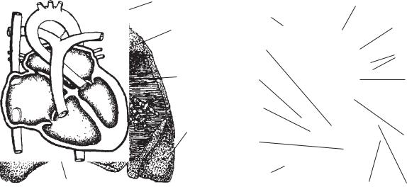
There are two phases of the circulation of the blood. These are: the general circulation which carries blood throughout the body with the exception of the lungs; the pulmonary circulation which carries blood from the heart to the lungs; the blood circulates throughout the body very rapidly. A complete tour of the blood only takes about one minute (Fig. 2).
The heart is a hollow, muscular pump which keeps the blood circulating through the blood vessels to all parts of the body. To gain some appreciation of this vital organ, do you realize that in a 70-year life span, the heart pumps enough blood to fill 70 thousand railroad tank
facial |
temporal |
|
|
|
carotid |
|
aorta |
|
|
|
heart |
cava |
|
superior |
brachial |
|
abdominal |
||
|
|
||
vena |
|
|
aorta |
|
|
radial |
|
|
|
inferior |
|
|
|
|
THE HEART
Objective:
• To understand the structure and function of the heart
Our blood circulatory system, like other systems of the body, is a marvel of efficiency. The main organ responsible for this efficiency is the heart, a tough, simply constructed muscle about the size of your closed fist.
femoral |
popliteral |
|
dorsalis pedis
Fig. 2. General plan of circulation
9
cars? And were you aware that this tremendous muscle is seven times as strong as it needs to be to do its job?
The heart is conical in shape and is located between the lungs in the center and lower part of the chest cavity. It is enclosed in a sac called the pericardium. The smooth lining of the heart is called the endocardium. This tissue forms the valves. It closes and opens like an umbrella (Fig. 3).
The heart muscle is the myocardium. The heart or cardiac (myocardium) muscle contracts rhythmically in order to perform its duty as a forceful pump. The control of the contractions of the heart muscle is found in the sinoauricular node located at the opening of the superior vena cava into the right atrium (auricle). The node is called the pacemaker of the heart. A recently invented device called the electric cardiac pacemaker has successfully maintained a normal heartbeat under conditions of cardiac arrhythmia or complete heart block.
The heart is composed of four chambers called: the right auricle, the right ventricle, the left auricle or atrium and the left ventricle.
The heart has valves which permit the blood to flow in one direction only. These valves are located as follows:
a.Between each auricle and ventricle.
b.At the orifice of the pulmonary artery (right ventricle) and the aorta leaving the left
ventricle.
These valves prevent the blood from returning to the previous chambers of the heart (Fig. 4).
FUNCTION AND PATH OF
GENERAL CIRCULATION
Objective:
• To get familiar with the path of the general circulation and with its many functions
The functions of the general circulation are fourfold; it carries nourishment, oxygen, water and secretions to all tissues (except the lungs) and back to the heart; it carries away from the tissues waste products such as carbon dioxide and other dissolved wastes; it helps equalize the body temperature; it aids in protecting the body from harmful bacteria.
The general circulation leaves the heart from the left thick-walled ventricle, by way of the aorta, the largest artery in the body. The aorta sends its many branches into the head, arms, trunk and legs. Following its tour of the body, this blood returns to the opposite side of the heart from which it left. It enters the right side of the heart by way of the vena cava (Fig. 5).
PULMONARY CIRCULATION
Objective:
• To discuss the route of the pulmonary circulation and what function it performs
The pulmonary circulation carries blood from the heart to the lungs and back to the heart. The pulmonary artery carries blood which is a darker red than when it leaves the lungs because it has an accumulation of waste products in it.
|
windpipe |
superior |
|
|
vena cava |
right |
aorta |
pulmonary |
lung |
|
semilunar |
|
|
valve |
|
peri- |
right |
|
cardium |
auricle |
|
|
tricuspid |
|
left |
valve |
|
|
|
|
lung |
|
|
|
right |
|
|
ventricle |
Right
Left
aorta
pulmonary artery
pulmonary veins
left auricle
 vitral valve
aortic semilunar valve
heart |
inferior vena |
endocardium |
left |
cava |
ventricle |
||
Fig. 3. Location of the heart |
Fig. 4. The heart and its valves |
||
10
to brain
aorta
pulmonary artery to lungs
vena cava
to liver
to stomach
to intestines
to extremities
Fig. 5. General circulation
One of these waste products, carbon dioxide, will be exchanged for a new supply of oxygen in the lungs. The blood will then become a bright red.
The pulmonary circulation starts its tour by leaving the right ventricle of the heart through the pulmonary artery. This artery carries the blood to the lungs where the exchange of carbon dioxide for oxygen takes place. The blood freshly supplied with oxygen, returns to the heart. It enters the left auricle of the heart, the opposite side from which it left the heart. It is now ready to make its tour throughout the body by way of the general circulation to distribute its fresh supply of oxygen.
BLOOD VESSELS
Objective:
• To learn how the various types of blood vessels are adapted to their particular function
The heart pumps the blood to all parts of the body through a remarkable system of three types of blood vessels:
The arteries carry: blood away from the heart. They have elastic walls which expand and contract to the pumping beat of the heart. The aorta, the artery leading directly from the heart, is the largest blood vessel of the body. Blood carried by the aorta is bright red because it has just
received a fresh supply of oxygen from the lungs. The blood carried by the pulmonary artery, however, is darker red because it is heavily laden with carbon dioxide which is to be exchanged for oxygen in the lungs.
The capillaries are very small; they must be seen through a microscope. They connect smaller arteries with smaller veins like a canal system. Their walls are extremely thin so that nourishment can pass out through them to the surrounding tissues and waste products from the tissues are absorbed back into the blood stream in the same way. This distribution and absorption is called capillary action.
The veins carry blood back to the heart. They are much less elastic than arteries. Since the pumping action of the heart is much diminished by the time blood reaches the veins for its return journey, the veins have valves which allow the blood to flow only in the direction of the heart.
PARTS OF THE BLOOD
Objectives:
•To become familiar with the important parts of the blood
•To learn the contribution of each part to the general health
The blood is the nourishing fluid of the body. It carries nutrients and oxygen to the tissues to nourish and refresh them. The average adult has four to five quarts of blood in his body. Loss of more than one quart at any one time may be serious. Blood generally has a slightly alkaline reaction.
The plasma is the straw-colored liquid part of the blood. It is the carrier of nutrients and waste products. It carries fibrinogen which helps in the clotting of the blood. It transports hormones as well as gamma globulin. Gamma globulin is used to prevent or alleviate some of the communicable diseases.
Erythrocytes (red blood cells) contain a red coloring substance called hemoglobin which consists of globin, a protein compound and hematin, an iron-containing pigment. The hemoglobin is the oxygen-carrying part of the blood. Erythrocytes also contain an antibody called the Rh factor which must be considered in prenatal care and in giving blood transfusions. The Rh factor is inherited.
Erythrocytes are made in the red bone marrow of the long bones. They live about 120 days and are probably broken up in the capillaries, spleen and liver. The hemoglobin again breaks
11
down into globin and hematin. The iron content of hematin is used to make new red blood cells.
A healthy person has 4,500,000 to 5,000,000 red blood cells per cubic millimeter of blood. The hemoglobin count is 14 to 16 grams per 100 cubic centimeters of blood.
Leukocytes (white blood cells) help to protect the body against infections. One type of leukocyte, called a phagocyte, goes to the site of an infection where germs are surrounded, engulfed and destroyed. Most of the leukocytes are made in the bone marrow. Our normal leukocyte count averages 7,000 to 9,000 cells per cubic millimeter of blood. Leukocytes vary in shape and size.
Thrombocytes (blood platelets), are plateshaped cells which help the blood to clot. They are formed in the bone marrow and disintegrate in the bone marrow or lungs. The mode of disintegration is still questionable. The normal blood platelet count is 200,000 to 400,000 for each cubic millimeter of blood.
There are different kinds of blood groups called “types”. An individual inherits his blood type from his parents.
The various blood types are:
O: Universal donor which can give blood to types A, AB, B and 0 but can only receive type 0.
AB: Universal recipient which can give only to type AB, but can receive blood from types A, B, AB and 0.
A: Can give blood to types A and AB and can receive from types A and 0.
B: Can give to types B and AB and can receive from types B and 0.
Blood must be cross-matched before a transfusion is administered as a safety measure for the patient.
Additional physiological norms are listed for your reference:
Test |
Normal range |
Bleeding range |
1 to 3 min |
Coagulation time |
6 to 12 min |
Hemoglobin count |
14 to 16 g/s per 100 cc |
Platelet count |
200,000 to |
|
400,000 per mm3 |
Prothrombin time |
10 to 15 sec. |
(Quick) |
|
Sedimentation rate |
Men — 0 to 12 mm |
in first hour |
Women — 0 to 20 mm |
Two of the above norms may need explanation. Prothrombin is a factor in blood plasma needed for coagulation. Before and after the administration of vitamin K, a test is made to determine the prothrombin concentration in the
blood plasma. If such concentration takes longer than the time shown to appear, it indicates liver damage or failure to absorb vitamin K.
Sedimentation rate is the rate at which erythrocytes settle to the bottom of an upright tube at room temperature. It indicates if disease is present. The Westergren method shows the rate for women normally slightly higher than for men.
LYMPHATIC SYSTEM
Objective:
• To become familiar with the lymphatic system and its importance in cell nourishment and excretion
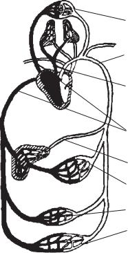
The lymphatic system has no pump similar to the heart in the blood circulatory system. The lymph, the fluid in the lymphatics, is kept moving by the contraction of the smooth muscles on the vessels.
The lymph is a fluid formed from blood plasma. It filters its way through the walls of the capillaries into the lymphatics. It bathes all portions of the body not reached by the blood. The lymph acts as a middleman between the blood and the tissues carrying and supplying food and oxygen to the cells and removing the wastes.
The lymph re-enters the general circulation through the thoracic duct at the left shoulder. In this way the waste products get back into the general circulation and are transported to the organs of excretion (Fig. 6).
DISORDERS OF CIRCULATORY SYSTEM
Objective:
• To become familiar with some of the disorders common to the heart, the blood vessels and the blood
DISEASES OF THE HEART
Acute rheumatic heart disease is an infection of the membrane lining of the heart, usually caused by the streptococcus organism.
red blood cells |
white blood cells |
cell |
lymph |
Fig. 6. Lymph circulation
12
Auricular fibrillation is a condition in which the atria (auricles) are never completely emptied of blood and their walls quiver instead of giving the usual contraction of a normal heartbeat.
Congenital heart disease is a condition in which the heart did not develop properly during fetal life.
Endocarditis is an inflammation of the membrane lining the heart.
Myocarditis is an infection of the heart muscle.
Pericarditis is an inflammation of the membrane covering the heart.
Angina pectoris is a condition in which the chief symptom is a severe pain down the arm, probably due to nerve spasm. The term is currently identified with coronary insufficiency. The heart is not usually damaged, but it is not receiving sufficient oxygen.
Murmurs may indicate some imperfection in the valves of the heart. They may take the form of gurgling and “hissing” sounds as the valves fail to close properly.
DISEASES OF THE BLOOD VESSELS
Arteriosclerosis is a thickening of the walls of the arteries and loss of elasticity — “hardening of the arteries.”
Gangrene is the death of body tissue due to an insufficient blood supply. Hemorrhoids are varicose veins in the walls of the rectum.
Phlebitis is an inflammation of the lining of a vein accompanied by clotting of blood in the vein.
Varicose veins are the result of loss of elasticity in walls, causing a slowing up of circulation and a “backing up” of blood over the valves.
Cerebral hemorrhage is a hemorrhage within the brain due to arterial sclerosis or injury.
Aneurysm is a sac caused by an enlarging of the blood vessel with the thinning of the vessel wall.
DISEASES OF THE BLOOD
Anemia is a deficiency of red blood cells or hemoglobin content.
Embolism is an infected clot which is carried by the blood stream until it reaches an artery too small for it to pass through.
Hemophilia is a condition in which the blood clots very slowly. It is inherited by the male but transmitted by the female.
Leukemia is a condition in which there is a great increase in the number of white blood cells. (Sometimes this is known as cancer of the blood.)
Thrombosis is a blood clot which forms in an artery and remains in the same place in which it was formed.
DISORDERS OF BLOOD PRESSURE
Hypertension is high blood pressure in which the systolic reading stays above 140 mm Hg.
Hypotension is low blood pressure, usually under 100 mm Hg.
TEST YOURSELF
1.What is the Rh factor? a) hemoglobin
b) antibody c) serum d) plasma e) titre
2.Structures in the heart and veins which permit the blood to flow in one direction only:
a) chambers b) valves
c) septum d) ventricles e) atria
3.Discoverer of circulation of blood:
a)Hippocrates
b)Vesalius
c)W. Harvey
d)M. Pyrogov
e)I. Pavlov
4.Red coloring matter of blood: a) erythrocytes
b) hemoglobin c) lymphocytes d) leucocytes e) thrombocytes
5.White blood cells which absorb and destroy harmful bacteria:
a) erythrocytes b) phagocytes c) lymphocytes d) leucocytes e) thrombocytes
6.Circulation through the kidneys:
a)portal
b)lymphatic
c)renal
d)pulmonary
e)cardiac
13
7.Lining of the heart: a) endocardium
b) pericardium c) cardiac muscle d) myocardium e) mesothelium
8.Arteries which nourish the heart: a) cardiac
b) lymphatic c) coronary d) renal
e) pulmonary
9.Covering of the heart:
a)endocardium
b)pericardium
c)valve
d)auricle
e)mesothelium
10.The membrane lining the chest cavity: a) plasma
b) serum c) pleura
d) pulmonary membrane e) pericardium
11.Varicose veins in the walls of the rectum: a) embolism
b)hemorrhoids
c)adenitis
d)rectitis
e)colitis
12.Severe pain down the arm: a) atherosclerosis
b) angina pectoris c) embolism
d) coronary thrombosis e) endocarditis
13.A condition in which the blood clots very slowly:
a) thrombosis b) embolism c) hemophilia d) anemia
e) leukemia
14.An inflammation of the membrane covering the heart:
a)myocarditis
b)endocarditis
c)pericarditis
d)congenital heart disease
e)pleurisy
15.An inflammation of the membrane lining the heart:
a) myocarditis b) endocarditis c) pericarditis
d) congenital heart disease e) pleurisy
16.An infection of the heart muscle:
a)gangrene
b)myocarditis
c)angina pectoris
d)embolism
e)pleurisy
17.Lymph is a fluid formed from … . a) cytoplasm
b) water
c) blood plasma d) serum
e) sarcoplasma
18.A complete tour of the blood throughout the body is about:
a) 1 second b) 1 minute
c) several minutes d) half an hour e) 15 minutes
19.How many red blood cells are there in a healthy person?
a) about 3,000,000 m3
b) 3,000,000–4,000,000 m3 c) 4,500,000–5,000,000 m3 d) 5,000,000–6,000,000 m3 e) 7,000,000 m3
20.How many white blood cells are there in a healthy person?
a) 4,000–5, 000 m3 b) 5,000–6,000 m3 c) 7,000–9,000 m3 d) over 9,000 m3 e) 10,000 m3
14
Lesson 3
HOW DO WE BREATHE?
INTRODUCTION
Objective:
• To understand the meaning and purpose of respiration
Breathing in man commonly refers to the inhalation of oxygen and the exhalation of carbon dioxide. This process takes place in the lungs.
True respiration refers to the oxidation of food within the body. Food, in order to be used by the body cells, must be burned or oxidized. Just as coal or wood when burned gives off energy in the form of heat, so does food when it is burned with the oxygen we inhale. As coal or wood burns, we further notice that they give off waste products; similarly, food when oxidized gives off wastes including carbon dioxide and water vapor. These are transported from the cell through the circulatory system and are removed from our bodies when we exhale. Respiration is controlled by the stimulation of carbon dioxide on the respiratory center in the medulla, a part of the brain.
ORGANS OF BREATHING
Objective:
• To study the organs which help us to breathe
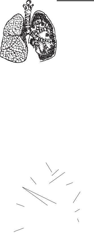
The organs of respiration are the lungs and a number of hollow organs which serve as a passageway to the lungs. These include the nose, pharynx, larynx or voice box, trachea or windpipe, and bronchi.
Air enters through the nose hairs (cilia) on the inside of the nostrils, filter out dust and dirt. The mucous membrane lining moistens the air and warms it. The air then passes through the
larynx or voice box where the vocal cords are located on through the windpipe and into the bronchi, two tubes which lead into the lungs. Inside the lungs, these bronchi divide and subdivide into smaller and smaller tubes (bronchiole) until they finally become bunches of tiny air sacs called alveoli. By this time they are microscopic in size. The walls of the alveoli or lung tissue are richly supplied with blood. Here the exchange of oxygen and carbon dioxide takes place. The oxygen absorbed by the blood is carried back to the heart by the pulmonary vein and then is pumped through the general circulation. The carbon dioxide, brought to the lungs by the pulmonary artery, escapes through the walls of the alveoli and is exhaled.
As a result of respiration, man is constantly being refreshed and is provided with the warmth and energy needed to do work (Fig. 7).
epiglottis |
voice box |
superior |
(larynx) |
windpipe |
|
lobe |
(trachea) |
|
superior |
bronchi |
lobe |
cluster
of air middle sacs lobe
bronchiole
inferior lobe
inferior lobe
Fig. 7. The lungs and windpipe (cut-a-way section)
15
BREATHING MOVEMENTS |
CHECK YOURSELF |
Objective:
• To understand how the diaphragm and ribs aid in exhalation and inhalation
Ventilation of the lungs is due to changes in pressure which occur within the chest cavity. This variation in pressure is brought about by movements of the chest wall and by contractions of the diaphragm.
When we inhale, the ribs are raised by contraction of the intercostal muscles, increasing the size of the chest cavity. At the same time, the diaphragm contracts and becomes flattened, moving downward. This serves to increase the space in the chest cavity in a vertical direction. Air rushes in to fill the extra space which has just been created, resulting in inhalation.
When we exhale, just the opposite takes place. The intercostal muscles and diaphragm relax, the ribs moving down and the diaphragm moving upwards. These two movements decrease the space within the chest cavity. This increased pressure forces the air out, resulting in exhalation.
SOME RESPIRATORY AILMENTS
Objective:
• To become familiar with some of the respiratory ailments which may affect the body
Our greatest loss in man-hours each year is due to the common cold. This respiratory infection spreads rapidly through the classroom or through a factory or business office. It is also very often the basis for more serious respiratory disease because it lowers our resistance and makes us open to infection. The direct cause of a cold is a virus; the indirect or contributing causes are lowered body resistance, chilling, fatigue, insufficient nourishment and rest. When one has a cold, one should remain in bed, drink warm liquids, take fruit juice, rest, and eat wholesome nourishing foods.
Other respiratory ailments include laryngitis, sinusitis, pharyngitis, tonsilitis, diphtheria, bronchitis, tuberculosis, influenza, and pneumonia. Each of these is caused by either a bacterium or a virus. More serious respiratory ailments include emphysema and cancer of the lung and larynx.
1.What do we inhale? a) carbon dioxide
b) nitric oxide c) oxygen
d) hydrogen e) smoke
2.What do we exhale? a) carbon dioxide
b) nitric oxide c) oxygen
d) hydrogen e) water vapor
3.Respiration is controlled in the respiratory center in the … .
a) pituitary b) medulla c) cerebellum d) brainstem
e) hypothalamus
4.The organs of respiration does not include … .
a) pharynx b) larynx
c) esophagus d) windpipe e) lungs
5.During inhalation our ribs are … by contraction of the rib muscles.
a) lowered b) raised c) flattened
d) moving forward e) moving backward
6.The microscopic air sacs in the lungs are called … .
a) capillaries b) bronchioles c) alveoli
d) bronchi e) windpipe
7.The most common respiratory ailment
is … .
a) bronchitis b) tracheitis c) sinusitis d) a cold
e) cancer
8.When we inhale, the diaphragm moves … . a) downwards
b) forwards c) upwards d) backwards e) sideward
16
9.When we exhale, the diaphragm moves … a) downwards
b) forwards c) upwards d) backwards e) sideward
10.The walls of the alveoli and lung tissue are richly supplied with … .
a) lymph b) plasma c) blood
d) erythrocytes e) water
11.What organ is not included into passageway to the lungs?
a) the nose
b) the pharynx c) the larynx d) the tongue e) the windpipe
12.Air enters through ... .
a)the windpipe
b)the pharynx
c)the larynx
d)the mouth
e)the esophagus
13.What filters out dust and dirt in the nose? a) mucosa
b) cilia c) skin
d) subcutaneous fat e) nostrils
14.What moistens the air and warm it in the
nose?
a) mucosa b) cilia
c) skin
d) subcutaneous fat e) nostrils
15.The bunches of tiny air sacs are called … a) bronchi
b) bronchiole c) alveoli
d) arteries e) lungs
16.Exchange of oxygen and carbon dioxide takes place in … .
a) bronchi b) bronchiole c) alveoli
d) arteries e) lungs
17.Ventilation of the lungs is due to … .
a)inhalation
b)changes in pressure
c)exhalation
d)heart contraction
e)muscle contraction
18.The principal waste product given off in exhalation is … .
a) hydrogen b) oxygen c) nitrogen
d) carbon dioxide e) vitamins
19.Drugs used to destroy microscopic organisms are called ... .
a) laxatives
b) tranquilizers c) antibiotics d) vitamins
e) minerals
20.Diphtheria is caused by ... .
a)spores
b)fungus
c)virus
d)dust
e)vapor
17

Lesson 4
HOW DO WE DIGEST OUR FOOD?
INTRODUCTION |
body since they are composed largely of miner- |
|
|
al salts of calcium and phosphorus. An adult |
|
Objectives: |
has 32 teeth which include incisors for biting food, |
|
• To gain a view of the digestive system |
canines for tearing it, bicuspids and molars for |
|
• To understand how the mouth and teeth con- |
crushing and chewing. The shape of each is par- |
|
tribute to digestion |
ticularly adapted to its purpose (Fig. 8A). |
|
|
Each tooth has a crown, a root and a neck |
|
All food which we eat must be prepared with- |
(Fig. 8B). The crown is covered with a substance |
|
in the body for final use by the cells. This means |
called enamel and the root is covered with ce- |
|
that certain physical and chemical changes must |
ment. The bulk of the tooth is made up of den- |
|
take place to change the food into a liquid which |
tine. The blood provides nourishment to the cells |
|
can be transported by the blood to all cells and |
of the tooth. |
|
be absorbed by them. The process of changing |
The gingivae, or gums, serve as protection |
|
solid food into a liquid form which can be ab- |
for the teeth. They are made up of fleshy tissue |
|
sorbed by the body cells is called digestion. |
covered with mucous membrane which sur- |
|
The organs which perform this change make |
rounds the teeth and covers the upper and low- |
|
up the digestive system, consisting of the alimen- |
er jaws. To maintain mouth health, it is as im- |
|
tary canal and accessory organs. The alimenta- |
portant to stimulate the circulation in the gingi- |
|
ry canal includes the mouth, pharynx, gullet, |
vae and keep them free of food particles as to |
|
stomach, small intestine and rectum. It is a 30 |
clean the teeth. Chronic infection of the gingi- |
|
to 40 foot long canal through which food passes |
vae is a frequent cause of tooth extraction. |
|
during digestion. |
The tongue lies on the floor of the mouth with- |
|
The accessory or helping organs are the teeth, |
in the curve of the lower jaw, which is also called |
|
the tongue, the salivary glands, the pancreas and |
the mandible. The tongue is the principal organ |
|
the liver. |
of taste. It assists in chewing and swallowing |
|
|
food. On the surface of the tongue are projec- |
|
THE MOUTH AND DIGESTION |
tions called papillae. Some of these papillae con- |
|
tain taste buds which respond to food flavors. |
||
|
||
The role of the mouth in the digestive proc- |
A few taste buds are also found in other areas |
|
of the mucous membrane lining the mouth cavi- |
||
ess is to break up food into smaller particles by |
||
ty. |
||
chewing. The salivary glands manufacture an |
||
The hard palate is a bony structure which forms |
||
enzyme, ptyalin, which helps to change starch |
||
the roof of the mouth. The soft palate is a curtain- |
||
to sugar as a preliminary step in digestion. We |
||
will begin our study of digestion by examining |
like muscular fold of membrane adjacent to the hard |
|
the structures of the mouth. |
palate in the back of the mouth. A small, soft struc- |
|
ture called the palatine uvula is suspended from the |
||
It may be divided into two parts. The vesti- |
||
bule consists of the area between the lips and |
center edge of the soft palate. The uvula is composed |
|
the teeth. The mouth cavity extends from the hard |
of mucous membrane, muscle and connective tissue |
|
and soft palate on top to the tongue on the bot- |
(Fig. 8C). |
|
tom. The vestibule and the mouth cavity is cov- |
Behind the soft palate is the pharyngopala- |
|
tine arch. This arch separates the mouth cavity |
||
ered with mucous membrane. |
||
from the pharynx. The tonsils are two promi- |
||
The teeth are the hardest structure in the |
||
nent masses located on either side of the mouth |
||
|
18