

ECHO 2013 / The Role of Vascular Imaging in Cardiovascular Risk Assessment
.pdfCase 2
43 year old female
Family history of premature CHD
Father CABG at age 49, brother MI at age 50
No hypertension, no DM
Non-smoker
TC 192, HDL 52, LDL 122, TG 92
Framingham Risk < 1%

Carotid Ultrasound: No Plaque

Carotid Ultrasound: No Plaque


Obtain cIMT measurement in plaque-free region
High-res image along include 1 cm length of arterial segment
cIMT can be measured in the CCA, bulb, and origin of the ICA
cIMT should be measured preferably on the far wall
Edge detection software provides accurate measurements of cIMT

Clinical CIMT Measurement
ASE Task Force Recommendation
3 imaging planes
Distal 1 cm of far wall of each CCA
4 cm depth, no zoom
3-5 beat loop gated to R wave

CIMT Measurement: Use of Border Detection Software
Mean value of 150 measurements performed on a 10-mm segment of CCA and max value within that segment

ARIC Study: CIMT and Plaque in
CHD Risk Prediction
13,145 subjects free of CHD or stroke (45-65yr)
Mean follow up 15.1 years
1,812 incident CHD events
Risk Prediction Models (10-yr CHD risk)
Traditional Risk Factors (TRF)
TRF + CIMT
TRF + Plaque
TRF + CIMT + Plaque
Nambi et al. JACC 2010;55:1600-7.
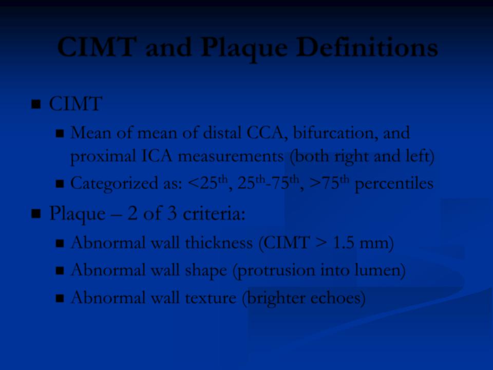
CIMT and Plaque Definitions
CIMT
Mean of mean of distal CCA, bifurcation, and proximal ICA measurements (both right and left)
Categorized as: <25th, 25th-75th, >75th percentiles
Plaque – 2 of 3 criteria:
Abnormal wall thickness (CIMT > 1.5 mm)
Abnormal wall shape (protrusion into lumen)
Abnormal wall texture (brighter echoes)

ARIC: Reclassification of Subjects
TRF + CIMT + Plaque resulted in reclassification of ~23% of subjects
More subjects reclassified to lower risk group
12.4% vs. 10.8%
No subjects were reclassified from low to high risk
No subjects reclassified from high to low risk
Results similar in FRS-based TRF model
www.ARICnews.net (CHD risk calculator)