

ECHO 2013 / The Role of 3D Echo in Valvular Heart Disease
.pdfThree-dimensional echocardiography for assessment of EROA.
Shanks M et al. Circ Cardiovasc Imaging 2010;3:694-700
Copyright © American Heart Association

Number of the patients classified in the same versus different mitral regurgitation severity grade: comparison between 2D TEE, 3D TEE, and MRI.
Shanks M et al. Circ Cardiovasc Imaging 2010;3:694-700
Copyright © American Heart Association

Are the raw images helpful?

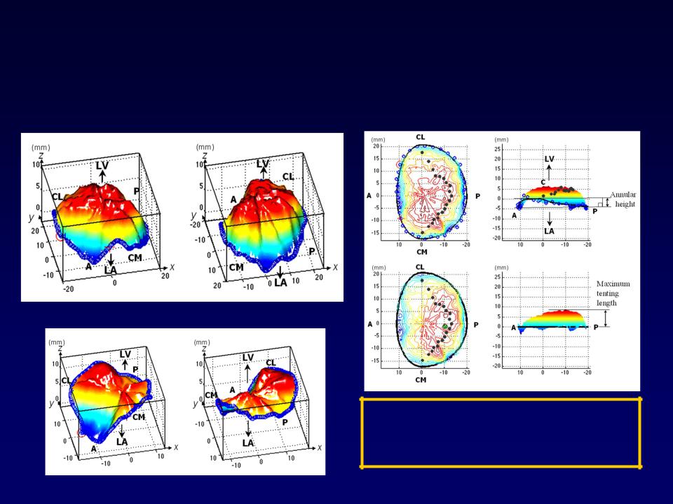
Quantitation of Mitral Valve Tenting in Ischemic MR by Transthoracic Real-time 3D Echo
normal
Tenting volume: 4.6ml
Tenting height: 9.2mm
Watanabe N, Ogasawara Y, Yoshida K et. al J Am Coll Cardiol 45:763-9, 2005

Does this impact clinical
decision making?
Perhaps not yet
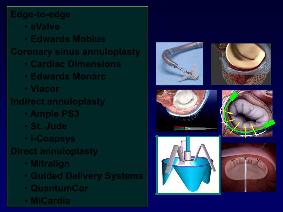
Edge-to-edge
•eValve
•Edwards Mobius Coronary sinus annuloplasty
•Cardiac Dimensions
•Edwards Monarc
•Viacor
Indirect annuloplasty
•Ample PS3
•St. Jude
•i-Coapsys Direct annuloplasty
•Mitralign
•Guided Delivery Systems
•QuantumCor
•MiCardia
Percutaneous
MV Repair

3D is essential for Mitra-Clip
Investigational in US
CE mark overseas
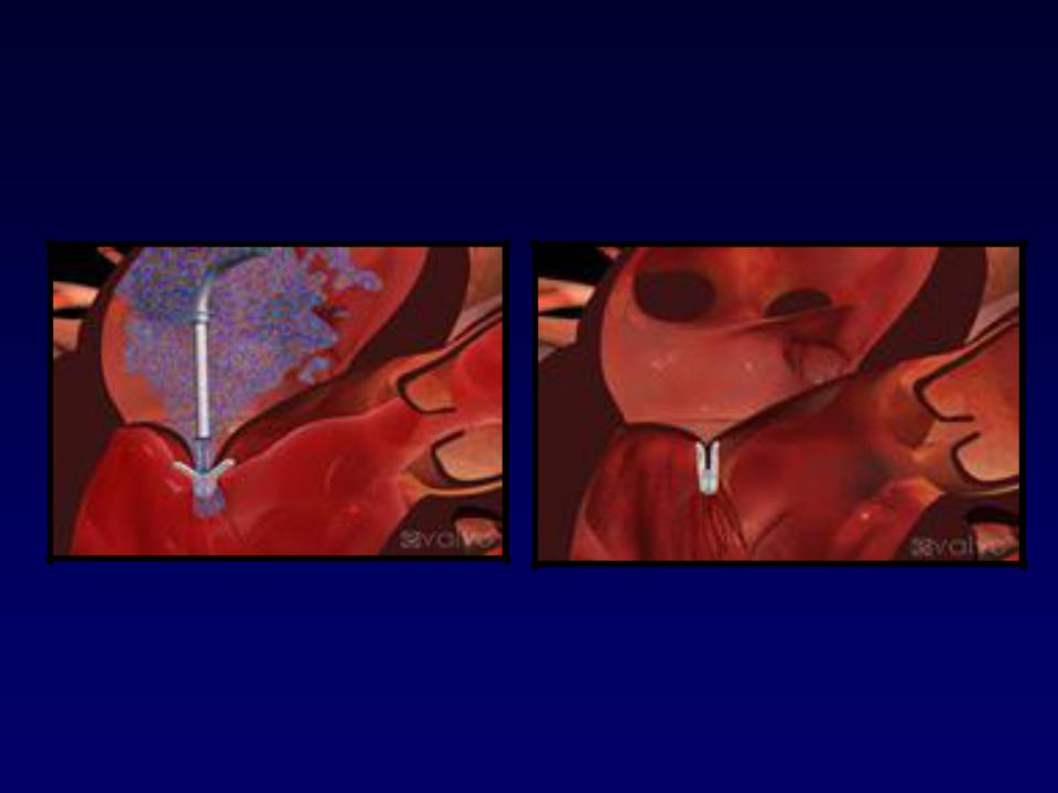

Mitra-clip
•Selection criteria for degenerative mitral regurgitation
–P2, A2
–Calcification
•Three-D invaluable in intraprocedural decision making