

Primer on molecular genetics (Human Genome Project, DOE)(44s)
.pdf(b) Constructing an Overlapping Clone Library.
A collection of clones of chromosomal DNA, called a library, has no obvious order indicating the original posit-
ions of the cloned pieces on (b) the uncut chromosome.
To establish that two particular clones are adjacent to each other in the genome, libraries of clones containing partly overlapping regions must be constructed. These clone libraries are ordered by dividing the inserts into smaller fragments and determining which clones share common DNA sequences.
Overlapping
Fragments
Cut vector DNA with a restriction enzyme
Restriction Enzyme Cutting Sites
Chromosomal DNA
Partially cut chromosomal DNA with a frequent-cutter restriction enzyme (controlling the conditions so that not all possible sites are cut on every copy of a specific
sequence) to generate a series of overlapping fragments representing every cutting site in the original sample
Join chromosomal fragments to vector, using the enzyme DNA ligase
Vector DNA
 Chromosomal DNA
Library of
Overlapping
Genomic Clones
Vector DNA
21
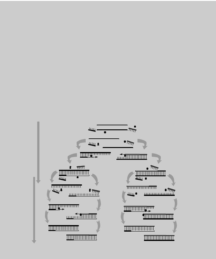
PCR (in vitro DNA amplification)
Described as being to genes what Gutenberg’s printing press was to the written word, PCR can amplify a desired DNA sequence of any origin (virus, bacteria, plant, or human) hundreds of millions of times in a matter of hours, a task that would have required several days with recombinant technology. PCR is especially valuable because the reaction is highly specific, easily automated, and capable of amplifying minute amounts of sample. For these reasons, PCR has also had a major impact on clinical medicine, genetic disease diagnostics, forensic science, and evolutionary biology.
PCR is a process based on a specialized polymerase enzyme, which can synthesize a complementary strand to a given DNA strand in a mixture containing the 4 DNA bases and 2 DNA fragments (primers, each about 20 bases long) flanking the target sequence. The mixture is heated to separate the strands of doublestranded DNA containing the target sequence and then cooled to allow (1) the primers to find and bind to their complementary sequences on the separated strands and (2) the polymerase to extend the primers into new complementary strands. Repeated heating and cooling cycles multiply the target DNA exponentially, since each new double strand separates to become two templates for further synthesis. In about 1 hour, 20 PCR cycles can amplify the target by a millionfold.
DNA Amplification Using PCR
Reaction mixture contains target |
|
|
|
|
|
|
|
|
|
TARGET DNA |
|
|
|
|
|
|
|
|||||||||||||
DNA sequence to be amplified, |
|
|
|
|
|
|
|
|
|
|
|
|
|
|
|
|
||||||||||||||
|
|
|
|
|
|
|
|
|
|
|
|
|
|
|
|
|
|
|
|
|
|
|
|
|
|
|
|
|
||
two primers (P1, P2), and |
|
|
|
|
|
|
|
|
|
|
|
|
|
|
|
|
|
|
|
|
|
|
|
|
|
|
|
|
|
|
heat-stable Taq polymerase |
|
|
|
|
|
|
|
|
|
|
|
|
|
|
|
|
|
|
|
|
|
|
|
|
|
|
|
|
|
|
|
|
|
|
|
|
|
|
|
|
|
|
|
|
|
|
|
|
|
|
|
|
|
|
|
|
|
|
|
||
P1 |
|
|
|
|
|
|
|
|
|
|
|
Taq |
|
|
P2 |
|||||||||||||||
Reaction mixture is heated |
|
|
|
|
|
|
|
|
|
|
|
|
|
|
|
|
|
|
|
|
|
|
|
|
||||||
|
|
|
|
|
|
|
|
|
|
|
|
|
|
|
|
|
|
|
|
|
|
|
|
|
|
|
|
|
||
tp 95°C to denature target |
|
|
|
|
|
|
|
|
|
|
|
|
|
|
|
|
|
|
|
|
|
|
|
|
|
|
|
|
|
|
DNA. Subsequent cooling |
|
|
|
|
|
|
|
|
|
|
|
|
|
|
|
|
|
|
|
|
|
|
|
|
|
|
|
|
|
|
|
|
|
|
|
|
|
|
|
|
|
|
|
|
|
|
|
|
|
|
|
|
|
|
|
|
|
|
|
||
to 37°C allows primers to |
|
|
|
|
|
|
|
|
|
|
|
|
|
|
|
|
|
|
|
|
|
|
|
|
|
|
|
|
|
|
hybridize to complementary |
|
|
|
|
|
|
|
|
|
|
|
|
|
|
|
|
|
|
|
|
|
|
|
|
|
|
|
|
|
|
|
|
|
|
|
|
|
|
|
|
|
|
|
|
|
|
|
|
|
|
|
|
|
|
|
|
|
|
|
||
sequences in target DNA |
|
|
|
|
|
|
|
|
|
|
|
|
|
|
|
|
|
|
|
|
|
|
|
|
|
|
|
|
|
|
FIRST CYCLE
SECOND CYCLE
When heated to 72°C, Taq polymerase extends complementary strands from primers
First synthesis cycle results in two copies of
target DNA sequence
DENATURE
DNA
HYBRIDIZE
PRIMERS
EXTEND
NEW DNA
STRANDS
Second synthesis cycle results in four copies of target DNA sequence
Source: DNA Science, see Fig. 11.
22
Current Sequencing Technologies
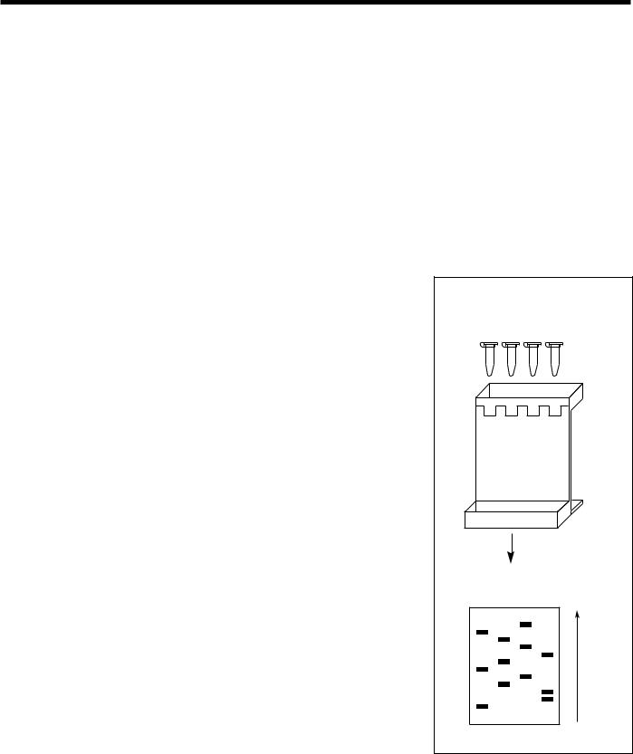
The two basic sequencing approaches, Maxam-Gilbert and Sanger, differ primarily in the way the nested DNA fragments are produced. Both methods work because gel electrophoresis produces very high resolution separations of DNA molecules; even fragments that differ in size by only a single nucleotide can be resolved. Almost all steps in these sequencing methods are now automated. Maxam-Gilbert sequencing (also called the chemical degradation method) uses chemicals to cleave DNA at specific bases, resulting in fragments of different lengths. A refinement to the Maxam-Gilbert method known as multiplex sequencing enables investigators to analyze about 40 clones on a single DNA sequencing gel. Sanger sequencing (also called the chain termination or dideoxy method) involves using an enzymatic procedure to synthesize DNA chains of varying length in four different reactions, stopping the DNA replication at positions occupied by one of the four bases, and then determining the resulting fragment lengths (Fig. 12).
These first-generation gel-based sequencing technologies are now being used to sequence small regions of interest in the human genome. Although investigators could use existing technology to sequence whole chromosomes, time and cost considerations make large-scale sequencing projects of this nature impractical. The smallest human chromosome (Y) contains 50 Mb; the largest (chromosome 1) has 250 Mb. The largest continuous DNA sequence obtained thus far, however, is approximately 350,000 bp, and the best available equipment can sequence only 50,000 to 100,000 bases per year at an approximate cost of $1 to $2 per base. At that rate, an unacceptable 30,000 work-years and at least $3 billion would be required for sequencing alone.
Fig. 12. DNA Sequencing. Dideoxy sequencing (also called chain-termination or Sanger method) uses an enzymatic procedure to synthesize DNA chains of varying lengths, stopping DNA replication at one of the four bases and then determining the resulting fragment lengths. Each sequencing reaction tube (T, C, G, and A) in the diagram contains
•a DNA template, a primer sequence, and a DNA polymerase to initiate synthesis of a new strand of DNA at the point where the primer is hybridized to the template;
•the four deoxynucleotide triphosphates (dATP, dTTP, dCTP, and dGTP) to extend the DNA strand;
•one labeled deoxynucleotide triphosphate (using a radioactive element or dye); and
•one dideoxynucleotide triphosphate, which terminates the growing chain wherever it is incorporated. Tube A has didATP, tube C has didCTP, etc.
For example, in the A reaction tube the ratio of the dATP to didATP is adjusted so that each tube will have a collection of DNA fragments with a didATP incorporated for each adenine position on the template DNA fragments. The fragments of varying length are then separated by electrophoresis (1) and the positions of the nucleotides analyzed to determine sequence. The fragments are separated on the basis of size, with the shorter fragments moving faster and appearing at the bottom of the gel. Sequence is read from bottom to top (2). (Source: see Fig. 11.)
ORNL-DWG 91M-17368
1.Sequencing reactions loaded onto polyacrylamide gel for fragment separation
T C G A
T C G A |
2.Sequence read (bottom to top) from gel autoradiogram
T C G A
G
T
C
G
A
C
T
G
C
A
A
T
23

Primer on Molecular Genetics
Sequencing Technologies Under Development
A major focus of the Human Genome Project is the development of automated sequencing technology that can accurately sequence 100,000 or more bases per day at a cost of less than $.50 per base. Specific goals include the development of sequencing and detection schemes that are faster and more sensitive, accurate, and economical. Many novel sequencing technologies are now being explored, and the most promising ones will eventually be optimized for widespread use.
Second-generation (interim) sequencing technologies will enable speed and accuracy to increase by an order of magnitude (i.e., 10 times greater) while lowering the cost per base. Some important disease genes will be sequenced with such technologies as (1) highvoltage capillary and ultrathin electrophoresis to increase fragment separation rate and
(2) use of resonance ionization spectroscopy to detect stable isotope labels.
Third-generation gel-less sequencing technologies, which aim to increase efficiency by several orders of magnitude, are expected to be used for sequencing most of the human genome. These developing technologies include (1) enhanced fluorescence detection of individual labeled bases in flow cytometry, (2) direct reading of the base sequence
on a DNA strand with the use of scanning tunneling or atomic force microscopies,
(3) enhanced mass spectrometric analysis of DNA sequence, and (4) sequencing by hybridization to short panels of nucleotides of known sequence. Pilot large-scale sequencing projects will provide opportunities to improve current technologies and will reveal challenges investigators may encounter in larger-scale efforts.
Partial Sequencing To Facilitate Mapping, Gene
Identification
Correlating mapping data from different laboratories has been a problem because of differences in generating, isolating, and mapping DNA fragments. A common reference system designed to meet these challenges uses partially sequenced unique regions (200 to 500 bp) to identify clones, contigs, and long stretches of sequence. Called sequence tagged sites (STSs), these short sequences have become standard markers for physical mapping.
Because coding sequences of genes represent most of the potentially useful information content of the genome (but are only a fraction of the total DNA), some investigators have begun partial sequencing of cDNAs instead of random genomic DNA. (cDNAs are derived from mRNA sequences, which are the transcription products of expressed genes.) In addition to providing unique markers, these partial sequences [termed expressed sequence tags (ESTs)] also identify expressed genes. This strategy can thus provide a means of rapidly identifying most human genes. Other applications of the EST approach include determining locations of genes along chromosomes and identifying coding regions in genomic sequences.
24

End Games: Completing Maps and
Sequences; Finding Specific Genes
Starting maps and sequences is relatively simple; finishing them will require new strategies or a combination of existing methods. After a sequence is determined using the methods described above, the task remains to fill in the many large gaps left by current mapping methods. One approach is single-chromosome microdissection, in which a piece is physically cut from a chromosomal region of particular interest, broken up into smaller pieces, and amplified by PCR or cloning (see DNA Amplification). These fragments can then be mapped and sequenced by the methods previously described.
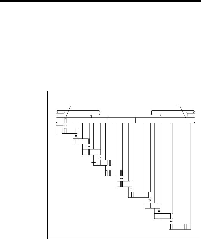
Chromosome walking, one strategy for filling in gaps, involves hybridizing a primer of known sequence to a clone from an unordered genomic library and synthesizing a short complementary strand (called “walking” along a chromosome). The complementary strand is then sequenced and its end used as the next primer for further walking; in this way the adjacent, previously unknown, region is identified and sequenced. The chromosome is thus systematically sequenced from one end to the other. Because primers must be synthesized chemically, a disadvantage of this technique is the large number of different primers needed to walk a long distance. Chromosome walking is also used to locate specific genes by sequencing the chromosomal segments between markers that flank the gene of interest (Fig. 13).
The current human genetic map has about 1000 markers, or 1 marker spaced every
3 million bp; an estimated 100 genes lie between each pair of markers. Higher-resolution genetic maps have been made in regions of particular interest. New genes can be located by combining genetic and physical map information for a region. The genetic map basically describes gene order. Rough information about gene location is sometimes available also, but these data must be used with caution because recombination is not equally likely at all places on the chromosome. Thus the genetic map, compared to the physical map, stretches in some places and compresses in others, as though it were drawn on a rubber band.
The degree of difficulty in finding a disease gene of interest depends largely on what information is already known about the gene and, especially, on what kind of DNA alterations cause the disease. Spotting the disease gene is very difficult when disease results from a single altered DNA base; sickle cell anemia is an example of such a case, as are probably most major human inherited diseases. When disease results from a large DNA rearrangement, this anomaly can usually be detected as alterations in the physical map of the region or even by direct microscopic examination of the chromosome. The location of these alterations pinpoints the site of the gene.
Identifying the gene responsible for a specific disease without a map is analogous to finding a needle in a haystack. Actually, finding the gene is even more difficult, because even close up, the gene still looks like just another piece of hay. However, maps give clues on where to look; the finer the map’s resolution, the fewer pieces of hay to be tested.
25
Primer on Molecular
Once the neighborhood of a gene of interest has been identified, several strategies can be used to find the gene itself. An ordered library of the gene neighborhood can be constructed if one is not already available. This library provides DNA fragments that can be screened for additional polymorphisms, improving the genetic map of the region and further restricting the possible gene location. In addition, DNA fragments from the region can be used as probes to search for DNA sequences that are expressed (transcribed to RNA) or conserved among individuals. Most genes will have such sequences. Then individual gene candidates must be examined. For example, a gene responsible for liver disease is likely to be expressed in the liver and less likely in other tissues or organs. This type of evidence can further limit the search. Finally, a suspected gene may need to be sequenced in both healthy and affected individuals. A consistent pattern of DNA variation when these two samples are compared will show that the gene of interest has very likely been found. The ultimate proof is to correct the suspected DNA alteration in a cell and show that the cell’s behavior reverts to normal.
Fig. 13. Cloning a Disease Gene by Chromosome Walking.
After a marker is linked to within 1 cM of a disease gene, chromosome walking can be used to clone the disease gene itself. A probe is first constructed from a genomic fragment identified from a library as being the closest linked marker to the gene. A restriction fragment isolated from the end of the clone near the disease locus is used to reprobe the genomic library for an overlapping clone. This process is repeated several times to walk across the chromosome and reach the flanking marker on the other side of the disease-gene locus. (Source: see Fig. 11.)
|
|
ORNL-DWG 91M-17370 |
|
|
LINKED FLANKING |
LINKED FLANKING |
|
5' |
MARKER |
MARKER |
3' |
|
|
||
DISEASE GENE
Probe from 5'flanking marker is
used to identify an overlapping fragment from a
genomic library
GENOMIC DNA FRAGMENT
PROBE 

Probes from the 3'ends
of cloned fragments are used to identify successive overlapping cloned fragments
Chromosome walking continues until a clone is identified that contains the 3'flanking marker
26

Model Organism Research
Most mapping and sequencing technologies were developed from studies of nonhuman genomes, notably those of the bacterium Escherichia coli, the yeast Saccharomyces cerevisiae, the fruit fly Drosophila melanogaster, the roundworm Caenorhabditis elegans, and the laboratory mouse Mus musculus. These simpler systems provide excellent models for developing and testing the procedures needed for studying the much more complex human genome.
A large amount of genetic information has already been derived from these organisms, providing valuable data for the analysis of normal gene regulation, genetic diseases, and evolutionary processes. Physical maps have been completed for E. coli, and extensive overlapping clone sets are available for S. cerevisiae and C. elegans. In addition, sequencing projects have been initiated by the NIH genome program for E. coli,
S. cerevisiae, and C. elegans.
Mouse genome research will provide much significant comparative information because of the many biological and genetic similarities between mouse and man. Comparisons of human and mouse DNA sequences will reveal areas that have been conserved during evolution and are therefore important. An extensive database of mouse DNA sequences will allow counterparts of particular human genes to be identified in the mouse and extensively studied. Conversely, information on genes first found to be important in the mouse will lead to associated human studies. The mouse genetic map, based on morphological markers, has already led to many insights into human biology. Mouse models are being developed to explore the effects of mutations causing human diseases, including diabetes, muscular dystrophy, and several cancers. A genetic map based on DNA markers is presently being constructed, and a physical map is planned to allow direct comparison with the human physical map.
Informatics: Data Collection and Interpretation
Collecting and Storing Data
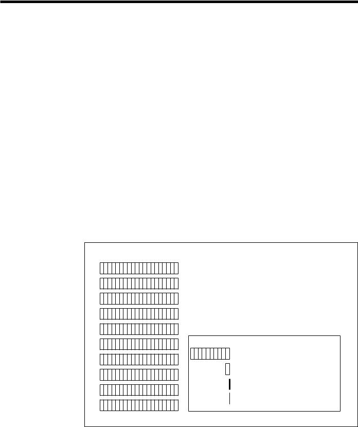
The reference map and sequence generated by genome research will be used as a primary information source for human biology and medicine far into the future. The vast amount of data produced will first need to be collected, stored, and distributed. If compiled in books, the data would fill an estimated 200 volumes the size of a Manhattan telephone book (at 1000 pages each), and reading it would require 26 years working around the clock (Fig.14).
Because handling this amount of data will require extensive use of computers, database development will be a major focus of the Human Genome Project. The present challenge is to improve database design, software for
HUMAN GENETIC DIVERSITY:
The Ultimate Human Genetic Database
 Any two individuals differ in about 3 x 10⁶ bases (0.1%).
 The population is now about 5 x 10⁹.
 A catalog of all sequence differences would require 15 x 1015 entries.
 This catalog may be needed to find the rarest or most complex disease genes.
27
Primer on Molecular Genetics
database access and manipulation, and data-entry procedures to compensate for the varied computer procedures and systems used in different laboratories. Databases need to be designed that will accurately represent map information (linkage, STSs, physical location, disease loci) and sequences (genomic, cDNAs, proteins) and link them to each other and to bibliographic text databases of the scientific and medical literature.
Interpreting Data
New tools will also be needed for analyzing the data from genome maps and sequences. Recognizing where genes begin and end and identifying their exons, introns, and regulatory sequences may require extensive comparisons with sequences from related species such as the mouse to search for conserved similarities (homologies). Searching a database for a particular DNA sequence may uncover these homologous sequences in a known gene from a model organism, revealing insights into the function of the corresponding human gene.
Correlating sequence information with genetic linkage data and disease gene research will reveal the molecular basis for human variation. If a newly identified gene is found to code for a flawed protein, the altered protein must be compared with the normal version to identify the specific abnormality that causes disease. Once the error is pinpointed, researchers must try to determine how to correct it in the human body, a task that will require knowledge about how the protein functions and in which cells it is active.
Fig. 14. Magnitude of Genome Data. If the DNA sequence of the human genome were compiled in books, the equivalent of 200 volumes the size of a Manhattan telephone book (at 1000 pages each) would be needed to hold it all. New data-analysis tools will be needed
for understanding the information from genome maps and sequences.
ORNL-DWG 91M-17472
HUMAN GENOME 200 Telephone Books
(1000 pages each)
Model Organism Genomes
Drosophila (fruit fly) |
10 books |
yeast |
1 book |
E. coli (bacterium) |
300 pages |
yeast chromosome 3 |
14 pages |
(longest continuous sequence now known)
28

Correct protein function depends on the three-dimensional |
|
|
|
|
|
|
|
|
|
|
|
|
|
|
|
|
|
|
ORNL-DWG 91M-17473 |
|
|||
(3D), or folded, structure the proteins assume in biological |
|
|
|
|
|
|
|
|
|||
|
|
|
|
|
|
|
|
|
|
||
|
GENE |
|
|
|
PROTEIN |
||||||
environments; thus, understanding protein structure will be |
|
|
|
|
|||||||
|
|
|
|
||||||||
essential in determining gene function. DNA sequences |
|
|
|
|
|
|
|
|
|
|
|
|
|
|
|
|
|
|
|
|
|
||
will be translated into amino acid sequences, and re- |
|
|
|
|
|
|
|
|
|
|
|
searchers will try to make inferences about functions either |
|
|
|
|
|
|
|
|
|
|
|
by com-paring protein sequences with each other or by |
|
|
|
|
|
|
|
|
|
|
|
comparing their specific 3-D structures (Fig. 15). |
|
|
|
|
|
|
|
|
|
|
|
Because the 3-D structure patterns (motifs) that protein |
|
|
|
|
|
|
|
|
|
|
|
molecules assume are much more evolutionarily con- |
|
|
|
|
|
|
|
|
|
|
|
|
|
|
|
|
|
|
|
|
|
||
served than amino acid sequences, this type of homology |
|
FUNCTION |
|
|
STRUCTURE |
||||||
search could prove more fruitful. Particular motifs may |
|
|
|
||||||||
|
|
|
|||||||||
serve similar functions in several different proteins, infor- |
|
|
|
|
|
|
|
|
|
|
|
mation that would be valuable in genome analyses. |
|
|
|
|
|
Fig. 15. Understanding |
|||||
Currently, however, only a few protein motifs can be recognized at the sequence level. |
|||||||||||
Gene Function. |
|||||||||||
Continued development of analytic capabilities to facilitate grouping protein sequences |
|||||||||||
Understanding how |
|||||||||||
into motif families will make homology searches more successful. |
|
|
|
|
|||||||
|
|
|
|
genes function will |
|||||||
|
|
|
|
|
|
require analyses of the |
|||||
Mapping Databases |
|
|
|
|
3-D structures of the |
||||||
|
|
|
|
proteins for which the |
|||||||
|
|
|
|
genes code. |
|||||||
The Genome Data Base (GDB), located at Johns Hopkins University (Baltimore, Maryland), provides location, ordering, and distance information for human genetic markers, probes, and contigs linked to known human genetic disease. GDB is presently working on incorporating physical mapping data. Also at Hopkins is the Online Mendelian Inheritance in Man database, a catalog of inherited human traits and diseases.
The Human and Mouse Probes and Libraries Database (located at the American Type Culture Collection in Rockville, Maryland) and the GBASE mouse database (located at Jackson Laboratory, Bar Harbor, Maine) include data on RFLPs, chromosomal assignments, and probes from the laboratory mouse.
Sequence Databases
Nucleic Acids (DNA and RNA)
Public databases containing the complete nucleotide sequence of the human genome and those of selected model organisms will be one of the most useful products of the Human Genome Project. Four major public databases now store nucleotide sequences: GenBank and the Genome Sequence DataBase (GSDB) in the United States, European Molecular Biology Laboratory (EMBL) Nucleotide Sequence Database in the United Kingdom, and the DNA Database of Japan (DDBJ). The databases collaborate to share sequences, which are compiled from direct author submissions and journal scans. The four databases now house a total of almost 200 Mb of sequence. Although human sequences predominate, more than 8000 species are represented. [Paragraph updated July 1994]
29

Primer on Molecular Genetics
Proteins
The major protein sequence databases are the Protein Identification Resource (National Biomedical Research Foundation), Swissprot, and GenPept (both distributed with GenBank). In addition to sequence information, they contain information on protein motifs and other features of protein structure.
Impact of the Human Genome Project
The atlas of the human genome will revolutionize medical practice and biological research into the 21st century and beyond. All human genes will eventually be found, and accurate diagnostics will be developed for most inherited diseases. In addition, animal models for human disease research will be more easily developed, facilitating the understanding of gene function in health and disease.
Researchers have already identified single genes associated with a number of diseases, such as cystic fibrosis, Duchenne muscular dystrophy, myotonic dystrophy, neurofibromatosis, and retinoblastoma. As research progresses, investigators will also uncover the mechanisms for diseases caused by several genes or by a gene interacting with environmental factors. Genetic susceptibilities have been implicated in many major disabling and fatal diseases including heart disease, stroke, diabetes, and several kinds of cancer. The identification of these genes and their proteins will pave the way to more-effective therapies and preventive measures. Investigators determining the underlying biology of genome organization and gene regulation will also begin to understand how humans develop from single cells to adults, why this process sometimes goes awry, and what changes take place as people age.
New technologies developed for genome research will also find myriad applications in industry, as well as in projects to map (and ultimately improve) the genomes of economically important farm animals and crops.
While human genome research itself does not pose any new ethical dilemmas, the use of data arising from these studies presents challenges that need to be addressed before the data accumulate significantly. To assist in policy development, the ethics component of the Human Genome Project is funding conferences and research projects to identify and consider relevant issues, as well as activities to promote public awareness of these topics.
30