

Solid Support Oligosaccharide Synthesis
.pdf280SOLID-PHASE SYNTHESIS OF BIOLOGICALLY IMPORTANT GLYCOPEPTIDES
29.Bayer, E., and Rapp, W., Chem. Pept. Protein 3, 3 (1986).
30.Seitz, O., and Kunz, H., Angew. Chem., Int. Ed. Engl. 34, 803 (1995).
31.Habermann, J., and Kunz, H., Tetrahedron Lett. 39, 4797 (1998).
32.Berndt, P., Fields, G. B., and Tirrell, M., J. Am. Chem. Soc. 117, 9515 (1995).
33.Itzkowitz, S. H., Yuan, M., Montgomery, C. K., Kjeldsen, T., Takahashi, H. K., Bigbee,
W.L., and Kim, Y. S., Cancer Res. 49, 197 (1989).
34.Itzkowitz, S. H., Bloom, E. J., Kolcal, W. A., Modin, G., Hakomori, S.-J., and Kim, Y. S., Cancer 66, 1960 (1990).
35.Carraway, K. L., and Hull, S. R., Glycobiology 1, 131 (1991).
36.Cooley, A. A., Pisano, A., and Williams, K. L., Trends Glycosci. Glycotechnol., 6, 328 (1994).
37.Hansen, J.-E. S., Clausen, H., Nielsen, C., Teglbjaerg, L. S., Hansen, L. L., Nielsen, C. M., Dabelsteen, E., Mathiesen, L., Hakomori, S.-J., and Nielsen, J. O.,J. Virol. 64, 2833 (1990).
38.Hansen, J.-E. S., Nielsen, C., Arendrup, M., Olofsson, S., Mathiesen, L., Nielsen, J. O., and Clausen, H., J. Virol. 65, 6461 (1991).
39.Liebe, B., and Kunz, H., Helv. Chim. Acta 80, 1473 (1997).
40.Marra, A. and Sinay, P., Carbohydr. Res. 187, 35 (1989).
41.Elofsson, M., Salvador, L. A., and Kihlberg, J., Tetrahedron 53, 369 (1997).
42.Nakahara, Y., Ito, Y., and Ogawa, T., Carbohydr. Res. 309, 287 (1998).
43.Sprengard, U., Kretzschmar, G., Bartnik, E., Hüls, C., and Kunz, H.,Angew. Chem., Int. Ed. Engl. 34, 990 (1995).
44.Rosen, T., Lico, I. M., and Chu, D. T. W., J. Org. Chem. 53, 1580–1585 (1988).
45.Ciommer, M., and Kunz, H., Synlett 593 (1991).
46.Ramage, R., and Green, J., Tetrahedron Lett. 28, 2287 (1987).
47.Fujii, N., Otaka, A., Ikemura, O., Akaji, K., Funakoshi, S., Hayashi, Y., Kuroda, Y., and Yajima, H., J. Chem. Soc., Chem. Commun. 274 (1987).
48.Broddefalk, J., Bäcklund, J., Almqvist, F., Johansson, M., Holmdahl, R., and Kihlberg, J., J. Am. Chem. Soc. 120, 76 (1998).
49.Broddefalk, J., Bergquist, K.-E., and Kihlberg, J., Tetrahedron Lett. 37, 3011 (1996).
50.Springer, T. A., Cell 76, 301 (1994).
51.Moore, K. L., Patel, K. D., Bruehl, R. E., Fugang, L., Johnson, D. A., Lichtenstein, H. S., Cummings, R. D., Bainton, D. F., and Mc Ever, R. P., J. Cell Biol. 128, 661 (1995).
52.Buerke, M., Weyrich, A. S., Zheng, X., Gaeta, F. C. A., Forrest, M. J., and Lefer, A. M.,
J.Clin. Invest. 1140 (1994); Giannis, A., Angew. Chem., Int. Ed. Engl. 33, 178 (1994).
53.Peilstöcker, K., and Kunz, H., Synlett, 820 and 823 (2000).
54.Garegg, P. J., Hultberg, H., and Wallin, S., Carbohydr. Res. 108, 97 (1982).
55.Sato, S., Mori, M., Ito, Y., and Ogawa, T., Carbohydr. Res. 155, C6–C10 (1986).
56.Lemieux, R. U., and Driguez, H., J. Am. Chem. Soc. 97, 4063 (1975).
57.(a) Xia, M.-Q., Tone, M., Packman, L., Hale, G., and Waldmann, H., Eur. J. Immunol. 21, 1677 (1991); (b) Xia, M.-Q., Hale, G., Lifely, M. R., Ferguson, M. A. J., Campbell, D., Packman, L., and Waldmann, H.,Biochem. J. 293, 633 (1993); Treumann, A., Lifely,
M.R., Schneider, P., and Ferguson, M. A. J., J. Biol. Chem. 270, 6088 (1995).
REFERENCES 281
58.(a) Matsuo, I., Nakahara, Y., Ito, Y., Nukada, T., Nakahara, Y., and Ogawa, T.,Bioorg. Med. Chem. 3, 1455 (1995); (b) Guo, Z.-W., Nakahara, Y., Nakahara, Y., and Ogawa, T., Bioorg. Med. Chem. 5, 1917 (1997); (c) Guo, Z.-W., Nakahara, Y., Nakahara, Y., and Ogawa, T., Angew. Chem., Int. Ed. Engl. 36, 1464 (1997).
59.(a) Baudry, D., Ephritikhine, M., and Felkin, H., J. Chem. Soc., Chem. Commun. 694 (1978); (b) Oltvoort, J. J., van Boeckel, C. A. A., de Konig, J. H., and van Boom, J. H., Synthesis 305 (1981).
60.Meinjohanns, E., Meldal, M., Paulsen, H., Dwek, R. A., and Bock, K., J. Chem. Soc. Perkin Trans. 1 549 (1998).
61.Likhosherstov, L. M., Novikova, O. S., Derevitskaja, V. A., and Kochetkov, N. K.,
Carbohydr. Res. 146, C1–C5 (1986).
62.(a) Cohen-Anisfeld, S. T., and Lansbury, P. T., J. Am. Chem. Soc. 115, 10531 (1993);
(b)Wong, S. Y. C., Guile, G. R., Rademacher, T. W., and Dwek, R. A., Glycoconj. J. 10, 227 (1993); (c) Sprengard, U., Schudok, M., Schmidt, W., Kretzschmar, G., and Kunz, H., Angew. Chem., Int. Ed. Engl. 35, 321 (1996).
63.Schleyer, A., Meldal, M., Renil, M., Paulsen, H., and Bock, K., Angew. Chem., Int. Ed. Engl. 36, 1976 (1997).
64.(a) Roberge, J. Y., Beebe, X., and Danishefsky, S. J., J. Am. Chem. Soc. 120, 3915 (1998); (b) Roberge, J. Y., Beebe, X., and Danishefsky, S. J., Science 269, 202 (1995);
(c)Savin, K. A., Woo, J. C. G., and Danishefsky, S. J., J. Org. Chem. 64, 4183 (1999).
65.Kunz, H., and März, J., Synlett 591 (1992).
66.Lampe, T. F. J., Weitz-Schmidt, G., and Wong, C.-H., Angew. Chem., Int. Ed. Engl. 37, 1707 (1998).
67.Graves, B. J., Crowther, R. L., Chandran, C., Rumberger, J. M., Li, S., Huang, K.-S., Presky, D. H., Familletti, P. C., Wolitzky, B. A., and Burnes, D. K., Nature 367, 532 (1994).
68.Ichikawa, Y., Lin, Y. C., Dumas, D. P., Shen, G. J., Garcia-Junceda, E., Williams, M. A., Bayer, R., Ketcham, C., Walker, L. E., Paulson, J. C., and Wong, C.-H.,J. Am. Chem. Soc., 114, 9283 (1992).
69.(a) Cooke, R. M., Hale, R. S., Lister, S. G., Shah, G., and Weir, M. P.,Biochemistry 33, 10591 (1994); (b) Scheffler, K., Ernst, B., Katopodis, A., Magnani, J. L., Wang, W. T., Weisemann, R., and Peters, T., Angew. Chem., Int. Ed. Engl. 34, 1841 (1995); (c) Poppe, L., Brown, G. S., Philo, J. S., Nikrad, P. V., and Shah, B. H.,J. Am. Chem. Soc. 119, 1727 (1997).
70.Bradley, B. K., Kiso, M., Abbas, S., Nikrad, P., Strivastava, O., Foxall, C., Oda, Y., and Hasegawa, A., Glycobiology 3, 633 (1993).
71.Stahl, W., Sprengard, U., Kretzschmar, G., and Kunz, H., Angew. Chem., Int. Ed. Engl. 33, 2096 (1994).
72.Bayer, E., Angew. Chem., Int. Ed. Engl. 30, 113 (1991).
73.Kunz, H., and Unverzagt, C., Angew. Chem., Int. Ed. Engl. 23, 436 (1984).

Solid Support Oligosaccharide Synthesis and Combinatorial Carbohydrate Libraries. Edited by Peter H. Seeberger Copyright © 2001 John Wiley & Sons, Inc.
ISBNs: 0-471-37828-3 (Hardback); 0-471-22044-2 (Electronic)
14 Preparation and Screening of
Glycopeptide Libraries
PHAEDRIA M. ST. HILAIRE, KOEN M. HALKES, and
MORTEN MELDAL
Department of Chemistry, Carlsberg Laboratory, Valby, Denmark
14.1 PARALLEL ARRAYS VERSUS LIBRARIES OF COMPOUNDS
Combinatorial methodology has been a driving impetus in many areas of research and has gained considerable ground since its introduction in 1989 or 1990. This progress, however, has not been reflected in the field of oligosaccharides primarily because of the difficulty in their synthesis and analysis. As an alternative, the readily available glycopeptide libraries have been exploited as potential functional mimetics of oligosaccharides. These libraries can be essential tools in the development of
Combinatorial synthesis
Parallel synthesis
Figure 14.1 The conceptual difference between the synthesis of a combinatorial library with exponential increase in number of compounds and the synthesis of parallel arrays of discrete compounds.
283
284 PREPARATION AND SCREENING OF GLYCOPEPTIDE LIBRARIES
carbohydrate-based therapeutics, such as tumor vaccines, xenotransplantation aids, and antiinfectives.
Here, we present an overview of the aspects of the design, synthesis, analysis, and screening of combinatorial solid-phase glycopeptide libraries as compared to parallel glycopeptide arrays.
Combinatorial libraries have been defined as assemblies of compounds that are derived through a real combinatorial step leading to exponentially increasing numbers of products relative to the number of reagents used (Fig. 14.1). This staggering increase in products may be achieved either by reacting mixtures of reagents to form product mixtures or by introducing mixing of intermediates in solution or compartmentalized on solid phase, such as by the split–combine approach. Because of the ease with which this process is carried out on solid phase, the potential of solid-phase libraries by far exceeds that of solution libraries; therefore, we focus on solid-phase libraries in the present review.
Parallel synthesis of glycopeptides is not described in great detail since such synthesis was reviewed previously and may be regarded as an extension of conventional glycopeptide synthesis.
14.2 CARBOHYDRATE BINDING PROTEINS
Carbohydrate binding proteins (CBPs) located in cellular membranes or recirculating in the cytosol or serum are involved in a variety of important biological functions, including communication and intercellular adhesion, adhesion of bacteria or viruses, activation of the innate immune system, leukocyte rolling, hepatic clearing of aged serum proteins, and sorting of newly synthesized glycoproteins.1,2 Mammalian CBPs have been divided into three major groups according to their mode of binding: the C-type, the S-type and the P-type lectins.3
The calcium-dependent C-type lectins include the E-, L- and P-selectins; the galectins are calcium-independent S-type lectins. The latter have a binding site sized for tight interaction with dior trisaccharides. The former bind their ligands mainly through coordination of two vicinal hydroxyl groups of a single sugar moiety to a bound calcium ion in the carbohydrate recognition domain (CRD), and the surrounding sugars of the oligosaccharide ligand add to the binding specificity through relatively weak additional interactions. Because of the simple nature of this interaction, the specificity of selectin binding is quite broad and can be mimicked by simple monosaccharide bearing compounds.4,5 However, the biological activity of ligands is structure-dependent and complicated by a range of factors such as multivalency and clustering.6
The calcium-independent receptors of the P-type involved in the clearing and sorting of glycoproteins include the mannose 6-phosphate receptors,7 the hepatic Gal/GalNAc receptors,8 and the Man/GlcNAc receptors isolated from liver extracts.9 These receptors are truly multivalent and bind to the termini of bi-, trior tetraantennary N-linked oligosaccharides. Although most known receptors recognize
14.3 THE CARBOHYDRATE LIGANDS 285
Gal/GalNAc or Man/GlcNAc, there are also receptors (e.g., the macrophage sialic acid receptor) for the recognition of sialylated complex glycans.10
The collectins, which are involved mostly in the innate immune system,11 are large surface or serum proteins composed of bundles of structural collagen stalks with trimer heads containing a calcium dependent CRDs and may be considered C-type by structural similarity. The binding to simple mannose oligosaccharides is relatively weak, and activation of the complement cascade is effected only when the protein interacts multivalently with large polymannans on foreign cell surfaces, creating an activation template for complement processing enzymes.12 Because of the unspecific nature of this multivalent interaction and the long distance between the sites of interaction (53 Å),13 multivalent synthetic glycopeptides for these proteins are difficult to design and prepare. However, each CRD may be targeted with high-affinity monovalent ligands.
14.3 THE CARBOHYDRATE LIGANDS
Mammalian glycoproteins consist of 5–90% glycan structures, and these fall into several major groups. There are the N-glycosylations including high-mannose, complex, and hybrid oligosaccharides and O-glycosylations, including the mucins, the blood group determinants, and the proteoglycans (Fig. 14.2). Glycolipids represent another group of oligosaccharide signaling molecules. The high-mannose oligosaccharides are based on 1→2-α-mannosylation of the mannose pentasaccharide core, α-D-Man-(1→3)-{α-D-Man-(1→3)-[α-D-Man-(1→6)-]α-D-Man-(1→6)}-β-D- Man-(1→4)-β-D-GlcNAc-(1→4)-β-D-GlcNAc-(1→N), and these oligosaccharides exist as a mixture of conformational populations around the 1→6 bonds as depicted in Figure 14.3. Either of these conformers may be the active one in contact with a receptor.
The complex antennary glycans are structures obtained by attachment of β-(1→4)-linked lactosamine unit to 2, 4, and 6 positions of terminal mannoses in a α-D-Man-(1→3)-{α−D-Man-(1→6)-}β-D-Man-(1→4)-β-D-GlcNAc-(1→4)-β-D-Glc NAc-(1→N) core. The terminal galactoses are often sialylated at the 3 or 6 position.
The tetraantennary oligosaccharide illustrated in Figure 14.2 has been shown to present an umbrellalike structure as one of the major conformational populations.14,15 The distance between terminal sugar residues interacting with receptors may vary from ~8 to ~30 Å. In the case of the phosphorylated high-mannose ligands for the MPR, the distance between phosphates is ~15 Å.16
The mucin oligosaccharides do not appear to have many mammalian receptors. However, bacteria in the gastrointestinal tract often display receptors that adhere to the mucin-coated epithelia.17 Recognition of the aberrantly glycosylated forms present in
malignant tissue is essential for the rejection of tumors by the immune system.18–20 The most thoroughly investigated ligands are analogs of the SLex antigen4,21,22
involved in leukocyte rolling, adhesion, and shedding by interaction with the E-, P-, or L-selectins for which the optimal ligands and mode of recognition for biological activity are still sought. The fucose residue, one hydroxyl group from the central
286 PREPARATION AND SCREENING OF GLYCOPEPTIDE LIBRARIES
|
|
|
|
|
|
HO |
OH |
|
|
|
|
|
|
|
|
|
|
|
||
|
|
|
|
|
|
HO O |
|
OH |
|
|
|
|
|
|
|
|
|
|
||
|
|
|
|
|
|
|
HO O |
|
|
|
|
|
|
|
|
|
|
|
|
|
HO |
OH |
HO |
NHAc |
AcHN |
O |
OH |
|
|
|
|
|
|
|
|
|
|
||||
|
O |
|
|
|
|
|
|
|
|
|
|
|
|
|
|
|||||
|
O |
O |
O |
O |
|
|
|
|
|
|
|
|
|
|
|
|
||||
HO |
|
|
|
|
|
|
|
|
|
|
|
|
|
|||||||
OH |
|
OH |
|
|
|
|
|
|
|
|
|
|
|
|
|
|
|
|||
|
|
|
|
|
|
|
|
|
|
|
|
|
|
|
|
|
|
|
||
|
|
HO |
|
O |
|
|
|
|
|
|
|
|
|
|
|
|
|
|
||
|
|
|
|
|
|
|
|
|
|
|
|
|
|
|
|
|
|
|||
|
|
|
HO |
|
|
|
|
|
|
|
|
|
|
|
|
|
|
|
|
|
|
|
|
|
|
|
O |
|
|
|
|
|
OH |
|
|
|
|
|
|
||
|
|
|
|
|
|
OH |
|
|
NHAc |
|
|
|
|
|
|
|||||
|
|
|
HO |
|
O |
HO |
|
O |
|
O |
|
|
NH |
|
|
|
||||
|
|
|
|
|
|
O |
|
O |
HO |
|
|
|
|
|
|
|
||||
|
|
|
|
O |
|
|
|
|
|
|
NHAc |
|
|
|
|
|||||
|
|
|
|
|
|
|
|
|
|
OH |
|
|
|
O |
|
|
|
|||
|
|
|
OH |
|
|
|
OH |
|
|
|
|
|
|
|
|
|
|
|
||
|
|
|
|
|
|
|
|
|
|
|
|
|
|
|
|
|
|
|||
|
HO |
|
|
|
|
O |
|
|
|
|
|
|
|
|
OH |
|
|
|||
|
O |
O O |
|
|
|
|
|
|
H2N |
|
|
|
||||||||
HO |
O |
|
|
OH |
|
|
|
|
|
|
|
|
|
|
||||||
HO |
|
O |
|
|
O |
|
NHAc |
|
|
|
|
|
|
O |
|
|
|
|||
HO |
OH |
NHAc HO |
|
|
|
|
|
|
|
|
|
|
||||||||
|
|
|
|
|
|
|
|
|
|
|
|
|
||||||||
|
|
|
|
|
|
|
|
|
OH |
|
|
|
OH |
|
|
|
|
|
||
|
|
|
|
|
|
|
|
O |
|
HO |
|
|
|
|
|
|
||||
|
|
|
|
|
|
|
|
|
|
|
|
|
|
|
|
|
|
|
|
|
|
|
|
|
|
|
|
HO |
|
O OH |
|
AcHN |
|
|
O |
OH |
|
|
|
||
N-Linked complex oligosaccharides |
|
|
|
|
|
|
|
|
|
|
|
|||||||||
|
|
OH |
|
|
|
|
O |
|
|
|
|
|
||||||||
|
|
|
|
|
|
|
|
HO |
|
HO |
|
|
|
AcHN OR |
||||||
|
|
|
|
|
|
|
|
|
|
|
|
O |
|
|||||||
|
|
|
|
|
|
|
|
|
|
|
|
HO |
|
|
|
O |
||||
|
|
|
|
|
|
|
|
|
|
|
|
|
|
|
|
|
|
|||
|
|
|
|
|
|
|
|
|
|
|
|
|
|
|
|
|
|
|
||
|
|
|
|
|
|
|
|
|
|
|
|
|
|
|
|
|
O |
|
||
|
|
|
|
|
HO |
|
|
|
|
|
|
|
|
|
OH |
|
||||
|
|
|
|
|
|
|
|
|
|
|
|
|
|
O |
|
|
||||
|
|
|
|
|
|
|
|
|
|
|
|
|
|
|
HO |
|
|
|||
|
|
|
|
HO |
CH3 |
|
|
|
|
|
|
|
|
|
O NHAc |
|||||
OH |
CO2H |
|
|
|
|
|
|
|
|
|
|
|
|
|
||||||
|
|
|
O |
|
|
|
|
|
|
|
|
|
|
HO |
|
|
||||
HO |
|
HO |
OH HO |
|
|
|
|
|
|
|
|
|
|
|
|
|
||||
AcHN |
O |
O |
O |
O |
O |
|
|
|
|
|
|
|
|
|
|
|
HO |
OH |
||
|
NHAc |
|
|
|
|
|
|
|
||||||||||||
HO HO |
|
OH |
|
|
|
|
|
|
|
|
|
|
|
|||||||
|
|
|
O |
|
|
|
|
|
|
|
|
|
|
|
|
|
||||
|
|
SLex |
|
|
OH |
OR |
|
O-Linked elongated core 2 mucin oligosaccharide |
||||||||||||
|
|
|
|
|
|
|||||||||||||||
|
OH |
|
|
|
|
|
|
|
H3C |
|
|
OH |
|
|
|
|
|
|||
|
|
|
|
|
|
|
|
HO |
|
|
|
|
|
|
|
|||||
HO |
|
|
|
|
|
|
|
|
O O |
|
|
|
|
|
|
|||||
|
|
|
|
|
|
|
|
|
|
|
|
|
|
|
|
|||||
|
|
OH |
|
|
|
|
|
|
|
|
HO |
|
|
|
OH |
|
|
|
|
|
|
|
|
|
|
|
|
|
|
|
|
OH |
|
|
|
|
|
|
|||
|
|
O |
NHAc |
|
|
|
|
|
|
|
|
|
O |
|
|
|
|
|
||
H3C |
HO |
O |
|
|
|
|
|
|
|
HO |
O |
NHAc |
|
|
||||||
|
|
OR |
|
|
|
|
|
|
H3C |
|
|
|
|
|
|
|||||
O |
O |
O |
|
|
|
|
|
|
|
|
O |
|
O |
OR |
|
|||||
HOOH |
|
|
|
|
|
|
|
|
|
|
O |
|
|
|
|
|
|
|
||
|
|
OH |
|
|
|
|
|
|
|
|
|
|
|
|
OH |
|
|
|
||
OH |
Le |
a |
|
|
|
|
HOOH |
|
OH |
|
Le |
b |
||||||||
|
|
|
|
|
|
|
|
|
|
|
|
|||||||||
|
|
|
|
|
|
|
|
|
|
|
|
|
|
|
|
|
||||
|
Chondroitins |
|
|
|
|
|
|
|
|
|
|
|
|
|
|
|
|
|
||
|
Dermatan sulfate |
|
|
|
HO |
OH |
HO |
OH |
|
HO |
|
OH |
|
|
|
|||||
|
|
|
|
|
|
|
|
|
|
O |
|
O |
|
|
|
|
|
|
||
|
Heparan sulfate |
|
|
|
HO |
|
O |
O |
|
|
O |
O |
|
|
||||||
|
|
|
|
|
|
OH |
|
|
|
|
|
|
||||||||
|
Heparin |
|
|
|
|
|
|
|
OH |
|
|
|
|
|
|
|
|
|
||
|
|
|
|
|
|
|
|
|
|
|
|
|
|
H2N |
OH |
|||||
|
|
|
|
|
|
|
|
|
|
Proteoglycans |
|
|
|
|||||||
|
|
|
|
|
|
|
|
|
|
|
|
|
|
|
|
|||||
O
Figure 14.2 Representative oligosaccharide structures found on mammalian glycoproteins and glycolipids. The complex oligosaccharides may be bi-, tri-, or tetra-antennary; the branches may be more or less elongated with 1→4 linked lactosamine units, and they may or may not be sialylated. The SLex, Lea, and Leb structures represent the different blood group determinants often present on lipids, and the elongated core 2 structure is a mucin-type glycosylation. Proteoglycans have a common core to which a variety of linear acidic polysaccharides are attached.

14.3 THE CARBOHYDRATE LIGANDS 287
Figure 14.3 Structure models of four different conformer populations (2 for each 1→6 link) of a triantennary high-mannose undecasaccharide M9. Terminal mannoses are highlighted as CPK models.12
Figure 14.4 Bound conformation of the SLex tetrasaccharide compared to that of an efficient fucosyl glycopeptide mimetic compound.81 Tightly bound hydroxyl groups are shown as spheres.
288 PREPARATION AND SCREENING OF GLYCOPEPTIDE LIBRARIES
galactose unit and the carboxyl group of the sialic acid are essential for the interaction (Fig. 14.4).
The structural investigations of receptors and their carbohydrate ligands indicate that binding of the ligand to the receptor often involves only a few terminal residues. It is therefore anticipated that these receptors will bind to carbohydrate mimetics that present these essential functional groups in the optimal binding orientation. The use of glycopeptide combinatorial libraries that are either randomly generated or carefully designed on the basis of knowledge of the receptor and/or ligand will greatly facilitate the discovery of mimetics of complex oligosaccharide ligands.
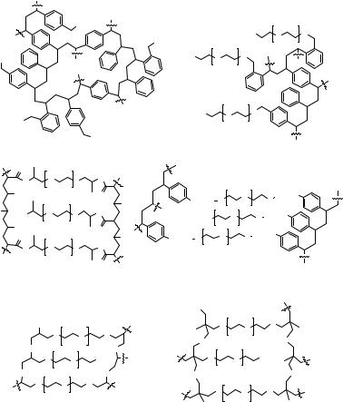
14.4 SUPPORTS FOR SOLID-PHASE LIBRARIES
Parallel arrays as well as libraries of glycopeptides can be synthesized in solution or on solid phase. The advantages of using solid phase methods23 are well documented, and detailed descriptions have been published.24,25 The choice of solid support is crucial and depends on the types of reactions to be carried out as well as the screening methods to be employed. The two most commonly used types of solid supports are based on polystyrene (PS) (e.g., Merrifield,23 including commonly used Wang resin26 and TentaGel27/ArgoGel) or polyethylene glycol (PEG) (e.g., PEGA,28 POEPOP,29 POEPS-3,30 and SPOCC31) (Fig. 14.5). Polystyrene resins are comprised of mainly 1–2% divinylbenzene crosslinked backbones with short linkers (Fig. 14.5a). Polystyrene based gels are generally unsuitable for direct on-bead screening either because of loss of enzymatic activity in the polymer or because of their poor swelling in polar media and consequent exclusion of biomolecules from the hydrophobic core of the polymer. This is also true for PS resins grafted with long PEG chains such as ArgoGel or TentaGel (Fig. 14.5b),32 although swelling in water is improved, and some access to the interior has been achieved.33,34 Furthermore, the polystyrene material absorbs light, thus interfering with some fluorescence assays, and the hydrophobic core may result in nonspecific protein binding.
PEG-based resins, on the other hand, are crosslinked with long-chain PEG macromonomers, which also present the amino or hydroxyl functional groups. Therefore, the mechanical and chemical properties of the resins are strongly influenced by the nature of the PEG chains.24 Additionally, PEG chains are highly miscible with most solvents creating a quasi-homogenous reaction medium. PEG
based resins swell tremendously in aqueous media, thus allowing biomolecules access to the entire bead.35,36 These resins are therefore suitable for protein ligation,37
enzymatic reactions, and screening for enzymatic activity and inhibition.34,38–43 Since PEG-based polymers confer the advantage of a support that is amenable to
both synthesis as well as screening of enzymatic activity and protein binding, efforts have been made to generate such polymers with superior properties in both areas. They may be obtained by radical polymerization of long-chain PEG macromonomers to give PEG-polyacrylamide (PEGA; Fig. 14.5c) copolymer28,35 or PEG-crosslinked oligostyrene (POEPS-3; Fig. 14.5d).30 Two novel types of gel supports obtained by anion catalyzed bulk polymerization of PEG, derivatized with epichlorohydrin
|
|
|
|
|
|
14.4 |
SUPPORTS FOR SOLID-PHASE LIBRARIES 289 |
|||||
|
|
|
|
Cl |
|
|
|
|
|
HO |
|
O |
|
|
|
|
|
|
|
Cl |
|
|
O |
||
|
|
|
|
|
|
|
|
|
|
m |
||
|
|
|
|
|
|
|
|
|
|
|
||
Cl |
|
|
|
|
|
|
HO |
O |
|
O |
|
|
|
|
|
|
|
|
|
m |
|
|
|||
|
|
|
|
|
|
|
|
|
|
|
||
|
|
|
|
|
|
|
HO |
|
O |
O |
|
|
|
Cl |
|
|
|
|
|
|
|
m |
|
|
|
|
|
|
|
|
|
|
|
|
|
|
||
|
|
a) CM-PS |
|
Cl |
|
|
|
|
|
|
|
|
|
|
|
|
|
|
b) TentaGel |
|
|
||||
|
O |
|
O |
|
H |
|
|
|
|
|
|
|
|
N |
|
O |
N |
|
|
|
|
|
|
|
|
|
R H |
|
|
n |
O |
|
|
|
|
O |
(CH2)3 |
|
R |
|
|
O |
|
H |
R |
(CH2)3 |
O |
|
n |
O |
|
H2N |
|
O |
N |
|
HO |
|
O |
O |
(CH2)3 |
|
||
|
|
|
|
n |
|
|
|
|
|
|
||
|
R |
|
|
|
O |
|
|
|
|
n |
|
|
|
O |
|
O |
|
H |
R |
(CH2)3 O |
O |
n |
O (CH2)3 |
|
|
|
N |
|
O |
N |
|
|
|
|
|
|
||
|
H |
|
|
n |
O |
|
|
|
|
|
|
|
|
|
|
|
|
|
|
|
|
|
|
|
|
|
c) PEGA, R: CON(CH3)2 or CONH2, |
|
d) POEPS-3 |
|
|
|||||||
|
|
|
|
|
|
|
HO |
|
|
|
|
|
|
|
HO |
|
|
|
|
|
O |
|
O n |
O |
|
|
|
|
O |
O |
O |
|
O |
|
|
O |
|
|
|
O |
|
n |
O |
|
|
|
|
||||
|
|
|
|
O |
|
|
OH |
|
||||
|
|
|
O |
O n |
OH |
|
|
O |
|
|||
|
O |
|
|
O |
|
O |
|
n |
O |
|
||
|
|
|
|
|
|
|
|
|
||||
|
|
O |
O |
n |
O |
|
|
O |
|
|
O |
|
|
|
|
|
|
|
|
O n |
|
||||
|
|
|
|
|
|
|
|
|
|
|
|
|
|
|
|
e) POEPOP |
|
|
f) SPOCC |
|
|
||||
Figure 14.5 The chemical structure of TentaGel and PEG-based resins. The open structure and the inert nature of PEG in biological systems confer ideal properties for bioassays to the PEG-based resins.24
(POEPOP; Fig. 14.5e)29 or by cation-catalyzed bulk polymerization of PEG, derivatized with oxetane (SPOCC, Fig. 14.5f)31 have been introduced. The inert character of the polymers allows the application of harsh organic reactions. Because of the excellent swelling in aqueous buffers, all the resins mentioned above have been used in bioassays for enzymes. PEGA resin was further investigated and showed no nonspecific binding in protein-binding studies.44
For the solid-phase synthesis of glycopeptides, both polystyrene and PEG-based resins have been successfully used. Experiments that compare the rates of reactions on various resins have revealed that the rate of reaction completely depends on the nature of the reaction itself.45 Some reactions perform better on hydrophobic resins, while others are better on hydrophilic resins.
290 PREPARATION AND SCREENING OF GLYCOPEPTIDE LIBRARIES
As of this writing, there are presently only two examples of successful synthesis of glycopeptide libraries in the literature, and these have been generated using PEG-based resins (PEGA and POEPOP), thus enabling rapid solid phase screening of the library (see Sections 14.8 and 14.7.2).
14.5 ANALYTICAL TOOLS FOR GLYCOPEPTIDE LIBRARIES
For solution-phase libraries that are composed of mixtures of compounds, the difficulty of analysis escalates with increasing numbers of compounds. Typically, large mixtures of compounds are not analyzed before screening, whereas small ones may be analyzed for reaction completeness using mass spectrometry, HPLC, NMR, or combinations thereof. The identification and analysis of active compounds from these mixtures is painstakingly tedious, and often complete characterization is possible only after deconvolution procedures and resynthesis of the active compound. For solid-phase libraries, the methods currently developed are discussed below.
14.5.1 Tagging Techniques for Libraries
The number of methods for analyzing nonpeptide libraries is increasing, and the methods generally fall into two categories: direct methods, usually based on mass spectrometry and NMR spectroscopy; and indirect methods, employing encoding, chemical,46–48 chemoluminescent,49 or other procedures.50–52 Many of the methods of chemical encoding are restricted by the additional synthetic effort required and the need to design orthogonal reaction conditions required for the two sets of syntheses. The currently most successful method utilizes a carbene insertion reaction to attach polyhalogenated aromatic tags that are photochemically released and then analyzed by GCMS.48
14.5.2 Analysis by Mass Spectrometry
The development glycopeptide libraries obtained by the split–mix method is severely hampered by the lack of concurrent development of a general, facile separation and characterization technology. Some headway has been made with chemical coding of the libraries, but very few direct methods of analysis exist. One promising method that could be applied to the direct characterization of both types of libraries is mass spectrometry. More specifically, post-source-decay matrix-assisted laser
desorption/ionization–time-of-flight mass spectrometry (PSD-MALDI-TOF-MS) and CID-FAB/MS/MS have been used to characterize glycopeptides.53–55
The analysis of glycopeptides has been carried out using primarily PSD-MALDI-TOF-MS. Two strategies are employed: (1) the glycan portion is first cleaved enzymatically or by base-catalyzed β-elimination54,56 and separately characterized by other means (MS or NMR) while the peptide is sequenced using PSD, or (2) the entire glycopeptide is fragmented by PSD and the spectra