

Flow Cytometry - First Principles (Second Edition)
.pdfFlow Cytometry: First Principles, Second Edition. Alice Longobardi Givan Copyright 2001 by Wiley-Liss, Inc.
ISBNs 0-471-38224-8 (Paper); 0-471-22394-8 (Electronic)
Flow
Cytometry
First Principles
Second Edition

Flow
Cytometry
First Principles
Second Edition
Alice Longobardi Givan
The Herbert C. Englert Cell Analysis Laboratory
of the Norris Cotton Cancer Center
and Department of Physiology
Dartmouth Medical School
Lebanon, New Hampshire
A John Wiley & Sons, Inc., Publication New York . Chichester . Weinheim . Brisbane . Singapore . Toronto
Designations used by companies to distinguish their products are often claimed as trademarks. In all instances where John Wiley & Sons, Inc., is aware of a claim, the product names appear in initial capital or all capital letters. Readers, however, should contact the appropriate companies for more complete information regarding trademarks and registration.
Copyright 2001 by Wiley-Liss, Inc. All rights reserved.
No part of this publication may be reproduced, stored in a retrieval system or transmitted in any form or by any means, electronic or mechanical, including uploading, downloading, printing, decompiling, recording or otherwise, except as permitted under Sections 107 or 108 of the 1976 United States Copyright Act, without the prior written permission of the Publisher. Requests to the Publisher for permission should be addressed to the Permissions Department, John Wiley & Sons, Inc., 605 Third Avenue, New York, NY 10158-0012, (212) 850-6011, fax (212) 850-6008, E-Mail: PERMREQ @ WILEY.COM.
This publication is designed to provide accurate and authoritative information in regard to the subject matter covered. It is sold with the understanding that the publisher is not engaged in rendering professional services. If professional advice or other expert assistance is required, the services of a competent professional person should be sought.
ISBN 0-471-22394-8
This title is also available in print as ISBN 0-471-38224-8.
For more information about Wiley products, visit our web site at www.Wiley.com.
The First Edition of this book was dedicated to my parents, Violet Litwin Longobardi and Vincent Longobardi, Jr., with gratitude for the example they set,
with pride in their achievements, and with love.
The Second Edition is dedicated to Curt, Ben, and Becky
(not only because I promised them that their turn would come).
Contents
Preface xi
Acknowledgments (First Edition) xv
Acknowledgments (Second Edition) xvii
1
2
3
4
The Past as Prologue 1
Setting the Scene 11
Instrumentation: Into the Black Box 15
Illumination of the Stream 16
Centering Cells in the Illuminating Beam 18 Detection of Signals from Cells 25 Electronics 31
Information: Harnessing the Data 41
Data Storage |
41 |
Data Analysis |
43 |
5 Seeing the Light: Lasers, Fluorochromes, and
Filters 59
General Theory |
59 |
Lasers 61 |
|
Fluorochromes |
65 |
Partitioning the Signal with Lenses, Filters, and Mirrors 72
Spectral Compensation 76
vii
viii |
Flow Cytometry |
6Cells from Without: Leukocytes, Surface Proteins, and
the Strategy of Gating |
81 |
|
|
|||
Cells from Blood |
82 |
|
|
|
||
Staining for Surface Markers 87 |
|
|
||||
Controls |
90 |
|
|
|
|
|
Quantitation |
95 |
|
|
|
|
|
Sensitivity |
98 |
|
|
|
|
|
The Strategy of Gating |
99 |
|
|
|||
Gating on Fluorescence |
108 |
|
|
|||
7 Cells from Within: Intracellular Proteins |
115 |
|||||
Methods for Permeabilizing Cells |
116 |
|
||||
Examples of Intracellular Staining |
118 |
|
||||
8 Cells from Within: DNA in Life and Death |
123 |
|||||
Fluorochromes for DNA Analysis |
123 |
|
||||
Ploidy 125 |
|
|
|
|
|
|
Cell Cycle Analysis |
131 |
|
|
|||
Two-Color Analysis for DNA and Another |
||||||
Parameter |
142 |
|
|
|
|
|
Chromosomes |
147 |
|
|
|
||
Apoptosis |
150 |
|
|
|
|
|
Necrosis |
154 |
|
|
|
|
|
9
10
The Sorting of Cells |
159 |
|
|
Sorting Theory |
159 |
|
|
Characterization of Sorted Cells |
166 |
||
Alternative Methods for Sorting |
170 |
||
The Condition of Cells After Sorting 172 |
|||
Disease and Diagnosis: The Clinical Laboratory 175 |
|||
The Hematology Laboratory |
178 |
||
The Pathology Laboratory |
186 |
|
|
Solid Organ Transplantation |
189 |
||
Comments 192 |
|
|
|
|
Contents |
|
ix |
|
11 Out of the Mainstream: Research Frontiers |
195 |
|||
|
Functional Assays |
196 |
|
|
|
The Aquatic Environment |
202 |
|
|
|
Reporter Molecules |
207 |
|
|
|
Microbiology 211 |
|
|
|
|
Molecular Biology |
213 |
|
|
|
Multiplex Cytometry for Soluble Analytes |
218 |
||
|
Reproductive Technology |
220 |
|
|
12 Flowing On: The Future |
225 |
|
|
|
General References 229 |
|
|
|
|
Glossary |
235 |
|
|
|
Figure Credits 257 |
|
|
|
|
Index |
263 |
|
|
|
Preface
Although ¯ow cytometry is simply a technique that is useful in certain ®elds of scienti®c endeavor, there is, at the same time, something special about it. Few other techniques involve specialists from so many di¨erent backgrounds. Anyone working with ¯ow systems for any length of time will realize that computer bu¨s, electronics experts, mathematicians, optical and ¯uidics engineers, and organic chemists rub shoulders with biologists, physicians, and surgeons around the ¯ow cytometer bench.
And it is not just a casual rubbing of shoulders, in passing, so to speak. Many of the specialists involved in ¯ow cytometry might, if asked, call themselves ¯ow cytometrists because the second aspect of ¯ow cytometry that distinguishes it from many other techniques is that ¯ow cytometry has itself become a ``®eld.'' Indeed, it is a ®eld of endeavor and of expertise that has captured the imaginations of many people. As a result, there exists a spirit of camaraderie; ¯ow cytometry societies, groups, meetings, networks, websites, journals, courses, and books abound.
A third aspect of ¯ow cytometry (known sometimes simply with the acronym for ¯uorescence-activated cell sorter, FACS, or even more familiarly as just ¯ow) that distinguishes it from many other techniques is the way in which its wide and increasing usefulness has continued to surprise even those who consider themselves experts. What began as a clever technique for looking at a very limited range of problems is now being used in universities, in hospitals, within industry, at marine stations, on submersible buoys, and on board ships; plans have existed for use on board space ships as well. The applications of ¯ow cytometry have proliferated (and continue to proliferate) rapidly both in the direction of theoretical science, with
xi
xii |
Flow Cytometry |
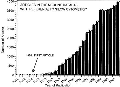
Fig. 1. Increasing reference to ``¯ow cytometry'' in the medical literature over the past three decades. The development of ¯ow cytometers antedates the use of the term itself.
botany, molecular biology, embryology, biochemistry, marine ecology, genetics, microbiology, and immunology, for example, all represented; and in the direction of clinical diagnosis and medical practice, with hematology, bacteriology, pathology, oncology, obstetrics, and surgery involved. We are, at present, living through what appears to be a rapid phase in ¯ow cytometry's growth curve (see Fig. 1).
Because ¯ow cytometry is an unusual ®eld, bringing together people with di¨ering scienti®c backgrounds at meetings, on editorial boards, in hospital wards, on advisory panels, and at laboratory benches and reaching increasing numbers of workers in new and unpredicted areas of endeavor, there is, as a result, a need to provide both recent and potential entrants into this diverse community with a common basis of knowledgeÐso that we can all understand the vocabulary, the assumptions, the strengths, and the weaknesses of the technology involved. I have for many years taught new and future users of ¯ow cytometers. My teaching attempts to present enough technical background to enable students, scientists, technologists, and
Preface |
xiii |
clinicians to read the literature critically, to evaluate the bene®ts of the technique realistically, and, if tempted, to design e¨ective protocols and interpret the results. I try to describe the theory of ¯ow cytometry in a way that also provides a ®rm (and accurate) foundation for those few who will go on to study the technique in greater depth. Details of protocols are avoided, but my teaching attempts to give enough information about applications to provide concrete examples of general concepts and to allow some appreciation of the range of practical goals that the instruments are able to achieve. With some expansion (but with little change in style or objectives), my classroom and workshop teaching is the basis for this book.
Notes on the Second Edition: This new edition, while similar to the ®rst edition in style and scope, has been modi®ed in many ways. The arrangement of some material has been altered to present, in my opinion, a more coherent pedagogical sequence, re¯ecting my changing thoughts on teaching. While all chapters have been re-written to a signi®cant extent, there are also some major expansions re¯ecting the progress of the ®eld. In particular, I have included more detail about the cell as it passes through the laser beam; the laser/¯uorochrome chapter has been expanded to include recently developed ¯uorochromes and multilaser options; a new chapter has been added on cytoplasmic staining; a discussion of apoptosis has been added to the chapter on DNA; the section on sorting has been expanded to a full chapter and includes high-speed sorting and alternative sorting methods as well as traditional technology; the clinical and research chapters have been updated and expanded considerably; the chapter on general references includes many of the recent excellent books in the ®eld; and the chapter on the future of ¯ow cytometry is now a subjective glimpse into the new decade from the vantage point of the year 2000.