

Flow Cytometry - First Principles (Second Edition)
.pdf180 |
Flow Cytometry |
Fig. 10.2. Surface antigen changes during hematopoiesis. The upper plot is of T-lymphocyte maturation and the lower plot of B cells. From Loken and Wells (2000).
treatment. In addition, the ¯ow phenotype of the malignancy can be used to look for the emergence of small numbers of these cells should relapse occur. This, of course, assumes that the relapse phenotype is identical to the abnormal clone de®ned at the initial diagnosis (Fig. 10.3).

Disease and Diagnosis |
181 |
|||||
|
|
|
|
|
|
|
|
|
|
|
|
|
|
|
|
|
|
|
|
|
|
|
|
|
|
|
|
|
|
|
|
|
|
|
|
|
|
|
|
|
|
|
|
|
|
|
|
|
Fig. 10.3. Clusters of cells indicating an abnormal (acute myeloid leukemia) leukemic population, at diagnosis, at remission, and after relapse. The leukemic phenotype is CD13, CD34, and CD33 positive and CD11b negative. Courtesy of Carleton Stewart.
HIV/AIDS
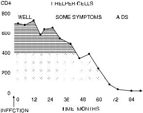
Another condition that involves analysis of peripheral blood leukocytes is AIDS. Early in the natural history of the disease (or at least in the natural history of immunologists' awareness of the disease), it was discovered that one subpopulation of T lymphocytes in particular was destroyed by the HIV virus; the cells destroyed are those that possess the CD4 protein on their surface. It is this CD4 protein that appears to be a receptor involved in virus targeting. Therefore, much of the diagnosis and staging of AIDS involves the enumeration of CD4-positive cells in the peripheral blood (Fig. 10.4).
These techniquesÐfor counting CD4-positive cells in connection with AIDS diagnosis and for phenotyping various populations of
182 |
|
|
|
|
|
|
|
|
|
|
|
|
|
|
|
|
|
|
|
Flow Cytometry |
|||||||||||||||||||||||||||||||||||||||||||||
|
|
|
|
|
|
|
|
|
|
|
|
|
|
|
|
|
|
|
|
|
|
|
|
|
|
|
|
|
|
|
|
|
|
|
|
|
|
|
|
|
|
|
|
|
|
|
|
|
|
|
|
|
|
|
|
|
|
|
|
|
|
|
|
|
|
|
|
|
|
|
|
|
|
|
|
|
|
|
|
|
|
|
|
|
|
|
|
|
|
|
|
|
|
|
|
|
|
|
|
|
|
|
|
|
|
|
|
|
|
|
|
|
|
|
|
|
|
|
|
|
|
|
|
|
|
|
|
|
|
|
|
|
|
|
|
|
|
|
|
|
|
|
|
|
|
|
|
|
|
|
|
|
|
|
|
|
|
|
|
|
|
|
|
|
|
|
|
|
|
|
|
|
|
|
|
|
|
|
|
|
|
|
|
|
|
|
|
|
|
|
|
|
|
|
|
|
|
|
|
|
|
|
|
|
|
|
|
|
|
|
|
|
|
|
|
|
|
|
|
|
|
|
|
|
|
|
|
|
|
|
|
|
|
|
|
|
|
|
|
|
|
|
|
|
|
|
|
|
|
|
|
|
|
|
|
|
|
|
|
|
|
|
|
|
|
|
|
|
|
|
|
|
|
|
|
|
|
|
|
|
|
|
|
|
|
|
|
|
|
|
|
|
|
|
|
|
|
|
|
|
|
|
|
|
|
|
|
|
|
|
|
|
|
|
|
|
|
|
|
|
|
|
|
|
|
|
|
|
|
|
|
|
|
|
|
|
|
|
|
|
|
|
|
|
|
|
|
|
|
|
|
|
|
|
|
|
|
|
|
|
|
|
|
|
|
|
|
|
|
|
|
|
|
|
|
|
|
|
|
|
|
|
|
|
|
|
|
|
|
|
|
|
|
|
|
|
|
|
|
|
|
|
|
|
|
|
|
|
|
|
|
|
|
|
|
|
|
|
|
|
|
|
|
|
|
|
|
|
|
|
|
|
|
|
|
|
|
|
|
|
|
|
|
|
|
|
|
|
|
|
|
|
|
|
|
|
|
|
|
|
|
Fig. 10.4. The number of T-helper (CD4-positive) lymphocytes ( 103=cm3) in peripheral blood of a patient after infection with HIV. Cells <400 are signi®cantly below the normal range; <100 indicates severe risk of clinical AIDS. Courtesy of LeÂonie Walker.
leukocytes for leukemia/lymphoma diagnosisÐcan be performed in hematology laboratories by the staining of cells with ¯uorochromeconjugated monoclonal antibodies followed by the visual identi®cation of di¨erent types of white cells and the counting of the ¯uorescent versus unstained cells under the microscope. Although the microscope has certain very de®nite advantages over the ¯ow cytometer, two advantages it does not have are those of speed and statistical reliability. Particularly as a result of their work load from the growing number of AIDS patients, hematologists' need for a way to count statistically reliable numbers of cells from large numbers of patients became increasingly urgent. Flow cytometry was the obvious answer to this need.
Erythrocytes and Platelets
Although the initial perceived need in the hematology laboratory for a ¯ow cytometer was to aid in the rapid processing of samples from leukemic and HIV-positive patients, the presence of the cytometer has
Disease and Diagnosis |
183 |
stimulated thought about new hematological applications. Although not yet in routine use for analyzing erythrocytes, ¯ow cytometers have been shown to be useful for looking at red-cell±bound immunoglobulin (as a result of autoimmune disease, sickle cell anemia, and thalassemia): The bound immunoglobulin on erythrocytes is detected by the use of ¯uorescent antibodies against human immunoglobulin. The staining of red cells for RNA content with a dye called thiazole orange has made possible the use of ¯ow cytometry to count the reticulocytes (immature erythrocytes) present in blood samples from anemic patients.
Hematologists have also extended the use of ¯ow analysis to plateletsÐthose particles with low forward scatter that usually are ignored in ¯ow cytometric applications because they fall well below the standard forward scatter threshold. Immunoglobulin bound to platelets can be measured with antibodies against human immunoglobulin (as in the detection of immunoglobulin on erythrocytes). Platelet-associated immunoglobulin (resulting from autoimmune disease) is determined by analyzing the patient's platelets in their natural state. In another clinical situation, antibodies in the serum with speci®cities for ``foreign'' platelets may develop after pregnancy or transfusions; these can be monitored by ¯ow cytometry if a patient's serum is incubated with a donor's platelets. Recently, the activation state of platelets has also been analyzed. Hyperactive platelets express P-selectin (CD62) on their surface; they have been primed to facilitate coagulation. Although not yet applied in routine diagnosis, research indicates that expression of CD62 may provide prognostic information in myocardial infarction and cardiopulmonary bypass surgery.
HLA-B27 Typing
Human cells express a series of proteins on their surface that are involved in self-recognition and in antigen presentation to e¨ector cells for the stimulation of immune reactions. These are called major histocompatibility (MHC) antigens, and each occurs in a variety of allelic forms. People, therefore, di¨er from each other in their MHC genotypes. As well as a¨ecting transplant compatibility, certain genotypes have been implicated in predisposition to autoimmune disease.
184 |
Flow Cytometry |
In particular, HLA-B27 (the 27th haplotype at the B locus of the human leucocyte antigens), has been associated with arthritic conditions (ankylosing spondylitis, juvenile rheumatoid arthritis, and Reiter's syndrome). While only about 20% of people with the HLAB27 genotype have ankylosing spondylitis, for example, a very high percentage (perhaps 90%) of people with this disease are HLA-B27 positive (compared with 4±8% in the general population). Antibodies against the HLA-B27 protein are available. Leukocytes can be incubated with ¯uorochrome-conjugated antibodies; positive staining indicates expression of the HLA-B27 protein. This assay can be performed with a microscope (using either ¯uorescent antibodies or complement-®xing antibodies that kill cells). Recently, increasing numbers of hematology laboratories have begun to use ¯ow cytometers for this assay.
CD34 Stem Cells
After a cancer patient has been treated with irradiation to destroy malignant cells, normal blood cells will also be depleted and the patient may require transplantation to regenerate these cells. Hematopoietic stem cells (classi®ed by possession of the CD34 antigen on their surface) are immature, undi¨erentiated cells that have the ability to di¨erentiate into all classes of blood cells. They exist, normally, in the bone marrow; detectable numbers can be found in the peripheral blood if the patient has been treated with blood cell growth factors. If enough of these stem cells can be harvested from the patient before irradiation, transplantation of these cells back into the patient after irradiation can replenish the immune system. The number of these CD34-positive cells given back after irradiation needs, therefore, to be assayed in order to ensure that the stem cell transplant is su½cient to return the patient to normal immune competency. Because these stem cells are uniquely characterized by the CD34 antigen, ¯ow cytometry can be used to con®rm the presence of su½cient stem cells in a cell suspension obtained from the patient before irradiation (Fig. 10.5). Because the actual number of these stem cells (rather than just their proportion in a 10,000 cell data ®le) is important, the volume ¯owing through the ¯ow cytometer can be calibrated by the addition of known numbers of beads to the sample.

|
|
|
Disease and Diagnosis |
185 |
||||||||
|
|
|
|
|
|
|
|
|
|
|
|
|
|
|
|
|
|
|
|
|
|
|
|
|
|
|
|
|
|
|
|
|
|
|
|
|
|
|
|
|
|
|
|
|
|
|
|
|
|
|
|
|
|
|
|
|
|
|
|
|
|
|
|
|
|
|
|
|
|
|
|
|
|
|
|
|
|
Fig. 10.5. Stem cells can be assayed by the use of antibodies against the CD34 and CD45 antigens. From R Sutherland as published in Gee and Lamb (2000).
Fetal Hemoglobin
Detection of fetal erythrocytes in the maternal circulation is important in diagnosing a cross-placental fetal±maternal hemorrhage that might have resulted from trauma with suspected maternal injury. In addition, in the case of Rh-blood group incompatibility between fetus and mother, rapid detection of fetal cells in the maternal circulation can allow early intervention and the prevention of Rh-incompatibility disease (erythroblastosis fetalis) in the newborn. Although detection of these fetal cells is di½cult because their proportion in the maternal circulation is low (less than 0.1% in a normal pregnancy; perhaps 0.6% in a pregnancy that needs therapeutic intervention), they can be assayed by the use of antibodies against the fetal form of hemoglobin (HbF). This procedure is typical of ¯ow protocols concerned with rare-event analysis (Fig. 10.6). De®ning the cells of interest (in this case the HbF-positive cells) by multiple parameters (forward scatter, side scatter, auto¯uorescence, HbF) reduces the likelihood of false positivity resulting from background noise.

186 |
Flow Cytometry |
Fig. 10.6. Detection of rare fetal erythrocytes in the maternal circulation. The fetal erythrocytes are HbF positive and low in auto¯uorescence. The cells in R1 are displayed and enumerated in the bottom right histogram. From Davis (1998).
THE PATHOLOGY LABORATORY
Tumor Ploidy
In the chapter about DNA analysis, I mentioned the large amount of work generated for ¯ow laboratories as a result of publication of the Hedley technique for analyzing the DNA content of para½nembedded pathology specimens. After the ®rst headlong rush of publications correlating DNA ploidy with long-term prognosis in
Disease and Diagnosis |
187 |
various types of cancer, the ®eld has now settled down a bit. As I have indicated, it would probably be fair to say that most (but not all) studies on disaggregated solid tumors (fresh, frozen, or ®xed) have shown some kind of correlation between abnormal DNA content and unfavorable long-term prognosis.
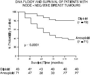
There is considerable debate in the literature about whether ¯ow cytometric analysis of ploidy gives any additional prognostic information that is independent of other known prognostic indicators, but most studies have indicated that it does. It appears to be useful, for example, in indicating, from all breast cancer patients without lymph node involvement, a group of women who are at risk for recurrence of disease (Fig. 10.7). Because any pathological material will be a mixture of both normal and abnormal cells, there is much current e¨ort expended on ways of selecting from among the cells of a disaggregated specimen those particular cells that are from the tumor itself. If those tumor cells can be stained selectively (as with a ¯uorescein-conjugated anti-cytokeratin antibody for epithelial cellderived tumors within nonepithelial tissue) and then the DNA con-
Fig. 10.7. Survival curves for breast cancer patients without lymph node involvement. Those with DNA aneuploid tumors (as diagnosed by propidium iodide ¯ow cytometry of 10-year-old para½n-embedded material) survived signi®cantly less long than those with DNA diploid tumors. Graph from Yuan et al. (1991).

188 |
Flow Cytometry |
tent of the ¯uorescein-stained cells determined, we may have a dual color ¯ow method of much improved sensitivity for detecting aneuploid cells from within a mixture of both malignant and normal components.
S-Phase Fraction
There is also evidence, in solid tumors as well as in the bone marrow plasma cells from patients with multiple myeloma, that determination of the proliferative potential of malignant cells is a better indicator of poor prognosis than simply the determination of the presence or absence of aneuploid cells. As discussed in the chapter on DNA, proliferation (the proportion of cells in S phase) can be assessed by using mathematical algorithms to analyze the shape of the DNA histogram. However, the presence of more than one cell type, with the G2/M peak from the diploid cell line overlapping the S phase of the aneuploid cells, makes mathematical analysis even more complex than with distributions resulting from single euploid cells or clones (Fig. 10.8). Nevertheless, correlations between high S-phase fraction and poor clinical prognosis appear relatively strong. Bromodeoxyuridine (BrdU) has been used in the research setting to give information about proliferation; it has been used both in vitro with fresh excised tumors and in vivo by infusion into patients at some time before
Fig. 10.8. Analysis of mixed diploid/aneuploid cells for the S-phase fraction of the aneuploid population.
Disease and Diagnosis |
189 |
excision of the tumor. Estimates of tumor doubling time, based on the rate of movement of BrdU-pulsed cells through the cell cycle and on the percentage of cells in the tumor that are dividing, have been shown to correlate with aggressiveness of the malignancy. There may in the future be more use of this technique for assaying malignancies for sensitivity to various cytotoxic drugs.
Ploidy and S-phase fraction determinations are, in most cases, the only ¯ow analyses that have achieved much routine application in the ®eld of solid tissue oncology. At the present time, it would appear that ¯ow cytometry has not settled quite so comfortably into the pathology laboratory as it has into the hematology setting. This may, I suspect, come from the crucial di¨erence between hematology and pathologyÐand it is a di¨erence that may remind us of one very de®nite limitation of ¯ow analysis. Pathological specimens come from solid tissue; to analyze them with a ¯ow cytometer, the solid tissue must be disaggregated in some way. This disaggregation process (either mechanical, with detergent, or with enzymes) not only has a good chance of compromising the integrity of some or many of the cells we want to analyze, but ¯ow cytometry will also, necessarily, lose any information that is contained in the structural orientation or pattern of the original cells and tissue, as viewed under the microscope.
A response to this dilemma has come, recently, in the development of laser-scanning cytometry (the LSC by CompuCyte). The LSC uses a laser to scan cells or tissues on slides and acquires data that can be presented much as any ¯ow cytometric ¯uorescence or light scatter information (in histograms or two-color plots). However, the LSC also acquires extra parameters that record the x/y position of each cell on the slide. In this way, cells that show aberrant ¯uorescence or scatter characteristics can be examined individually under the LSC's microscope to provide high-resolution images and con®rm malignant or otherwise interesting phenotype. In many ways, the LSC bridges the technologies of ¯ow cytometry and traditional microscopic pathology.
SOLID ORGAN TRANSPLANTATION
It has been known for many years that transplant recipients may possess serum antibodies that will react with and destroy cells from a transplanted organ. If these so-called cytotoxic antibodies are present at the time of transplantation, then an immediate and violent rejec-