7.2 ● Monosaccharides |
211 |
ALDOTRIOSE
1 CHO
|
|
|
|
|
|
|
|
|
|
|
|
|
|
|
|
|
|
|
|
Carbon 2 |
|
|
|
|
|
|
|
|
|
|
|
|
|
|
|
|
|
|
|
|
|
|
|
|
|
|
|
|
|
|
|
|
|
|
|
|
|
|
|
|
|
|
|
|
|
HCOH |
|
|
|
|
|
|
|
|
|
|
|
|
|
|
|
|
|
|
|
|
|
|
|
|
|
|
|
|
|
|
|
|
|
|
|
|
|
|
|
|
|
|
|
|
|
|
|
number |
|
|
|
|
|
|
|
|
|
|
|
|
|
|
|
|
|
|
|
|
|
|
|
|
|
|
|
|
|
|
|
|
|
|
|
|
|
|
|
|
|
|
|
|
|
|
|
|
|
|
|
|
|
3 |
|
|
CH2OH |
|
|
|
|
|
|
|
|
|
|
|
|
|
|
|
|
|
|
|
|
|
|
|
|
|
|
|
|
|
|
|
|
|
|
|
|
|
|
|
|
|
|
|
|
|
D-Glyceraldehyde |
|
|
|
|
|
|
|
|
|
|
|
|
|
|
|
|
|
|
|
|
|
|
|
|
|
|
1 |
|
|
CHO |
|
|
|
|
|
|
|
|
|
|
|
|
|
|
|
|
|
|
|
|
|
|
|
|
|
|
|
CHO |
|
|
|
|
|
|
|
|
|
|
|
|
|
|
|
|
|
|
|
|
|
|
|
|
|
|
|
|
|
|
|
|
|
|
|
|
|
|
|
|
|
|
|
|
|
|
|
|
|
|
|
|
|
|
|
|
|
|
|
|
|
|
|
|
|
|
|
2 |
|
HCOH |
|
|
|
|
|
|
|
|
|
|
|
|
|
|
|
|
|
|
|
|
|
|
|
|
HOCH |
|
|
|
|
|
|
|
|
|
|
|
|
|
|
|
|
|
|
|
Carbon |
|
|
|
|
|
|
|
|
|
|
|
|
ALDOTETROSES |
|
|
|
|
|
|
|
|
|
|
|
|
|
|
|
|
|
|
|
|
|
|
|
|
|
|
|
number 3 |
|
|
|
|
|
|
|
|
|
|
|
|
|
|
|
|
|
|
|
|
|
|
|
|
|
|
|
|
|
|
|
|
|
|
|
|
|
|
|
HCOH |
|
|
|
|
|
|
|
|
|
|
|
|
|
|
|
|
|
|
|
|
|
|
|
|
|
|
HCOH |
|
|
|
|
|
|
|
|
|
|
|
|
|
|
|
|
|
|
|
|
|
|
|
|
|
|
|
|
|
|
|
|
|
|
|
|
|
|
|
|
|
|
|
|
|
|
|
|
|
|
|
|
|
|
|
|
|
|
|
|
|
|
|
|
|
|
|
|
|
|
|
|
|
|
4 |
|
|
CH2OH |
|
|
|
|
|
|
|
|
|
|
|
|
|
|
|
|
|
|
|
|
CH2OH |
|
|
|
|
|
|
|
|
|
|
|
|
|
|
|
|
D-Erythrose |
|
|
|
|
|
|
|
|
|
|
|
|
|
|
|
|
|
|
|
D-Threose |
|
|
|
|
|
|
1 |
|
|
CHO |
|
|
|
|
|
|
|
CHO |
|
|
|
|
|
|
|
|
|
|
|
|
CHO |
|
|
|
|
|
|
|
|
|
|
|
|
|
CHO |
|
|
|
|
|
|
2 |
|
|
|
|
|
|
|
|
|
|
|
|
|
|
|
|
|
|
|
|
|
|
|
|
|
|
|
|
|
|
|
|
|
|
|
|
|
|
|
|
|
|
|
|
|
|
|
|
|
|
|
|
|
|
|
|
HCOH |
|
|
|
|
|
|
|
|
|
HOCH |
|
|
|
|
|
|
|
|
|
|
|
|
|
HCOH |
|
|
|
|
|
|
|
|
|
|
|
HOCH |
|
|
|
|
|
|
Carbon |
|
|
|
|
|
|
|
|
|
|
|
|
|
|
|
|
|
|
|
|
|
|
|
|
|
|
|
|
|
|
|
|
|
|
|
|
|
|
|
|
|
|
|
|
|
|
|
|
|
|
|
|
|
HCOH |
|
|
|
|
|
|
|
HCOH |
|
|
ALDOPENTOSES HOCH |
|
|
|
|
|
|
|
|
|
|
|
HOCH |
|
|
|
|
|
number 3 |
|
|
|
|
|
|
|
|
|
|
|
|
|
|
|
|
|
|
|
|
|
|
|
|
|
|
|
|
|
|
|
|
|
|
|
|
|
|
|
|
|
|
|
|
|
|
|
|
|
|
|
|
|
|
|
|
|
|
|
|
|
|
|
|
|
|
|
|
|
|
|
|
|
|
|
|
|
|
|
4 |
|
HCOH |
|
|
|
|
|
|
|
|
|
|
|
HCOH |
|
|
|
|
|
|
|
|
|
|
|
|
HCOH |
|
|
|
|
|
|
|
|
|
|
|
|
|
HCOH |
|
|
|
|
|
|
|
|
|
|
|
|
|
|
|
|
|
|
|
|
|
|
|
|
|
|
|
|
|
|
|
|
|
|
|
|
|
|
|
|
|
|
|
|
|
|
|
|
|
|
|
|
|
|
|
|
|
|
|
|
5 |
|
|
CH2OH |
|
|
|
|
|
|
|
CH2OH |
|
|
|
|
|
|
|
|
CH2OH |
|
|
|
|
|
CH2OH |
|
|
D-Ribose (Rib) |
|
|
|
|
D-Arabinose (Ara) |
|
|
|
|
|
|
D-Xylose (Xyl) |
|
|
D-Lyxose (Lyx) |
1 |
CHO |
|
|
|
|
|
CHO |
|
|
|
CHO |
|
CHO |
|
|
CHO |
|
CHO |
|
CHO |
|
CHO |
2 |
|
|
|
|
|
|
|
|
|
|
|
|
|
|
|
|
|
|
|
|
|
|
|
|
|
|
|
|
|
|
|
|
|
|
|
|
|
|
|
|
|
|
|
|
|
|
|
|
|
|
|
|
|
|
|
|
HCOH |
|
|
|
|
HOCH |
|
|
|
|
HCOH |
|
HOCH |
|
|
|
|
|
|
HCOH |
|
HOCH |
|
|
HCOH |
|
HOCH |
|
|
|
|
|
|
|
|
|
|
|
|
|
|
|
|
|
|
|
|
|
|
|
|
|
|
|
|
|
|
|
|
|
|
|
|
|
|
|
|
|
|
|
|
|
3 |
HCOH |
|
|
|
|
|
HCOH |
|
|
HOCH |
HOCH |
|
|
|
|
|
HCOH |
|
HCOH |
HOCH |
HOCH |
|
|
|
|
|
|
|
|
|
|
|
|
|
|
|
|
|
|
|
|
|
|
|
|
|
|
|
|
|
|
|
|
|
|
|
|
|
|
|
|
4 |
HCOH |
|
|
|
|
|
HCOH |
|
|
|
HCOH |
|
HCOH |
HOCH |
HOCH |
HOCH |
HOCH |
5 |
|
|
|
|
|
|
|
|
|
|
|
|
|
|
|
|
|
|
|
|
|
|
|
|
|
|
|
|
|
|
|
|
|
|
|
|
|
|
|
|
|
|
|
|
|
|
|
|
|
|
|
|
|
|
|
|
HCOH |
|
|
|
|
|
|
HCOH |
|
|
|
HCOH |
|
|
HCOH |
|
|
|
HCOH |
|
|
HCOH |
|
|
HCOH |
|
|
HCOH |
|
|
|
|
|
|
|
|
|
|
|
|
|
|
|
|
|
|
|
|
|
|
|
|
|
|
|
|
|
|
|
|
|
|
|
|
|
|
|
|
|
6 |
CH2OH |
|
|
|
|
CH2OH |
CH2OH |
|
CH2OH |
|
|
CH2OH |
|
CH2OH |
|
CH2OH |
|
CH2OH |
|
D-Allose |
|
|
|
|
|
D-Altrose |
D-Glucose |
|
D-Mannose |
|
|
D-Gulose |
|
D-Idose |
D-Galactose |
|
D-Talose |
|
|
|
|
|
|
|
|
|
|
|
|
|
|
|
(Glc) |
|
(Man) |
|
|
|
|
|
|
|
|
|
|
|
|
|
|
|
|
(Gal) |
|
|
|
|
|
ALDOHEXOSES
FIGURE 7.2 ● The structure and stereochemical relationships of D-aldoses having three to six carbons. The configuration in each case is determined by the highest numbered asymmetric carbon (shown in gray). In each row, the “new” asymmetric carbon is shown in red.
As noted in Chapter 4, the Fischer projection system is used almost universally for this purpose today. The structures shown in Figures 7.2 and 7.3 are Fischer projections. For monosaccharides with two or more asymmetric carbons, the prefix D or L refers to the configuration of the highest numbered asymmetric carbon (the asymmetric carbon farthest from the carbonyl carbon). A monosaccharide is designated D if the hydroxyl group on the highest numbered asymmetric carbon is drawn to the right in a Fischer projection, as in D-glyc- eraldehyde (Figure 7.1). Note that the designation D or L merely relates the
FIGURE 7.3
212 Chapter 7 ● Carbohydrates
● The structure and stereochemical relationships of D-ketoses having three to six carbons. The configuration in each case is determined by the highest numbered asymmetric carbon (shown in gray). In each row, the “new” asymmetric carbon is shown in red.
|
1 |
CH2OH |
|
Carbon |
|
|
|
|
|
|
|
2 |
C |
|
|
O |
KETOTRIOSE |
|
|
number |
|
|
|
|
|
|
|
|
|
|
|
|
|
|
|
|
|
|
|
|
|
|
|
|
|
3 |
CH2OH |
|
|
Dihydroxyacetone |
|
|
|
|
|
|
|
|
|
|
|
1 |
CH2OH |
|
|
|
|
|
|
|
|
|
Carbon |
2 |
C |
|
|
O |
KETOTETROSE |
|
|
|
|
|
|
|
|
|
|
|
number 3 |
|
|
|
|
|
|
HCOH |
|
|
|
|
|
|
|
|
|
|
|
4 CH2OH
D-Erythrulose
|
|
|
|
1 |
CH2OH |
|
|
|
|
|
|
CH2OH |
|
|
|
|
|
|
|
|
|
|
|
|
|
|
|
|
|
|
|
|
|
|
|
|
|
|
Carbon |
2 |
C |
|
|
O |
|
|
|
|
|
|
C |
|
O |
|
|
|
|
|
|
|
|
|
|
|
|
|
|
|
|
|
|
|
|
|
|
|
|
|
|
|
|
|
|
|
|
|
|
|
|
|
|
|
|
|
|
|
|
|
|
3 |
HCOH |
|
|
|
|
|
|
|
|
|
|
|
HOCH |
|
|
|
|
|
|
KETOPENTOSES |
number |
|
|
|
|
|
|
|
|
|
|
|
|
|
|
|
|
|
|
|
|
|
|
|
|
|
|
|
|
|
|
|
|
|
|
|
|
|
|
|
|
|
|
|
|
|
|
|
|
|
|
|
|
|
|
|
|
|
|
|
|
|
|
|
|
|
|
|
|
|
|
|
|
|
|
|
|
|
|
|
|
|
|
|
|
|
|
|
|
|
|
|
|
|
|
|
4 |
HCOH |
|
|
|
|
|
|
|
|
|
|
|
|
HCOH |
|
|
|
|
|
|
|
|
|
|
|
|
|
|
|
|
|
|
|
|
|
|
|
|
|
|
|
|
|
|
|
|
|
|
|
5 |
CH2OH |
|
|
|
|
|
|
CH2OH |
|
|
|
|
|
|
D-Ribulose |
|
|
|
|
|
|
D-Xylulose |
1 |
CH2OH |
|
|
|
|
|
CH2OH |
|
CH2OH |
|
CH2OH |
|
|
|
|
|
|
|
|
|
|
|
|
|
|
|
|
|
|
|
|
|
|
|
|
2 |
C |
|
O |
|
|
|
|
|
C |
|
O |
|
C |
|
O |
|
C |
|
O |
|
|
|
|
|
|
|
|
|
|
|
|
|
|
|
|
|
|
|
|
|
|
|
|
|
|
|
|
|
|
|
|
|
|
|
|
|
|
|
|
|
|
|
|
|
|
|
|
|
|
|
|
|
|
|
3 |
HCOH |
|
|
|
|
HOCH |
|
|
|
|
HCOH |
|
|
|
|
HOCH |
|
|
|
Carbon |
|
|
|
|
|
|
|
|
|
|
|
|
|
|
|
|
|
|
|
|
|
|
|
|
|
|
|
|
|
|
|
KETOHEXOSES |
|
|
|
|
|
|
|
|
|
|
|
|
|
|
|
|
|
|
|
|
|
|
|
|
|
|
|
|
|
|
|
number 4 |
|
|
|
|
|
|
|
|
|
|
|
|
|
|
|
|
|
|
|
|
|
|
|
|
|
|
|
|
|
|
|
HCOH |
|
|
|
|
|
HCOH |
HOCH |
HOCH |
|
|
|
|
|
|
|
|
|
|
|
|
|
|
|
|
|
|
|
|
|
|
|
|
|
|
|
|
|
|
|
|
|
5 |
HCOH |
|
|
|
|
|
|
HCOH |
|
|
HCOH |
|
|
|
|
|
HCOH |
|
|
|
|
|
|
|
|
|
|
|
|
|
|
|
|
|
|
|
|
|
|
|
|
|
|
|
6 |
CH2OH |
|
|
|
|
|
CH2OH |
|
CH2OH |
|
CH2OH |
|
D-Psicose |
|
|
|
|
|
D-Fructose |
|
D-Sorbose |
|
D-Tagatose |
configuration of a given molecule to that of glyceraldehyde and does not specify the sign of rotation of plane-polarized light. If the sign of optical rotation is to be specified in the name, the Fischer convention of D or L designations may be used along with a (plus) or (minus) sign. Thus, D-glucose (Figure 7.2) may also be called D( )-glucose because it is dextrorotatory, whereas D-fructose (Figure 7.3), which is levorotatory, can also be named D( )-fruc- tose.
All of the structures shown in Figures 7.2 and 7.3 are D-configurations, and the D-forms of monosaccharides predominate in nature, just as L-amino acids do. These preferences, established in apparently random choices early in evolution, persist uniformly in nature because of the stereospecificity of the enzymes that synthesize and metabolize these small molecules.
214 Chapter 7 ● Carbohydrates
1
CH2OH
2 C  O
O
HO 3 C H
H 4 C OH
H 5 C OH
6 CH2OH
D-Fructose
FIGURE 7.6
R''
C
R' O
Ketone
6
HOH2C
C 5
H
|
|
|
|
|
|
|
|
|
|
1 |
2 |
|
|
|
|
|
|
|
|
|
|
R |
|
O |
R'' |
|
|
|
|
|
|
|
|
|
|
|
|
|
|
|
|
|
|
|
|
|
|
|
|
|
|
|
|
|
|
|
|
C |
|
|
|
|
HOH2C |
|
C |
|
OH |
|
|
|
|
|
|
|
|
|
|
|
|
|
|
|
3 |
|
|
|
|
|
|
|
|
|
|
|
R' |
OH |
|
|
|
|
HO |
|
C |
|
H |
|
|
|
|
|
|
|
|
|
|
|
|
|
|
|
|
|
|
|
|
|
|
4 |
|
|
|
|
O |
|
|
|
Hemiketal |
|
|
|
|
|
|
|
|
|
|
|
|
|
|
|
|
|
|
|
|
|
|
|
|
|
|
|
|
|
|
|
H |
|
C |
|
OH |
|
|
|
|
|
|
|
|
|
|
|
|
|
|
|
|
|
|
|
|
|
|
|
|
|
|
|
|
|
|
|
|
H |
|
5 |
|
|
|
|
|
|
|
|
|
|
|
|
|
|
|
|
|
|
|
|
|
|
|
|
|
|
|
|
|
|
|
|
HOH2C |
O |
CH2OH |
|
C |
|
|
|
|
|
|
|
|
|
|
|
|
|
|
|
|
|
|
|
|
|
|
|
|
6 |
|
CH2OH |
|
|
|
|
|
|
H |
HO |
|
|
|
|
|
|
|
|
|
|
|
H |
|
|
α -D-Fructofuranose |
|
|
|
|
|
|
|
OH |
|
|
H |
|
|
|
|
OH |
H |
|
|
|
|
|
|
|
|
|
|
|
|
|
|
|
|
|
|
α -D-Fructofuranose |
|
|
|
|
|
|
|
|
|
|
|
|
O |
|
1 |
|
|
|
|
|
|
|
|
|
|
|
|
|
|
|
|
|
|
|
|
|
|
|
|
|
|
|
|
|
|
|
|
|
|
CH2OH |
|
|
|
|
|
|
|
|
|
|
|
|
|
|
|
|
H |
HO |
2 C |
Cyclization |
|
|
|
|
|
|
|
|
|
|
|
|
CH2OH |
|
|
|
|
|
HO |
|
|
C |
|
|
|
|
|
|
|
|
|
|
|
|
|
|
|
4 |
3 |
|
|
|
O |
|
|
|
|
|
|
|
|
|
|
|
|
|
|
|
|
|
|
|
|
|
|
|
|
|
|
|
|
|
|
|
|
|
|
|
C |
C |
|
|
|
|
|
|
|
|
HO |
|
|
|
|
|
|
|
H |
|
OH |
H |
|
|
|
|
|
|
|
|
|
C |
|
|
|
|
|
|
|
|
|
|
|
|
|
|
|
|
|
|
O |
|
|
|
|
|
|
|
|
|
|
|
|
O |
|
|
|
|
|
|
|
|
|
|
|
|
|
|
|
|
|
|
|
|
|
|
|
HOH2C |
OH |
|
H |
|
|
C |
|
|
OH |
|
O |
|
|
|
|
H |
HO |
|
|
|
|
|
|
|
|
|
|
|
H |
|
|
|
|
|
|
|
|
|
|
|
|
|
|
|
|
|
|
|
|
|
|
|
|
|
|
|
|
|
|
|
|
|
H |
CH2OH |
|
|
C |
|
|
|
|
|
|
|
|
|
|
|
|
|
|
|
|
|
|
|
|
|
|
|
|
|
|
|
|
|
|
|
|
|
|
|
|
|
|
|
|
|
|
|
|
|
OH |
H |
|
|
|
|
|
CH2OH |
Furan |
|
|
|
β -D-Fructofuranose |
β -D-Fructofuranose |
|
|
|
|
|
HAWORTH PROJECTION |
FISCHER PROJECTION |
|
|
|
|
|
|
FORMULAS |
|
|
FORMULAS |
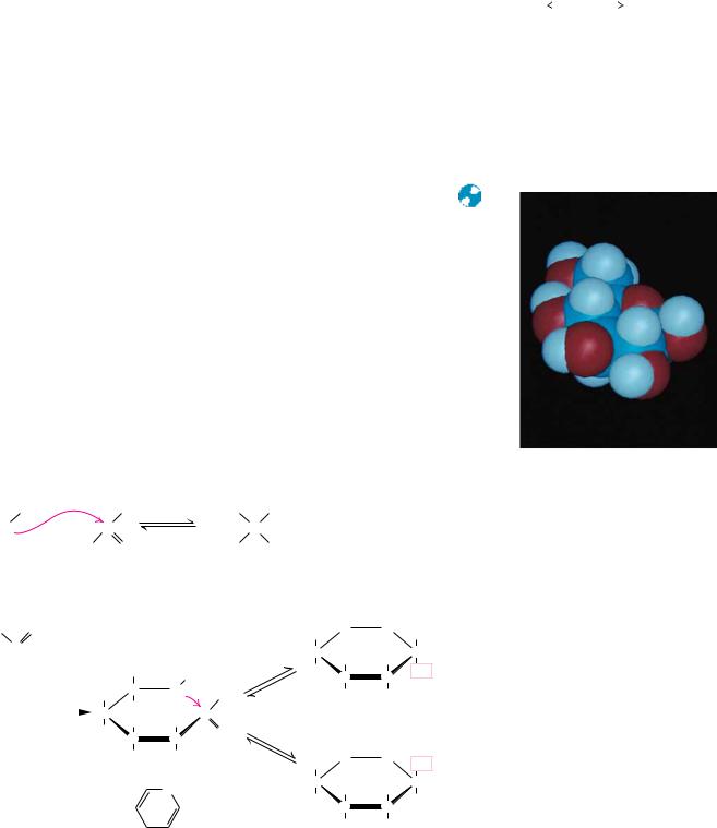
In a similar manner, ketones can react with alcohols to form hemiketals. The analogous intramolecular reaction of a ketose sugar such as fructose yields a cyclic hemiketal (Figure 7.6). The five-membered ring thus formed is reminiscent of furan and is referred to as a furanose. The cyclic pyranose and furanose forms are the preferred structures for monosaccharides in aqueous solution. At equilibrium, the linear aldehyde or ketone structure is only a minor component of the mixture (generally much less than 1%).
When hemiacetals and hemiketals are formed, the carbon atom that carried the carbonyl function becomes an asymmetric carbon atom. Isomers of monosaccharides that differ only in their configuration about that carbon atom are called anomers, designated as or , as shown in Figure 7.5, and the carbonyl carbon is thus called the anomeric carbon. When the hydroxyl group at the anomeric carbon is on the same side of a Fischer projection as the oxygen atom at the highest numbered asymmetric carbon, the configuration at the anomeric carbon is , as in -D-glucose. When the anomeric hydroxyl is on the opposite side of the Fischer projection, the configuration is , as in -D- glucopyranose (Figure 7.5).
The addition of this asymmetric center upon hemiacetal and hemiketal formation alters the optical rotation properties of monosaccharides, and the original assignment of the and notations arose from studies of these properties. Early carbohydrate chemists frequently observed that the optical rotation of glucose (and other sugar) solutions could change with time, a process called mutarotation. This indicated that a structural change was occurring. It was eventually found that -D-glucose has a specific optical rotation, [ ]D20, of 112.2°, and that -D-glucose has a specific optical rotation of 18.7°. Mutarotation involves interconversion of and forms of the monosaccharide with intermediate formation of the linear aldehyde or ketone, as shown in Figures 7.5 and 7.6.
FIGURE 7.8
|
|
CH2OH |
|
|
|
O |
OH |
|
|
|
|
|
OH |
|
|
CH2OH |
HO |
|
|
|
|
|
|
OH |
|
|
HC OH |
Pyranose form |
|
|
|
|
|
OH |
H |
|
|
OH |
C |
|
|
CH2OH |
|
|
|
O |
|
|
OH |
CHOH |
|
|
O |
|
|
D-Glucose |
OH |
|
|
|
|
OH |
|
|
|
OH |
|
|
|
Furanose form |
|
7.2 ● Monosaccharides |
215 |
FIGURE 7.7 ● D-Glucose can cyclize in two ways, forming either furanose or pyranose structures.
Haworth Projections
Another of Haworth’s lasting contributions to the field of carbohydrate chemistry was his proposal to represent pyranose and furanose structures as hexagonal and pentagonal rings lying perpendicular to the plane of the paper, with thickened lines indicating the side of the ring closest to the reader. Such Haworth projections, which are now widely used to represent saccharide structures (Figures 7.5 and 7.6), show substituent groups extending either above or below the ring. Substituents drawn to the left in a Fischer projection are drawn above the ring in the corresponding Haworth projection. Substituents drawn to the right in a Fischer projection are below the ring in a Haworth projection. Exceptions to these rules occur in the formation of furanose forms of pentoses and the formation of furanose or pyranose forms of hexoses. In these cases, the structure must be redrawn with a rotation about the carbon whose hydroxyl group is involved in the formation of the cyclic form (Figures 7.7 and 7.8) in order to orient the appropriate hydroxyl group for ring formation. This is
|
|
|
O |
OH |
|
|
|
|
|
|
HO |
|
|
CH2 OH |
|
OH |
OH |
|
OH |
H |
Pyranose form |
|
|
C |
|
|
|
|
O |
|
|
|
OH |
OH |
CH2OH |
O |
|
D-Ribose |
|
OH |
|
|
|
|
OH |
OH |
|
|
|
Furanose form |
|
● D-Ribose and other five-carbon saccharides can form either furanose or pyranose structures.
FIGURE 7.9
216 Chapter 7 ● Carbohydrates
merely for illustrative purposes and involves no change in configuration of the saccharide molecule.
The rules previously mentioned for assignment of - and -configurations can be readily applied to Haworth projection formulas. For the D-sugars, the anomeric hydroxyl group is below the ring in the -anomer and above the ring in the -anomer. For L-sugars, the opposite relationship holds.
As Figures 7.7 and 7.8 imply, in most monosaccharides there are two or more hydroxyl groups which can react with an aldehyde or ketone at the other end of the molecule to form a hemiacetal or hemiketal. Consider the possibilities for glucose, as shown in Figure 7.7. If the C-4 hydroxyl group reacts with the aldehyde of glucose, a five-membered ring is formed, whereas if the C-5 hydroxyl reacts, a six-membered ring is formed. The C-6 hydroxyl does not react effectively because a seven-membered ring is too strained to form a stable hemiacetal. The same is true for the C-2 and C-3 hydroxyls, and thus fiveand six-membered rings are by far the most likely to be formed from sixmembered monosaccharides. D-Ribose, with five carbons, readily forms either five-membered rings ( - or -D-ribofuranose) or six-membered rings ( - or - D-ribopyranose) (Figure 7.8). In general, aldoses and ketoses with five or more carbons can form either furanose or pyranose rings, and the more stable form depends on structural factors. The nature of the substituent groups on the carbonyl and hydroxyl groups and the configuration about the asymmetric carbon will determine whether a given monosaccharide prefers the pyranose or furanose structure. In general, the pyranose form is favored over the furanose ring for aldohexose sugars, although, as we shall see, furanose structures are more stable for ketohexoses.
Although Haworth projections are convenient for display of monosaccharide structures, they do not accurately portray the conformations of pyranose and furanose rings. Given COCOC tetrahedral bond angles of 109° and COOOC angles of 111°, neither pyranose nor furanose rings can adopt true planar structures. Instead, they take on puckered conformations, and in the case of pyranose rings, the two favored structures are the chair conformation and the boat conformation, shown in Figure 7.9. Note that the ring substituents
(a) |
Axis |
|
|
|
Axis |
|
|
|
|
|
|
|
|
109 a |
e |
|
|
a |
|
|
a |
e |
O |
|
e |
|
O |
e |
|
a |
|
|
a |
|
|
a |
|
|
|
|
|
e |
|
|
|
e |
|
|
|
e |
|
|
|
|
a |
e |
|
a |
a |
|
|
|
|
|
|
|
|
a |
|
|
|
|
|
Chair |
|
|
|
Boat |
|
|
a = axial bond |
|
|
|
|
|
|
e = equatorial bond |
|
|
|
|
|
(b) |
|
|
|
|
|
|
|
H |
CH2OH O |
|
|
CH2OH |
|
OH |
|
|
H |
|
|
H |
O |
|
HO |
H |
H |
OH |
OH |
H |
|
|
H |
H |
|
|
|
H |
OH |
|
|
H |
|
OH |
|
|
|
|
|
|
● (a) Chair and boat conformations of a pyranose sugar. (b) Two possible chair conformations of -D-glucose.
7.2 ● Monosaccharides |
217 |
in these structures can be equatorial, which means approximately coplanar with the ring, or axial, that is, parallel to an axis drawn through the ring as shown. Two general rules dictate the conformation to be adopted by a given saccharide unit. First, bulky substituent groups on such rings are more stable when they occupy equatorial positions rather than axial positions, and second, chair conformations are slightly more stable than boat conformations. For a typical pyranose, such as -D-glucose, there are two possible chair conformations (Figure 7.9). Of all the D-aldohexoses, -D-glucose is the only one that can adopt a conformation with all its bulky groups in an equatorial position. With this advantage of stability, it may come as no surprise that -D-glucose is the most widely occurring organic group in nature and the central hexose in carbohydrate metabolism.
Derivatives of Monosaccharides
A variety of chemical and enzymatic reactions produce derivatives of the simple sugars. These modifications produce a diverse array of saccharide derivatives. Some of the most common derivations are discussed here.
Sugar Acids
Sugars with free anomeric carbon atoms are reasonably good reducing agents and will reduce hydrogen peroxide, ferricyanide, certain metals (Cu2 and Ag ), and other oxidizing agents. Such reactions convert the sugar to a sugar acid. For example, addition of alkaline CuSO4 (called Fehling’s solution) to an aldose sugar produces a red cuprous oxide (Cu2O) precipitate:
|
|
B |
B |
RCO H 2 Cu2 5 OH |
RCO O Cu2O 3 H2O |
Aldehyde |
Carboxylate |
and converts the aldose to an aldonic acid, such as gluconic acid (Figure 7.10). Formation of a precipitate of red Cu2O constitutes a positive test for an aldehyde. Carbohydrates that can reduce oxidizing agents in this way are referred to as reducing sugars. By quantifying the amount of oxidizing agent reduced by a sugar solution, one can accurately determine the concentration of the sugar. Diabetes mellitus is a condition that causes high levels of glucose in urine and blood, and frequent analysis of reducing sugars in diabetic patients is an important part of the diagnosis and treatment of this disease. Over-the-counter kits for the easy and rapid determination of reducing sugars have made this procedure a simple one for diabetics.
Monosaccharides can be oxidized enzymatically at C-6, yielding uronic acids, such as D-glucuronic and L-iduronic acids (Figure 7.10). L --Iduronic acid is similar to D-glucuronic acid, except for having an opposite configuration at C-5. Oxidation at both C-1 and C-6 produces aldaric acids, such as D-glucaric acid.
Sugar Alcohols
Sugar alcohols, another class of sugar derivative, can be prepared by the mild reduction (with NaBH4 or similar agents) of the carbonyl groups of aldoses and ketoses. Sugar alcohols, or alditols, are designated by the addition of -itol to the name of the parent sugar (Figure 7.11). The alditols are linear molecules that cannot cyclize in the manner of aldoses. Nonetheless, alditols are characteristically sweet tasting, and sorbitol, mannitol, and xylitol are widely used to sweeten sugarless gum and mints. Sorbitol buildup in the eyes of dia-
218 |
|
Chapter 7 |
● Carbohydrates |
|
|
|
|
|
|
|
|
|
|
|
FIGURE 7.10 |
|
|
|
|
|
|
|
|
|
|
|
|
|
|
|
|
|
|
|
COOH |
|
|
|
|
|
|
|
|
|
|
|
|
|
|
|
|
|
|
|
|
|
|
|
|
|
|
|
|
|
|
|
|
|
|
|
|
|
|
|
|
|
|
|
|
|
H |
|
C |
|
OH |
|
|
|
|
|
|
|
|
|
|
|
|
|
|
|
|
|
|
|
|
HO |
|
|
|
|
|
|
|
|
|
|
|
|
|
|
|
|
|
|
C |
|
H |
|
|
|
|
|
|
|
|
|
|
|
|
|
|
|
|
|
|
|
|
|
|
|
|
|
|
|
|
|
|
|
|
|
|
|
|
|
|
|
|
H |
|
C |
|
OH |
|
|
|
|
|
|
|
|
|
|
|
|
|
|
|
|
|
|
|
|
|
|
|
|
|
|
|
|
|
|
|
|
|
|
|
|
|
|
|
|
|
H |
|
C |
|
OH |
|
|
|
|
|
|
|
|
|
|
|
|
|
|
|
|
|
|
|
|
|
|
|
|
|
|
|
|
|
|
|
|
|
|
|
|
|
Oxidation |
|
|
|
|
CH2OH |
|
|
|
|
|
|
|
|
D-Gluconic acid |
|
|
|
|
|
|
|
at C–1 |
|
|
|
|
|
|
|
|
|
|
|
|
|
|
|
|
|
|
|
O |
|
|
|
H |
|
|
|
|
|
|
|
|
|
|
|
|
|
|
|
|
C |
|
|
|
|
|
|
|
|
|
|
|
|
|
|
|
|
|
|
|
|
|
|
|
|
|
|
|
|
|
|
|
|
|
|
|
H |
|
|
C |
|
OH |
|
|
H |
|
|
|
|
|
|
|
|
|
|
|
|
|
|
|
|
|
|
|
|
|
|
|
|
|
|
HO |
COOH |
|
|
|
O |
|
|
|
|
|
|
|
|
|
|
|
|
HO |
|
|
C |
|
H |
|
|
H |
|
H |
|
|
|
|
|
|
|
H |
|
|
|
|
|
|
|
|
|
|
|
|
|
|
C |
|
OH |
|
|
|
|
|
|
|
|
|
Oxidation |
|
|
|
|
|
|
|
HO |
|
|
|
|
|
|
|
OH |
|
|
|
|
|
|
|
|
|
|
|
|
|
|
H |
|
|
C |
|
OH |
at C–6 |
|
|
|
H |
|
|
|
|
|
|
|
|
|
|
|
|
|
|
|
|
|
|
|
|
|
|
|
|
|
|
|
|
|
|
|
|
|
|
|
|
|
D-Glucuronic |
|
|
|
CH2OH |
|
|
|
|
|
D-Glucose |
|
|
|
|
|
acid (GlcUA) |
|
|
|
|
|
|
|
|
|
|
|
|
|
|
|
|
|
|
|
|
Oxidation at |
|
|
|
|
|
|
|
|
|
|
|
|
|
|
|
C–1 and C–6 |
|
|
|
|
|
|
|
|
|
|
|
|
|
|
|
|
|
|
|
|
|
COOH |
|
|
|
|
|
|
|
|
|
|
|
|
|
|
|
|
|
|
|
|
|
|
|
|
|
|
|
|
|
|
|
H |
|
|
C |
|
|
OH |
|
|
|
|
|
|
|
|
|
|
|
|
|
|
|
|
|
|
CH2OH |
|
|
|
|
|
|
|
|
|
CH2OH |
O |
|
|
|
|
|
|
|
|
|
|
|
|
H |
|
|
OH |
O |
|
|
|
H |
|
|
|
|
H |
|
|
C |
|
|
|
|
H |
|
|
+ OH– |
|
H |
H |
O |
|
|
|
|
|
|
|
|
OH |
|
|
|
|
OH |
|
|
O– |
|
|
|
|
|
|
|
HO |
|
|
|
|
|
|
|
HO |
|
|
|
|
|
|
|
|
|
|
|
|
|
|
|
H |
OH |
|
|
H |
|
OH |
|
|
|
|
|
|
|
D-Gluconic acid |
|
|
|
|
|
D-δ -Gluconolactone |
Note: D-Gluconic acid and other aldonic acids exist in equilibrium with lactone structures.
|
|
H |
|
|
|
HO |
H |
O |
|
|
|
|
|
|
H |
|
H |
|
|
H |
|
|
COOH |
OH |
|
HO |
OH OH |
|
|
|
|
|
|
|
H |
|
|
|
|
L-Iduronic acid |
|
|
|
|
(IdUA) |
|
HO C H
H C OH
H C OH
COOH
D-Glucaric acid
|
|
CH2OH |
|
|
CH2OH |
|
|
|
|
|
|
|
|
|
|
|
|
HO |
|
|
|
|
|
|
|
CH2OH |
H |
|
C |
|
OH |
|
C |
|
H |
|
|
|
|
|
|
|
|
|
|
|
|
|
|
|
|
|
|
|
|
|
|
|
|
|
HO |
|
C |
|
H |
HO |
|
C |
|
H |
H |
|
C |
|
OH |
|
|
|
|
|
|
|
|
|
|
|
|
|
|
|
|
|
|
HO |
|
|
|
|
|
H |
|
C |
|
OH |
H |
|
C |
|
OH |
|
C |
|
H |
|
|
|
|
|
|
|
|
|
|
|
|
|
|
|
|
|
|
|
|
|
|
|
|
H |
|
C |
|
OH |
H |
|
C |
|
OH |
H |
|
C |
|
OH |
|
|
|
|
|
|
CH2OH |
CH2OH |
CH2OH |
D-Glucitol |
D-Mannitol |
D-Xylitol |
(sorbitol) |
|
|
CH2OH
H C OH
CH2OH
D-Glycerol
FIGURE 7.11 ● Structures of some sugar alcohols.
|
|
|
|
|
|
|
|
|
|
|
CH2OH |
|
|
|
HO |
OH |
|
|
H |
|
|
|
|
|
|
|
|
|
|
|
C |
|
OH |
H |
|
|
3 |
2 |
|
OH |
|
|
|
|
|
|
|
|
|
|
|
|
|
|
|
4 |
H |
H |
1 |
|
H |
|
C |
|
OH |
|
|
|
|
|
|
|
OH |
H |
|
|
|
|
|
|
|
|
|
|
|
|
|
|
|
|
|
|
HO |
|
5 |
6 H |
H |
|
C |
|
OH |
|
|
|
|
|
|
H |
OH |
|
|
|
|
|
|
|
|
|
|
|
|
|
CH2OH |
|
|
|
myo-Inositol |
|
|
|
D-Ribitol |
betics is implicated in cataract formation. Glycerol and myo -inositol, a cyclic alcohol, are components of lipids (see Chapter 8). There are nine different stereoisomers of inositol; the one shown in Figure 7.11 was first isolated from heart muscle and thus has the prefix myo- for muscle. Ribitol is a constituent of flavin coenzymes (see Chapter 20).
7.2 ● Monosaccharides |
219 |
|
|
|
|
|
|
|
|
|
H |
|
|
|
|
HOH2C |
O H |
HO |
|
|
O OH |
|
CH3 |
|
|
H |
H |
|
|
|
H |
|
|
|
|
|
|
H |
|
|
H |
|
|
OH |
H |
|
|
|
H |
|
|
OH H |
|
|
OH |
OH |
|
2-Deoxy-α -D-Ribose |
α -L-Rhamnose (Rha) |
OH
OH CH3
HO
CH2
OH
|
|
H |
|
|
O |
|
HO |
|
|
O |
|
OH |
|
|
|
|
|
|
|
|
CH3 |
H |
|
|
|
|
|
|
|
|
|
|
H |
|
|
|
H |
|
|
|
|
H |
|
|
OH |
OH |
|
|
Ouabain |
H
HCH3 O OH H HO
HO H
OH H
α-L-Fucose (Fuc)
O
O
FIGURE 7.12 ● Several deoxy sugars and ouabain, which contains -L-rhamnose (Rha).
Hydrogen atoms highlighted in red are “deoxy” positions.
Deoxy Sugars
The deoxy sugars are monosaccharides with one or more hydroxyl groups replaced by hydrogens. 2-Deoxy-D-ribose (Figure 7.12), whose systematic name is 2-deoxy-D-erythropentose, is a constituent of DNA in all living things (see Chapter 11). Deoxy sugars also occur frequently in glycoproteins and polysaccharides. L-Fucose and L-rhamnose, both 6-deoxy sugars, are components of some cell walls, and rhamnose is a component of ouabain, a highly toxic cardiac glycoside found in the bark and root of the ouabaio tree. Ouabain is used by the East African Somalis as an arrow poison. The sugar moiety is not the toxic part of the molecule (see Chapter 10).
Sugar Esters |
|
|
|
|
|
|
|
|
|
|
|
|
|
|
|
|
|
|
|
|
|
|
|
|
|
|
|
|
|
|
|
|
|
|
|
|
|
Phosphate esters of glucose, fructose, and other monosaccharides are impor- |
|
|
|
|
|
|
|
|
|
|
|
|
|
|
|
|
|
tant metabolic intermediates, and the ribose moiety of nucleotides such as ATP |
|
|
|
|
|
|
|
|
|
|
|
|
|
|
|
|
|
and GTP is phosphorylated at the 5 -position (Figure 7.13). |
|
|
|
|
|
|
|
|
|
|
|
|
|
|
|
|
|
|
NH2 |
|
|
|
|
|
|
|
|
|
|
|
|
|
|
|
|
|
|
|
|
|
|
|
|
|
|
|
|
|
|
|
|
|
|
|
|
|
|
N |
|
|
N |
|
|
|
|
|
|
|
|
|
|
|
|
|
|
|
|
|
|
|
|
|
|
|
|
|
|
|
|
|
|
|
|
|
|
|
|
|
|
|
|
|
|
|
|
|
|
|
|
|
|
|
|
|
|
|
|
|
|
|
|
|
|
|
|
|
|
|
|
|
|
|
|
|
|
|
|
|
|
N |
N |
|
|
|
|
|
|
|
|
|
|
|
|
|
|
|
|
|
|
|
|
|
|
|
|
|
|
|
|
|
|
|
|
|
|
|
|
|
|
|
|
|
|
|
|
|
|
|
|
|
|
|
|
|
|
|
|
|
|
|
|
|
|
|
|
|
|
|
|
|
|
|
|
|
|
|
|
|
|
|
|
|
|
|
|
CH2OH |
|
|
|
|
|
|
|
|
|
|
|
|
|
|
|
|
|
|
O– |
|
|
O– |
|
|
O– |
|
|
|
|
|
|
|
|
|
|
|
|
|
|
|
|
|
|
|
|
|
O |
|
|
|
|
–O |
|
|
|
|
|
|
|
|
|
|
|
O |
|
|
|
O |
|
|
|
|
|
|
|
|
|
|
|
|
|
|
|
|
|
|
|
|
|
|
|
|
|
|
|
|
|
|
|
|
|
|
|
|
|
|
|
|
|
O |
|
|
|
|
|
|
|
|
|
|
|
|
|
|
|
P |
|
O |
|
P |
|
O |
|
P |
|
CH |
|
|
|
H |
|
H |
|
2– |
|
|
|
|
|
|
CH2 |
2– |
|
|
|
|
|
|
|
|
|
|
|
|
|
O3PO |
H2C |
|
|
OPO3 |
|
|
|
|
|
|
|
|
|
|
|
|
|
|
|
|
2 |
|
|
|
|
|
|
|
H |
|
|
|
|
|
|
|
|
|
|
|
|
|
|
|
|
|
|
|
|
|
|
|
|
|
|
|
|
|
|
|
|
|
|
|
|
|
|
|
|
|
|
|
|
|
|
|
|
|
|
|
|
|
|
|
|
|
|
|
|
|
|
|
|
|
|
|
|
|
|
|
|
|
|
|
|
|
|
|
OH |
H |
|
|
|
|
|
|
|
H |
HO |
|
|
|
|
|
|
O |
|
|
O |
|
|
O |
|
|
|
H |
H |
|
|
|
OPO32– |
|
|
|
|
|
|
|
|
|
|
|
HO |
|
|
|
|
H |
|
|
|
|
OH |
|
|
|
|
|
|
|
|
|
|
|
|
|
|
H |
|
|
|
|
H |
|
|
|
|
|
|
|
|
|
|
|
|
|
|
|
|
|
|
|
|
|
|
|
|
|
|
|
|
|
|
|
|
|
|
|
|
|
|
|
|
|
|
|
|
|
|
|
|
|
|
|
|
|
|
|
|
|
|
|
|
|
|
|
|
|
|
|
H |
OH |
|
|
|
|
|
|
|
OH H |
|
|
|
|
|
|
|
|
|
|
|
|
|
|
|
|
|
|
|
OH |
OH |
|
|
α -D-Glucose-1-phosphate |
|
|
α -D-Fructose-1,6-bisphosphate |
|
|
|
|
|
|
|
|
|
|
|
|
|
Adenosine-5'-triphosphate |
FIGURE 7.13 ● Several sugar esters important in metabolism.

220 Chapter 7 ● Carbohydrates
|
|
|
CH2OH |
|
|
|
|
|
CH2OH |
|
|
|
|
|
|
O |
|
|
|
|
|
|
O |
|
|
|
H |
|
OH |
HO |
H |
OH |
|
|
|
|
|
|
|
H |
|
|
|
|
|
|
|
|
|
|
|
|
OH |
H |
|
|
|
|
|
OH |
H |
|
|
HO |
|
|
H |
|
H |
|
|
H |
|
|
|
|
|
|
|
|
|
|
|
|
|
|
|
|
|
H |
NH2 |
|
|
|
|
|
H |
NH2 |
|
|
β -D-Glucosamine β -D-Galactosamine
FIGURE 7.14 ● Structures of D-glucosamine
and D-galactosamine.
Amino Sugars
Amino sugars, including D-glucosamine and D-galactosamine (Figure 7.14), contain an amino group (instead of a hydroxyl group) at the C-2 position. They are found in many oligoand polysaccharides, including chitin, a polysaccharide in the exoskeletons of crustaceans and insects.
Muramic acid and neuraminic acid, which are components of the polysaccharides of cell membranes of higher organisms and also bacterial cell walls, are glucosamines linked to three-carbon acids at the C-1 or C-3 positions. In muramic acid (thus named as an ami ne isolated from bacterial cell wall polysaccharides; murus is Latin for “wall”), the hydroxyl group of a lactic acid moiety makes an ether linkage to the C-3 of glucosamine. Neuraminic acid (an amine isolated from neur al tissue) forms a COC bond between the C-1 of N- acetylmannosamine and the C-3 of pyruvic acid (Figure 7.15). The N-acetyl and N-glycolyl derivatives of neuraminic acid are collectively known as sialic acids and are distributed widely in bacteria and animal systems.
|
|
CH2OH |
|
|
H |
O |
H |
|
|
|
H |
|
|
|
|
|
|
|
|
|
O |
H |
|
|
|
OH |
HO |
|
|
|
|
|
|
|
|
|
H |
NH2 |
|
|
CH3 CH COOH
Muramic acid
HOOC C OH
|
|
|
|
|
|
|
COOH |
|
|
|
|
|
|
|
|
|
|
|
|
|
|
|
|
Pyruvic acid |
|
|
|
|
|
|
|
|
|
|
|
|
|
|
|
|
|
|
|
|
|
CH2 |
|
|
|
|
|
O |
|
H |
|
|
C |
|
|
|
OH |
|
|
|
|
|
|
|
|
|
|
|
|
|
|
|
|
|
|
CH3 |
|
C |
|
N |
|
|
C |
|
|
|
H |
|
|
|
|
|
|
|
|
|
|
H |
|
|
|
|
|
|
|
|
|
|
|
|
HO |
|
|
C |
|
|
|
H |
|
|
|
|
|
|
|
|
|
|
|
H |
|
C |
|
OH |
|
|
|
|
H |
|
C |
|
OH |
|
|
|
|
|
|
|
CH2OH |
|
|
|
|
|
|
|
C |
|
O |
N-Acetylmannosamine
N- Acetyl-D-neuraminic acid (NeuNAc)
|
|
|
|
|
|
|
|
|
|
|
|
CH2 |
|
|
|
|
|
|
|
|
|
H |
|
|
|
|
|
|
|
|
|
|
|
|
|
|
|
|
|
|
|
|
|
|
|
|
|
|
|
|
|
|
|
|
|
O |
|
|
O |
|
|
|
|
|
|
|
|
|
|
|
|
|
|
|
|
O |
|
H |
|
C |
|
OH |
|
|
|
|
HO |
|
H |
|
H |
|
|
|
|
|
|
|
|
|
|
|
|
|
|
|
|
|
|
|
|
|
|
|
|
|
|
|
|
H |
HCOH |
|
|
|
|
|
|
|
|
COOH |
|
|
|
|
|
|
|
|
|
|
|
|
|
|
|
|
|
|
|
|
|
|
|
|
|
|
|
|
|
|
|
|
|
|
|
|
|
CH |
3 |
|
C |
|
N |
|
C |
|
H |
CH3 |
|
C |
|
N |
|
|
COOH |
HOH C |
|
|
|
|
|
|
|
|
|
|
|
|
|
|
|
|
|
|
|
|
|
|
|
|
|
|
|
|
|
|
|
|
|
|
|
H |
|
|
|
|
|
|
|
|
|
|
|
|
|
|
HCOH |
|
|
2 |
|
H |
OH H |
|
O |
|
|
O |
|
|
|
|
|
|
|
|
|
|
|
C |
|
H |
|
|
|
|
|
|
|
|
|
|
|
|
CH3 |
|
|
|
|
|
|
|
OH |
|
|
|
|
|
|
|
|
|
|
|
|
|
|
|
|
|
|
|
|
|
|
|
H |
CH2OH |
OH |
N |
|
|
|
|
|
|
|
|
|
|
|
|
|
|
|
|
|
|
|
|
|
|
|
|
|
|
|
|
|
|
|
|
|
|
H |
H |
C |
H |
|
HO |
|
|
|
|
|
|
|
|
|
|
|
|
|
|
|
H |
|
C |
|
OH |
|
|
|
|
|
|
|
|
H |
|
|
H |
|
|
|
|
|
|
|
|
|
|
|
|
|
|
|
|
|
|
|
|
|
|
|
|
|
|
|
|
|
|
|
|
|
|
|
|
|
|
|
|
|
|
|
|
|
|
|
|
|
|
|
|
|
|
|
|
|
|
|
|
|
|
|
|
|
|
|
|
|
|
|
|
|
|
|
|
|
|
|
|
|
|
|
|
|
|
|
|
|
|
|
|
|
|
|
|
|
O |
|
|
|
|
|
H |
|
|
|
|
|
|
|
|
|
|
|
|
H |
|
C |
|
OH |
|
|
|
|
|
|
|
|
OH |
H |
|
|
|
|
|
|
|
|
|
|
|
|
|
|
|
|
|
|
|
|
|
|
|
|
|
|
|
|
|
|
|
|
|
|
|
|
|
|
|
|
|
|
|
|
|
|
|
|
|
|
|
|
|
|
|
|
|
|
|
|
|
|
|
|
|
|
|
|
|
|
|
|
|
|
|
|
|
|
|
|
|
|
|
|
|
|
|
|
|
|
|
|
|
|
|
|
|
|
|
|
|
|
|
|
|
|
|
|
|
|
|
|
|
|
|
|
|
|
|
|
|
|
|
|
CH2OH |
|
|
|
|
|
|
|
|
|
|
|
|
|
|
|
|
|
|
|
|
|
|
|
|
|
|
|
Fischer projection |
|
|
|
|
|
|
|
|
Haworth projection |
|
|
|
|
Chair conformation |
|
|
N- Acetyl-D-neuraminic acid (NeuNAc), a sialic acid
FIGURE 7.15 ● Structures of muramic acid and neuraminic acid and several depictions of sialic acid.