

Genomic Imprinting and Uniparental Disomy in Medicine
.pdf251
TABLE 1 Imprinted Genes Identified in Mice and Humans
(The genes are grouped together based on map postion in the mouse to emphasize the clustered nature of imprinted genes in both mice and humans)
|
Expression |
|
Chromosome |
|
|
||||
|
|
|
|
|
|
|
|
|
|
Gene |
Mouse |
Human |
|
Mouse |
Human |
Description |
Reference |
||
|
|
|
|
|
|
|
|
||
Wt1a |
BAE |
mat=pat=BAE |
|
prox. 2 |
11p13 |
Zinc finger, tumor suppressor |
Jinno et al. (1995) |
||
Wt1as |
ND |
pat |
|
prox. 2 |
11p13 |
|
Malik et al. (2000) |
||
Nnat |
pat |
ND |
|
|
|
|
Neuronatin |
Kagitani et al. (1997) |
|
|
dist. 2 |
20q11-12 |
|||||||
Gnas |
mat |
mat |
|
dist. 2 |
20q13 |
Guanine nucleotide binding protein |
Hayward et al. (1998a); Hayward et al. |
||
|
|
|
|
|
|
|
|
(G protein), alpha-stimulating activity |
(1998b); Wroe et al. (2000); Yu et al. |
|
|
|
|
|
|
|
|
polypeptide 1 |
(1998) |
Gnasxi |
pat |
pat |
|
dist. 2 |
20q13 |
GNAS extralarge |
Peters et al. (1999); Wroe et al. (2000); |
||
|
|
|
|
|
|
|
|
|
Yu et al. (1998) |
Nesp |
mat |
mat |
|
dist. 2 |
20q13 |
Neuroendocrine secretory protein |
Peters et al. (1999); Wroe et al. (2000); |
||
|
|
|
|
|
|
|
|
|
Yu et al. (1998) |
Nespas |
pat |
pat |
|
dist. 2 |
20q13 |
Noncoding RNA |
Hayward and Bonthron (2000); Wroe et |
||
|
|
|
|
|
|
|
|
|
al. (2000) |
Arh1 |
ND |
pat |
4 |
1p31 |
Ras-related protein |
Yu et al. (1999) |
|||
Sgce |
pat |
pat |
|
6 cen. |
7q21-22 |
Sarcoglycan epsilon |
Piras et al. (2000) |
||
|
|
|
|
|
|
|
|
|
|
Peg1=Mest |
pat |
pat |
|
prox. 6 |
7q32 |
|
Hydrolase |
Kaneko-Ishino et al. (1995); Nishita et al. |
|
|
|
|
|
|
|
|
|
|
(1996) |
Copg2a |
mat |
pat |
|
prox. 6 |
7q32 |
|
Coatomer protein complex subunit |
Lee et al. (2000); Blagitko et al. (1999) |
|
|
|
|
|
|
|
|
|
Gamma |
Yamasaki et al. (2000) |
Copg2asa |
pat |
ND |
|
prox. 6 |
7q32 |
|
Noncoding RNA |
Lee et al. (2000) |
|
Mit1=lb9 |
pat |
pat |
|
prox. 6 |
7q32 |
|
— |
Lee et al. (2000) |
|
Zim1 |
mat |
ND |
|
prox. 7 |
19q13 |
Zinc finger transcription factor |
Kim et al. (1999) |
||
Peg3=Pw1 |
pat |
ND |
|
prox. 7 |
19q13 |
Zinc finger transcription factor |
Kuroiwa et al. (1996) |
||
Usp29 |
pat |
ND |
|
prox. 7 |
19q13 |
Ubiquitin-specific processing protease 29 |
Kim et al. (2000) |
||
Snrpn |
pat |
pat |
|
|
|
|
snRNP-associated polypeptide, splicing |
Cattanach et al. (1992); Glenn et al. |
|
|
cen. 7 |
15q12 |
|||||||
|
|
|
|
|
|
|
|
factor |
(1993b); Leff et al. (1992); Reed and |
|
|
|
|
|
|
|
|
|
Leff (1994) |
Magel2 |
pat |
pat |
|
cen. 7 |
15q12 |
|
MAGe-like 2 |
Boccaccio et al. (1999) |
|
|
|
|
|
|
|
|
|
|
|
|
|
|
|
|
|
|
|
|
|
(continued)
252
TABLE 1 |
(continued) |
|
|
|
|
|
|
|
|
|
|
|
|
|
|
|
|||
|
|
Expression |
Chromosome |
|
|
||||
|
|
|
|
|
|
|
|
|
|
Gene |
|
Mouse |
Human |
Mouse |
Human |
Description |
Reference |
||
|
|
|
|
|
|
|
|
|
|
Ndn |
|
pat |
pat |
|
|
|
Unknown |
Jay et al. (1997) |
|
cen. 7 |
15q12 |
||||||||
Zfp127 |
|
pat |
pat |
cen. 7 |
15q12 |
|
Zinc finger transcription factor |
Jong et al. 1993; Saitoh et al. (1996) |
|
Zfp127as |
|
pat |
pat |
cen. 7 |
15q12 |
|
Noncoding RNA |
|
|
Ipw |
|
pat |
pat |
cen. 7 |
15q12 |
|
Noncoding RNA |
Wevrick and Francke (1997); Wevrick |
|
|
|
|
|
|
|
|
|
|
et al. (1994) |
Par1 |
|
ND |
pat |
cen. 7 |
15q12 |
|
Noncoding RNA |
Sutcliffe et al. (1994) |
|
Par5 |
|
ND |
pat |
cen. 7 |
15q12 |
|
Noncoding RNA |
Sutcliffe et al. (1994) |
|
Ube3a |
|
mat |
mat |
cen. 7 |
15q12 |
|
Ubiquitin conjugating enzyme E3A |
Albrecht et al. (1997) |
|
H19 |
|
mat |
mat |
|
|
|
Noncoding RNA |
Bartolomei et al. (1991); Rainier et al. |
|
dist. 7 |
11p15 |
||||||||
|
|
|
|
|
|
|
|
|
(1993) |
Igf2 |
|
pat |
pat |
dist. 7 |
11p15 |
Insulinlike growth factor 2, mitogenic |
DeChiara et al. (1991); Giannoukakis |
||
|
|
|
|
|
|
|
|
growth factor |
et al. (1993); Ohlsson et al. (1993) |
Igf2as |
|
pat |
pat |
dist. 7 |
11p15 |
Noncoding RNA |
Okutsu et al. (2000) |
||
Ins2 |
|
pat |
BAE |
dist. 7 |
11p15 |
Insulin |
Giddings et al. (1994) |
||
Mash2 |
|
mat |
mat |
dist. 7 |
11p15 |
bHLH transcription factor |
Alders et al. (1997); Guillemot et al. |
||
|
|
|
|
|
|
|
|
|
(1995) |
Kvlqt1 |
|
mat |
mat |
dist. 7 |
11p15 |
Potassium channel involved in long |
Lee (1997) |
||
|
|
|
|
|
|
|
|
QT syndrome |
|
Kvlqt1-as |
|
pat |
pat |
dist. 7 |
11p15 |
Noncoding RNA |
Lee et al. (1999b) |
||
p57KIP2 |
|
mat |
mat |
dist. 7 |
11p15 |
CDK inhibitor |
Hatada et al. (1996); Matsuoka |
||
|
|
|
|
|
|
|
|
|
et al. (1996) |
Impt1 |
|
mat |
mat |
dist. 7 |
11p15 |
Solute carrier family 22 (organic |
Dao et al. (1999) |
||
|
|
|
|
|
|
|
|
cation transporter), member 1-Ilke |
|
Ipl=Tssc3 |
|
mat |
mat |
dist. 7 |
11p15 |
Tumor-suppressing subtransferable |
Schwlenbacher et al. (1998) |
||
|
|
|
|
|
|
|
|
candidate 3 |
|
Tssc4 |
|
mat |
BAE? |
dist. 7 |
11p15 |
— |
Lee et al. (1999a) |
||
Rasgrf1 |
pat |
pat |
9 |
15q24 |
Ras protein-specific guanine |
Plass et al. (1996) |
|
|
|
|
|
nucleotide-releasing factor 1 |
|
Zac1 |
pat |
pat |
10 |
6q24-25 |
Zinc finger transcription factor, |
Piras et al. (2000) |
|
|
|
|
|
putative tumor suppressor |
|
Hyma1 |
ND |
pat |
10 |
6q24-25 |
— |
Arima et al. (2000) |
Meg1=Grb10 |
mat |
mat |
|
|
Growth factor receptor-bound protein 10 |
Miyoshi et al. (1998); Yoshihashi et al. |
prox. 11 |
7p12 |
|||||
|
|
|
|
|
|
(2000) |
U2af1-rs1 |
pat |
BAE |
prox. 11 |
5q23-31 |
U2AF auxiliary factor small subunit, |
Hatada et al. (1995); Kitagawa et al. |
|
|
|
|
|
splicing factor |
(1995); Pearsall et al. (1996) |
|
|
|
|
|
|
|
Dlk |
pat |
pat |
dist. 12 |
14q32 |
Homologue of Drosophila deltalike |
Schmidt et al. (2000); Wylie et al. (2000) |
|
|
|
|
|
protein |
|
Gtl2 |
mat |
mat |
dist. 12 |
14q32 |
Unknown function |
Miyoshi et al. (2000); Schmidt et al. |
|
|
|
|
|
|
(2000); Wylie et al. (2000) |
|
|
|
|
|
|
|
Htr2a |
mat |
mat |
dist. 14 |
13q14-21 |
Serotonin receptor 2A |
Kato et al. (1996) |
Igf2r |
mat |
mat |
|
|
Lysosomal transport receptor |
Barlow et al. (1991); Smrzka et al. (1995) |
prox. 17 |
6q25.3 |
|||||
Air (Igf2ras) |
pat |
ND |
prox. 17 |
6q25.3 |
Noncoding RNA |
Lyle et al. (2000); Wutz et al. (1997) |
Impact |
pat |
BAE |
|
|
Unknown |
Hagiwara et al. (1998); Okamura et al. |
prox. 18 |
18q11 |
|||||
|
|
|
|
|
|
(2000) |
Ins1a |
pat |
— |
|
|
Insulin |
Giddings et al. (1994) |
19 |
No homologue |
|||||
Xist |
pat=ran |
pat=ran |
X |
X |
Induces X inactivation |
Goto et al. (1998); Kay et al. (1994) |
|
|
|
|
|
|
|
pat ¼ paternal expression; mat ¼ maternal expression; BAE ¼ biallelic expression. a The data concerning the imprinting status of these genes are conflicting.
253
254 GENOMIC IMPRINTING IN THE MOUSE
these regions and genomewide screens for imprinted genes may be expected to find more. The identification and analysis of imprinted gene function enable us to ask if individual genes are responsible for the phenotypes of UPD cases. For example, Igf2r = mice and the Tme mutant mice have a very similar phenotype, strongly arguing that this gene is responsible for the phenotypes observed for paternal UPD or maternal deletion of proximal mouse chromosome 17 (Barlow et al., 1991; Wang et al., 1994). Further examples are discussed later in the context of mouse models of Prader-Willi syndrome.
As more imprinted genes have been identified, it has become increasingly clear that they are clustered within the genome, which suggests that there may be a functional basis for grouping imprinted genes. In mice, deletion of the H19 gene or its enhancers results in disruption of imprinting of two upstream genes, Igf2 and Ins2 (Leighton et al. 1995a, 1995b). In humans, a postulated imprinting center (IC) has been suggested to control the imprinting of a number of genes within the PraderWilli syndrome region (Buiting et al., 1995; Sutcliffe et al., 1994) (see later discussion). In addition to these effects on the expression of genes, imprinted domains also show parental differences in replication timing (Kitsberg et al., 1993; Simon et al., 1999) and meiotic recombination (Paldi et al., 1995), and imprinted regions may also show the homologous association at certain stages of the cell cycle (LaSalle and Lalande, 1995). It is not known how these domain effects, which affect imprinted and nonimprinted genes within the regions, relate mechanistically to imprinting. However, these data taken together do suggest the existence of functional imprinted domains in both mice and humans.
THE IMPRINTING MECHANISM
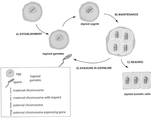
The ability to express one allele in a parent-specific manner in a diploid somatic cell implies that the alleles can be distinguished throughout development. Such a distinguishing mark has been termed the imprint. It is important to realize that imprinted gene expression can be considered in a number of separate stages: first, establishment of the imprint; second, maintenance of the imprint; third, reading of this imprint; fourth, imprint erasure (Figure 4).
The imprint must be established while the two parental genomes are separate, and this is most likely to occur during gametogenesis (Figure 4a). It may be predicted that certain sequences are imprinted in the male germ line and others imprinted in the female germ line, but such sequences remain elusive and what distinguishes a maternal from a paternal imprint remains unknown. The imprint must be stable and copied through DNA replication in somatic cells to maintain imprinted expression in daughter cells (Figure 4b). Reading the imprint by the transcriptional machinery (Figure 4c) can be considered a separate step since, e.g., in Igf2r (Sto¨ger et al., 1993), the imprint is present during preimplantation development but imprinted gene expression is not established until after implantation. And finally, since imprinting for a particular sequence is switched on passage through the germ line of the
255
Figure 4 Different stages in the imprinting mechanism. The example shown considers a maternally imprinted, paternally expressed gene. (A) Establishment of the imprint on the maternal chromosome during oogenesis. (B) Maintenance of the imprint throughout early development and in somatic cells. (C) Reading the imprint by the various mechanisms involved in imprinting, resulting in monoallelic expression. (D) Erasure of the imprint from the maternal chromosome during spermatogenesis.
256 GENOMIC IMPRINTING IN THE MOUSE
opposite sex, the imprint must be erased and then reestablished (Figure 4d). These criteria suggest that the imprint is maintained as an epigenetic mark on the DNA.
The best candidate so far identified for the imprint is methylation of cytosine within CpG dinucleotides [for a review of CpG methylation, see (Cross and Bird, 1995)]. The first indication of the importance of methylation in imprinting came with the observation that imprinted genes contain regions of allele-specific methylation [e.g., see (Sto¨ger et al., 1993)]. Indeed, all imprinted genes examined so far have such regions, showing that methylation is used to distinguish the two alleles. However, not all these differentially methylated regions (DMRs) can be considered the imprint in the sense that methylation is present in the gametes and maintained throughout development. Only for three genes, Igf2r (Sto¨ger et al., 1993), H19 (Bartolomei et al., 1993) and Snrpn (Shemer et al., 1997), have such candidate imprints been identified. Other DMRs are methylation-free during gametogenesis and early development, only acquiring methylation later, probably after imprinting gene expression has been established. Thus, DMRs may be primary imprints whose function is to determine the pattern of imprinted gene expression and secondary imprints that function to maintain this pattern during development.
The importance of methylation to imprinting was underlined by analyzing mice that had a targeted disruption of the maintenance methyltransferase gene (Dnmt1; Li et al., 1993); Dnmt1 = mice fail to maintain monoallelic expression of imprinted genes. Although these experiments established that methylation can meet the criteria for the imprint and is required to maintain imprinted expression, it is known that methylation is completely removed from these allele-specific methylated regions in germ-line passage through the opposite sex, indicating that mechanisms other than methylation are involved in establishing the imprint.
Although we are far from a complete understanding of the imprinting mechanism, and it is still not clear if all imprinted genes are regulated in the same way, some common themes are present. As mentioned above, all imprinted genes have regions of differential methylation. In addition, imprinted genes are always in groups of two or more and there is evidence for imprinting control regions that regulate the imprinted expression of one or more genes (e.g., SNRPN and LIT1). How the imprint is read to produce monoallelic expression is being studied in many genes and some examples are discussed below to illustrate the apparent themes in imprinted gene expression (Figure 5).
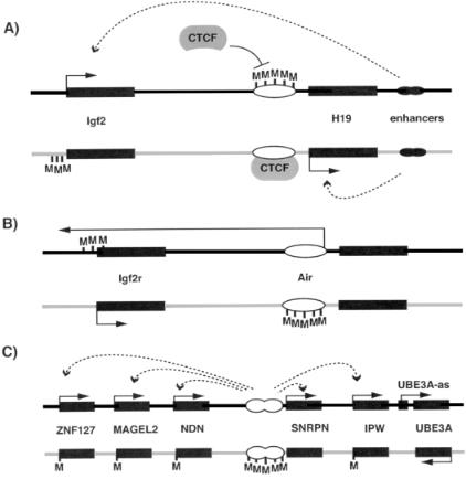
Methylation is an attractive candidate for the imprint since a great deal of work has shown that methylation represses transcription (Bird and Wolffe, 1999), thus providing an obvious direct link between the imprint itself and the outcome of reading the imprint, which is transcription. Interestingly, in Dnmt1 = mice the H19 gene becomes biallelically expressed and the Igf2r gene is completely silenced (Li et al., 1993). This implies that in some cases, methylation is repressing transcription, in other cases activating transcription. This apparent paradox can be understood if we consider that a methylated imprint may either repress transcription by modifying a cis-activating activator (e.g., a promoter or enhancer) or activate transcription by modifying a cis-acting repressor (e.g., a repressor-binding site; Figure 5).
THE IMPRINTING MECHANISM |
257 |
Figure 5 Examples of different mechanisms involved in imprinted gene expression. (A) At the Igf2=H19 locus, paternal methylation at the differentially methylated region (DMR) 50 to the H19 gene prevents the repressor CTCF binding; this, in turn, allows the enhancers to interact with the Igf2 promoter. Paternal H19 expression is presumed to be repressed because of the methylation at its promoter. On the maternal chromosome, CTCF can interact with the unmethylated DMR to set up a boundary of expression so that the enhancers can only interact with H19. Subsequent methylation of the Igf2 promoter helps to maintain this pattern of expression. (B) At Igf2r, an intronic DMR is known to be essential to repress Igf2r expression on the paternal chromosome. This DMR is also the promoter for an antisense RNA (Air), raising the possibilty of repression via an antisense RNA mechanism. On the maternal chromosome, this DMR shows imprinted methylation, Air is not expressed and Igf2r is expressed. (C) Imprinted expression of genes at the PWS region is controlled by an IC that is responsible for paternal gene expression throughout the region. How this long-range effect is achieved is not understood. Expression of an antisense gene UBE3A-AS, under control of the IC, may regulate the Angelman syndrome gene (UBE3A) in a manner similar to Igf2r=Air. Black oblongs represent genes; white ovals represent imprinting control regions (ICs)=imprinted differentially methylated regions (DMRs). Dark gray and light gray lines represent paternal and maternal chromosomes, respectively. CTCF, repressor; M, methylation; arrows with solid lines represent gene expression; arrrows with dotted lines indicate interactions between imprinting control regions (B and C) or enhancers
(A) with genes. See text for further explanation.
258 GENOMIC IMPRINTING IN THE MOUSE
This second mechanism is, in fact, suggested by recent work on H19 and Igf2 (Bell and Felsenfeld, 2000; Hark et al., 2000). Previous work showed that there is a region of paternal-specific methylation between the maternally expressed H19 and paternally expressed Igf2, and that both genes require access to enhancers downstream of H19 (Bartolomei et al., 1993) for expression. The reciprocal imprinting of this gene pair was originally suggested to be controlled by enhancer competition: the region of differential methylation silenced the promoter for H19, and thus Igf2 could gain access to the enhancers and was expressed (Figure 5a). However, it now appears that the differentially methylated region is a binding site for the repressor CTCF (Bell and Felsenfeld, 2000; Hark et al., 2000). Thus when methylated, CTCF cannot bind and Igf2 is expressed. The presence of potential CTCF binding sites at another imprinting gene pair (DLK1=GTL2; Wylie et al., 2000) raises the exciting possibility that boundary elements may be a common feature regulating imprinted gene expression.
Another well-studied imprinted gene pair that appears similar to Igf2=H19 is Igf2r=Air (Figure 5b). Igf2r and Air are reciprocally imprinted and both have differentially methylated CpG island promoters (Lyle et al., 2000; Sto¨ger et al., 1993). Region 2 is methylated throughout development and is the promoter for the noncoding RNA, Air. Deletion of region 2 in mice results in biallelelic Igf2r expression (Wutz et al., 1997), indicating that region 2=Air functions as a cis repressor of Igf2r expression, possibly via an antisense RNA mechanism (Lyle et al., 2000). The presence of many noncoding, antisense RNAs at other imprinted loci (Table 1) indicates that this is a common mechanism. For example, at the UBE3A locus, the paternally expressed antisense RNA (UBE3A-as) may repress paternal UBE3A expression (Rougeulle et al., 1998) in a similar way to Air=Igf2r. However, it remains to be shown whether these RNAs are the cause of the imprinting or are simply a secondary consequence of, e.g., local changes in chromatin structure.
Certain regions thus appear to control the imprinted expression of genes. This is illustrated very clearly in the PWS=AS region (as discussed in Chapter 6). Approximately 5% of PWS and AS patients have imprinting mutations, revealed as abnormal methylation and gene expression throughout the region (Glenn et al., 1993a). Around half these patients have small deletions in the first exon of SNRPN in the case of PWS (Buiting et al., 1995; Sutcliffe et al., 1994) and deletion upstream of the first exon in AS (Buiting et al., 1995; Dittrich et al., 1996; Saitoh et al., 1996) (Figure 5c). These patients have a normal biparental content in these region, but only the maternal epigenotype in PWS and only the paternal epigenotype in AS. Thus, there is a bipartite imprinting control (IC) center in the 50 region of SNRPN controlling the epigenetic status, and thus gene expression, over a 2-Mb region (Figure 5c).
In summary, imprinted expression of certain genes seems to be regulated by small imprinting control (IC) regions. What remains intriguing is the variety of mechanisms (boundary elements, antisense RNA, methylation) involved at these ICs. Is there a unifying mechanism underlying the expression of all imprinted genes?
USING MICE TO MODEL IMPRINTING |
259 |
USING MICE TO MODEL IMPRINTING
The clustering of imprinted genes and the existence of coordinate regulation may make it difficult to interpret genotype-phenotype correlations in UPD and imprinting syndromes. Only by analyzing the individual phenotypic effects of imprinted genes, either by targeted mutation in mice or identification of mutations in humans, can we begin to understand the complex phenotypes seen in UPD. Working with mice has the advantage that specific genetic changes can be introduced into the germ line. These can be broadly split into two categories: first, targeted mutations of single genes or control regions; second, deletions of much larger chromosomal regions containing many genes. The usefulness of these approaches is discussed below with reference to dissecting the phenotype of the Prader-Willi syndrome.
PWS has a complex phenotype with developmental, behavioral and mental aspects (see Chapter 6 for a more detailed discussion). Unlike for AS, where approximately 20% of cases are caused by mutation of a single gene (UBE3A; Kishino et al., 1997; Matsuura et al., 1997), no single gene defects have been found. This implies that PWS is a contiguous gene syndrome caused by alteration in the expression of a number of genes. Although patients may exist with mutations in single genes from the PWS region, they would not be recognized as PWS. Thus in order to dissect the contribution of single or multiple genes to the phenotype, and to gain insights into the imprinting mechanism in this region, the mouse has been used as a model system (Table 2).
The first mouse model of PWS (Cattanach et al., 1992) used translocation mouse strains to produce mice with UPD for the central region of mouse chromosome 7, the region homologous to human 15q11-13. Maternal UPD for this region is early postnatal lethal in these mice, clearly indicating an imprinting effect in this region. However, while the authors suggest that these mat UPD mice have a possibly reduced suckling activity reminiscent of the failure to thrive of PWS patients, the usefulness of this model is limited by the large number of genes that map within the translocated segment.
Three other studies have examined large deletions within this region to evaluate the contribution of multiple genes to PWS. A radiation-induced deletion (P30PUb) at the mouse p locus includes genes from p to Ipw, but does not show an imprinted early lethal phenotype (Johnson et al., 1995). Genes within this region are therefore unlikely to contribute, at least grossly, to the PWS phenotype. A transgene insertion into mouse chromosome 7C resulted in a large deletion encompassing Zfp127 to Herc2, thus including all genes within the PWS=AS region (Gabriel et al., 1999). Mice inheriting this deletion paternally had a much reduced growth rate and died within 1 week of birth. Using Cre=lox technology, Tsai et al. (1999) generated mice with a deletion from Snrpn to Ube3a. Paternal inheritance of this deletion resulted in severe growth retardation, hypotonia, poor feeding, decreased movement, and approximately 80% of pups died before weaning. Together, these data would suggest that a gene or genes lying between Snrpn and Ipw is responsible for the early lethality and feeding defect and at least three imprinted transcripts lie within this region (Lee and Wevrick, 2000). In humans, genes within this area may therefore be

260
TABLE 2 Mouse Models Created to Evaluate Contributions of Genes within PWS Region to the Phenotype
Type of Mutation |
Genetic Region |
Phenotype |
Reference |
|
|
|
|
Single gene targeted disruption |
Snrpn |
No obvious phenotype |
Yang et al. (1998) |
|
Snurf (Snrpn exon2) |
No obvious phenotype |
Tsai et al. (1999b) |
|
Ipw |
No obvious phenotype |
Goss et al. (unpublished)a |
|
Znp127 |
No obvious phenotype |
Carey et al. (unpublished)a |
|
Ndnb |
Straindependent failure to thrive, |
Ge´rard et al. (1999); |
|
|
respiratory defects; partial postnatal |
Tsai et al. (1999a) |
|
|
lethality on paternal transmission |
|
Deletion of IC |
IC and Snrpn |
Neonatal lethal, impaired feeding |
Yang et al. (1998) |
|
IC |
Failure to switch imprint |
Bielinska et al. (2000) |
Construction of UPD strains |
Central chromosome 7 |
Maternal UPD is postnatal lethal |
Cattanach et al. (1992) |
Deletion of multiple genes |
Deletion of p-Ipw |
No obvious phenotype |
Johnson et al. (1995) |
|
Deletion of Zfp127-Herc2 |
Early postnatal lethal |
Gabriel et al. (1999) |
|
Deletion of Snrpn-Ube3a |
Hypotonia, growth retardation, partial lethality |
Tsai et al. (1999b) |
a Unpublished data cited in (Nicholls, 1999).
b The data for Ndn are conflicting; see text for details. IC ¼ imprinting control center.