

Herz and Chen Nature Reviews Neuroscience 7, 850–859 (November 2006) | doi:10.1038/nrn2009

•Reelin modulates NMDA (N-methyl-D-aspartate) receptor (NMDAR) activity through SRC family tyrosine kinases (SFKs), which is a general mechanism utilized by several signalling pathways, including Eph-receptors (Box 1). G- protein-coupled receptors (GPCRs) and metabotropic glutamate receptor 5 (mGluR5) both signal through protein kinase C (PKC) to activate SFKs.
Tyrosine phosphorylation of the NMDAR modulates receptor gating properties. PKC129 and D1-type dopamine receptor (D1R)130-mediated signals have been shown to regulate the trafficking of NMDA receptors. APOER2, apolipoprotein E receptor 2; CAK, cell adhesion kinase-; VLDLR, very-low-density lipoprotein receptor.
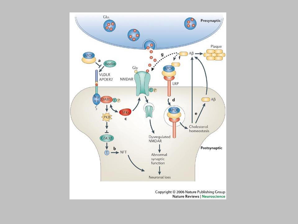

•Lipid-associated apolipoprotein E (APOE) could impede reelin-induced signalling by competing for lipoprotein receptor binding3, 88 (a). Impaired reelin signalling results in elevated phosphorylation4, which could in turn lead to the formation of neurofibrillary tangles (NFTs) (b). APOE might also inhibit reelin-mediated potentiation of NMDA (N- methyl-D-aspartate) receptor (NMDAR) activity and synaptic plasticity (c). After binding to low-density lipoprotein receptor (LDLR)-related proteins (LRP), APOE- containing lipoproteins undergo endocytosis (d). Different APOE isoforms might differentially affect intracellular trafficking131, 132 of the NMDAR (through association with APOE receptors84, 87) and thereby affect its functional availability. Cholesterol homeostasis has a profound impact on the production and trafficking of the amyloid-
(A) peptide6, 51, 133 (e). Extracellular A could associate with APOE and get cleared through receptor-mediated endocytosis (f). Extracellular A represses NMDAR activity
52 directly, but also by promoting the endocytosis of NMDARs120 (g). Reduced glutamatergic transmission could result in impaired synaptic plasticity and promote neuronal loss and dementia. APOER2, APOE receptor 2; DAB1, disabled 1; GSK3b;, gylcogen synthase kinase 3; PKB, protein kinase B; SFKs, SRC family tyrosine kinases; VLDLR, very-low-density lipoprotein receptor.
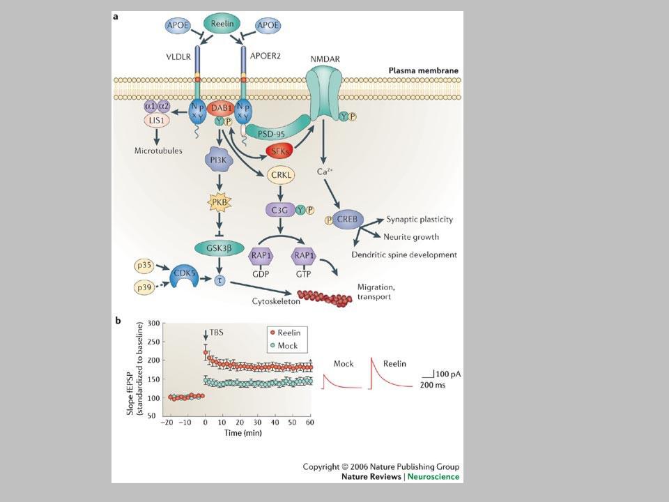

•a | Reelin binds to lipoprotein receptors, the VLDLR and the APOER2, with high affinity at the cell surface. Binding of reelin to the receptors induces feed-forward activation of DAB1, an adaptor protein that interacts with NPxY motifs in both receptor tails. The clustering of DAB1 activates SRC family tyrosine kinases (SFKs), which potentiates tyrosine phosphorylation of DAB1 (Refs 32, 33). Phosphorylated DAB1 further activates phosphatidylinositol-3-kinase (PI3K) and subsequently protein kinase B (PKB)121. PKB activation inhibits the activity of glycogen synthase kinase 3 (GSK3). As a result, phosphorylation of is reduced, promoting microtubule stability. Tyrosine- phosphorylated (YP) DAB1 also recruits CRK-like (CRKL), which induces phosphorylation of a guanine nucleotide exchange factor, C3G122. Activated C3G promotes the formation of RAP1-GTP, which controls actin cytoskeleton rearrangement. Lissencephaly 1 (LIS1) is another binding partner of tyrosine-phosphorylated DAB1. It associates with a-subunits to form a Pafah1b complex, which regulates microtubule dynamics35, 123, 124, 125, 126, 127. Cyclin-dependent kinase 5 (CDK5) acts in parallel with reelin on numerous substrates, including microtubules. p35 and p39 are activating subunits of CDK5. APOER2 associates with postsynaptic density protein 95 (PSD-95), an abundant scaffolding protein in the PSD, through an alternatively spliced exon. This interaction is crucial for the coupling of the reelin signalling complex to the NMDA (N-methyl-D-aspartate) receptor (NMDAR)84, 86. Reelin- activated SFKs tyrosine phosphorylate the NMDAR on NR2 subunits, resulting in the potentiation of NMDAR-mediated Ca2+ influx. Elevated intracellular Ca2+ can activate the transcription factor cyclic AMP-response element binding protein (CREB), thereby potentially initiating the expression of genes that are important for synaptic plasticity, neurite growth and dendritic spine development86. b | Reelin treatment potentiates long-term potentiation in wild-type hippocampal slices82. Reelin treatment also enhances NMDAR-mediated whole cell current in wild-type CA1 pyramidal neurons
86. Traces were recorded at a holding potential of +40 mV. fESP, field excitatory postsynaptic potential; TBS, theta burst stimulation. Panel a modified, with permission from Ref. 84 © (2005) Elsevier Science. Panels a and b (right) modified, with permission, from Ref. 82 © (2002) American Society for Biochemistry and Molecular Biology. Panel b (left) modified from Ref. 86 © (2005) Society for Neuroscience.
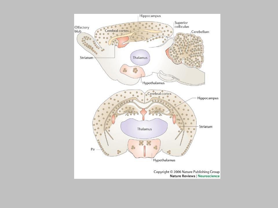

The expression pattern of reelin128 is represented by the yellow dots in the sagittal (top) and coronal (bottom) sections. Reelin is primarily expressed by GABA- containing interneurons throughout the neocortex, with the highest levels in layers I and V. In the hippocampus, reelin is present in the strata oriens and radiatum of CA1 and CA3, and in the hilus of the dentate gyrus. Reelin is also expressed in the mitral cells of the olfactory bulb and the granule cell layer of the cerebellar cortex. Some areas of the hypothalamus, striatum and superior colliculus also express reelin. Pir, piriform cortex.


GABAA receptor subunits are assembled into pentameric receptors as they pass
through the endoplasmic reticulum and the Golgi. GABARAP (GABAA receptor-
associated protein)13 and NSF (N-ethylmaleimide-sensitive factor)14 seem to be involved in the intracellular membrane trafficking of GABAA receptors. It is not
clear whether Plic-1 assists the surface expression of receptors synthesized de novo. Once GABAA receptors reach the cell surface, they are either localized
extrasynaptically or targeted to postsynaptic sites by a mechanism that requires the 2 subunit and gephyrin6 and possibly other factors yet to be identified.
Constitutive endocytosis of GABAA receptors via clathrin-coated pits is indicated by the interaction between GABAA receptors and the adapter protein AP2 and is
known to require the 2 subunit and dynamin12. The paper by Bedford et al. suggests that endosomal GABAA receptors are targeted for degradation or are
possibly held and recycled back to the surface with the help of Plic-1.