

Nanomaterials and Nanosystems for Biomedical Applications - M. Reza Mozafari
.pdf68 |
SAHIN |
The mechanism of vesicle formation upon use of nonionic surfactants is not completely clear. The most common theory is that nonionic surfactants form a closed bilayer in aqueous media based on their amphiphilic nature (Figure 1). Formation of this structure involves some input of energy, for instance by means of physical agitation (e.g. using the hand-shaking method; see Baillie et al 1985) or heat (e.g. using the heating method; see Mozafari 2005a). In this closed bilayer structure, hydrophobic parts of the molecule are oriented away from the aqueous solvent whereas the hydrophilic head comes in contact with the aqueous solvent. It resembles phospholipid vesicles in liposomes and hence, enables entrapment of hydrophilic drugs. The low cost, stability and resultant ease of storage of nonionic surfactants has led to the exploitation of these compounds as alternatives to phospholipids.
Niosomes can entrap hydrophilic drugs and other bioactives upon encapsulation or hydrophobic material by partitioning of these molecules into hydrophobic domains. These vesicles can be formulated either unilamellar or multilamellar in structure. Moreover, niosomes possess great stability, cost-effectiveness, and simple methodology for the routine and large-scale production without the use of hazardous solvents.
The superiorities and advantages of niosomes, compared to other micro and nano encapsulation technologies can be summarized as follows:
•Compared to phospholipid molecules used in liposome formulations, the surfactants used in the formation of niosomes are more stable;
•Simple methods are required for manufacturing and large–scale production of niosomes;
Figure 1. Schematic representation of a noisome. Dark circles represent polar head groups and lines are apolar tails of the surfactant molecules
NIOSOMES AS NANOCARRIER SYSTEMS |
69 |
•As the excipients and equipments used for production are not expensive, niosome manufacturing process is cost-effective;
•Niosomes possess longer shelf-life than liposomes and most other nanocarrier systems;
•Unlike liposomes, they are stable at room temperature and less susceptible to light.
2.FACTORS AFFECTING THE FORMATION OF NIOSOMES
2.1.Type of Surfactants
Type of the surfactants influences encapsulation efficiency, toxicity, and stability of niosomes. The first niosomes were formulated using cholesterol and single-chain surfactants such as alkyl oxyethylenes. The alkyl group chain length is usually from C12–C18. The hydrophiliclipophilic balance (HLB) is a good indicator of the vesicle forming ability of any surfactant. Uchegbu et al (1995, 1998) reported that the sorbitan monostearate (Span) surfactants with HLB values between 4 and 8 were found to be compatible with vesicle formation. Polyglycerol monoalkyl ethers and polyoxylate analogues are the most widely used single-chain surfactants. However, it must be noted that they possess less encapsulation efficiency in the presence of cholesterol. Etheric surfactants have also been used to form niosomes. These types of surfactants are composed of single-chain, monoalkyl or dialkyl chain. The latest ones are similar to phospholipids and possess higher encapsulation efficiency. Esther type amphyphilic surfactants are also used for niosome formulation. They are degraded by estherases, triglycerides and fatty acids. Although these types of surfactants are less stable than ether type ones, they possess less toxicity. Furthermore, glucosides of myristil, cethyl and stearyl alcohols form niosomes.
2.2.Surfactant/Lipid and Surfactant/Water Ratios
Other important parameters are the level of surfactant/lipid and the surfactant/water ratio. The surfactant/lipid ratio is generally 10–30 mM (1–2.5% w/w). If the level of surfactant/lipid is too high, increasing the surfactant/lipid level increases the total amount of drug encapsulated. Change in the surfactant/water ratio during the hydration process may affect the system’s microstructure and thus, the system’s properties.
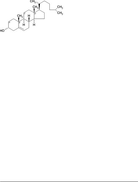
2.3.Cholesterol
Steroids are important components of cell membranes and their presence in membranes brings about significant changes with regard to bilayer stability, fluidity and permeability. Cholesterol, a natural steroid, is the most commonly used membrane additive (Figure 2) and can be incorporated to bilayers at high molar
70 |
SAHIN |
Figure 2. Chemical structure of cholesterol
ratios. Cholesterol by itself, however, does not form bilayer vesicles. It is usually included in a 1:1 molar ratio in most formulations to prevent vesicle aggregation by the inclusion of molecules that stabilize the system against the formation of aggregates by repulsive steric or electrostatic effects. It leads to the transition from the gel state to liquid phase in niosome systems. As a result, niosomes become less leaky.
2.4.Other Additives
As is the case with liposomes, charged phospholipids such as dicethylphosphate (DCP) and stearyl amine (SA) have been used to produce charge in niosome formulations. The former molecule provides negative charge to vesicles whereas the later one is used in the preparation of positively charged (cationic) niosomes.
2.5.Nature of the Drug
One of the overlooked factors is the influence of the nature of the encapsulated drug on vesicle formation (Table 1). The encapsulation of the amphipathic drug doxorubicin has been shown to alter the electrophoretic mobility of hexadecyl diglycerol ether (C16G2) niosomes in a pH dependent manner, indicating that the amphipathic drug is incorporated in the vesicle membrane.
Table 1. The effect of the nature of the drug on the formation of niosomes
Nature of the drug |
Leakage from the vesicle |
Stability |
Other properties |
|
|
|
|
Hydrophobic drug |
Decreased |
Increased |
Improved transdermal delivery |
Hydrophilic drug |
Increased |
Decreased |
– |
Amphiphilic drug |
Decreased |
– |
Increased encapsulation, |
|
|
|
altered electrophoretic |
|
|
|
mobility |
Macromolecular drug |
Decreased |
Increased |
– |
|
|
|
|
NIOSOMES AS NANOCARRIER SYSTEMS |
71 |
3.PREPARATION OF NIOSOMES
Niosomes can be prepared using non-ionic surfactants. As the number of double layers, vesicle size and its distribution, entrapment efficiency of the aqueous phase, and permeability of vesicle membranes are influenced by the way of preparation, these parameters should be taken into account while making a decision on selecting the optimum methodology for formulation.
Most of the experimental methods consist of the hydration of a mixture of the surfactant/lipid at elevated temperature followed by optional size reduction to obtain a colloidal dispersion. Subsequently, the unentrapped drug is separated from the entrapped drug by centrifugation, gel filtration or dialysis. Only a couple of methods could be found in the literature on the preparation of niosomes on an industrial scale (Novasome®, heating method). In the Novasome® method, niosomes are prepared upon injection of the melted surfactants/lipids into a large volume of well-agitated, heated aqueous solutions. The novel heating method and other well-known procedures for niosome preparation are summarized below.
3.1.Ether Injection Method
This method is essentially based on slow injection of an ether solution of niosomal ingredients into an aqueous medium at high temperature. Typically a mixture of surfactant and cholesterol (150 μmol) is dissolved in ether (20 mL) and injected into an aqueous phase (4 mL) using a 14-gauge needle syringe. Temperature of the system is maintained at 60°C during the process. As a result, niosomes in the form of large unilamellar vesicles (LUV) are formed (Baillie et al 1985; Vyas and Khar 2002).
3.2.Film Method
The mixture of surfactant and cholesterol is dissolved in an organic solvent (e.g. diethyl ether, chloroform, etc.) in a round-bottomed flask. Subsequently, the organic solvent is removed by low pressure/vacuum at room temperature, for example using a rotary evaporator. The resultant dry surfactant film is hydrated by agitation at 50–60°C and multilamellar vesicles (MLV) are formed (Baillie et al 1985; Varshosaz et al 2003).
3.3.Sonication
Typically the aqueous phase is added into the mixture of surfactant and cholesterol in a scintillation vial. Then, it is homogenized using a sonic probe. The resultant vesicles are of small unilamellar (SUV) type niosomes (Baillie et al 1986). The SUV type niosomes are larger than SUV liposomes (i.e. SUV niosomes are >100 nm in diameter while SUV liposomes are <100 nm in diameter).
72 |
SAHIN |
It is possible to obtain SUV niosomes by sonication of MLV type vesicles, obtained for example through the film method explained above. For small volume samples probe type sonicator is used while for larger volume samples bath type sonicator is more appropriate.
3.4.Method of Handjani–Vila
Equivalent amounts of synthetic non-ionic lipids are mixed with the aqueous solution of the active substance to be encapsulated and a homogenous lamellar film is formed by shaking. The resultant mixture is homogenized employing ultracentrifugation and agitation at a controlled temperature (Handjani-Vila 1990).
3.5.Reverse Phase Evaporation
Reverse phase evaporation technique is being used to prepare different carrier systems including archaeosomes, liposomes, nanoliposomes and niosomes. Typically surface-active agents are dissolved in chloroform, and 0.25 volume of phosphate saline buffer (PBS) is emulsified to get water in oil (w/o) emulsion. The mixture is then sonicated and subsequently chloroform is evaporated under reduced pressure. The lipid or surfactant first forms a gel and then hydrates to form niosomal vesicles (Kiwada et al 1985a, 1985b; Vyas and Khar 2002).
Alternatively, hydrogenated or nonhydragenated egg phosphatidylcholine (ePC) is dissolved in chloroform and PBS. The mixture is sonicated under low pressure, forming a gel. The gel is subsequently hydrated. Free drug or other bioactives to be encapsulated (un-entrapped material) is generaly removed by dialysis or centrifugation. Protamine is added prior to centrifugation process to achieve phase separation.
3.6.Heating Method
This is a non-toxic, scalable and one-step method and is based on the patented procedure of Mozafari (2005b). Mixtures of non-ionic surfactant, cholesterol and/or charge inducing molecules are added to an aqueous medium (e.g. buffer, distilled H2O, etc.) in the presence of a polyol such as glycerol. The mixture is heated while stirring (at low shear forces) until vesicles are formed (Mozafari 2005b).
3.7.Post-Preparation Processes
The main post-preparation processes in the manufacture of niosomes are downsizing and separation of unentrapped material. After preparation, size reduction of niosomes is achieved using one of the methods given below:
1.Probe sonication results in the production of the niosomes in the 100–140 nm size range.
2.Extrusion through filters of defined pore sizes.
NIOSOMES AS NANOCARRIER SYSTEMS |
73 |
3.Combination of sonication and filtration has also been used to obtain niosomes in the 200nm size range (e.g. doxorubicin niosomes).
4.Microfluidization yielding niosomes in sub-50 nm sizes.
5.High-pressure homogenisation also yields vesicles of below 100nm in diameter. As in most cases 100% of the bioactive agent cannot be encapsulated in the niosomal vesicles, the unentrapped bioactive agent should be separated from the entrapped ones (Kiwada et al 1985a, 1985b). In some instances, this provides an advantage since this drug delivery system (or generally speaking bioactive carrier system) gives an initial burst to initiate therapy followed by a sustained maintenance dose.
Most commonly used methods for separating unentrapped material from niosomes are
as follows:
• Dialysis;
• Gel filtration (e.g. Sephadex G50);
• Centrifugation (e.g. 7000 × g for 30 min for the niosomes prepared by hand-
shaking and ether injection methods);
• Ultracentrifugation (150000 × g for 1.5 h).
4.ENTRAPMENT EFFICIENCY
Both the yield and the entrapment efficiency of niosomes depend on the method of preparation. Niosomes prepared by ether injection method have better entrapment efficiency than those prepared by the film method or sonication. Addition of cholesterol to non-ionic surfactants with singleor dialkyl-chain significantly alters the entrapment efficiency. However, surfactants of glycerol type lead to reduction in entrapment capacity as the amount of cholesterol increases.
Employing film method and a subsequent sonication results in formation of liquid crystal and gel type niosomes. Niosomes in the form of liquid crystals possess better entrapment efficiency than gel type vesicles as observed in liposomes as well. Urea niosomes are the best example for gel type niosomes and exhibit 10% entrapment capacity. This can be improved by the addition of cholesterol.
5.STABILITY OF NIOSOMES
Vesicles are stabilized based upon formation of 4 different forces:
1.van der Waals forces among surfactant molecules;
2.repulsive forces emerging from the electrostatic interactions among charged groups of surfactant molecules;
3.entropic repulsive forces of the head groups of surfactants;
4.short-acting repulsive forces.
Electrostatic repulsive forces are formed among vesicles upon addition of charged surfactants to the double layer, enhancing the stability of the system.
Biological stability of the niosomes prepared with alkyl glycosides was investigated by Kiwada et al (1985a, 1985b). They reported that niosomes were not stable
74 |
SAHIN |
enough in plasma. This may be due to single–chain alkyl surfactants. SUVs were found to be more stable.
Niosomes in the form of liquid crystal and gel can remain stable at both room temperature and 4°C for 2 months. No significant difference has been observed between the stability of these two types of niosomes with respect to leakage. Even though no correlation between storage temperature and stability has been found, it is recommended that niosomes should be stored at 4°C. Ideally these systems should be stored dry for reconstitution by nursing staff or by the patient and when rehydrated should exhibit dispersion characteristics that are similar to the original dispersion.
Simulation studies conducted to investigate physical stability of these niosomes during transportation to the end-user revealed that mechanical forces didn’t have any influence on physical stability. It is assumed that the reason behind the stability of niosomes may be due to the prevention of aggregation caused by steric interactions among large polar head groups of surfactants.
The factors which affect the stability of niosomes are as following:
•type of surfactant;
•nature of encapsulated drug;
•storage temperature;
•detergents;
•use of membrane spanning lipids;
•the interfacial polymerization of surfactant monomers in situ;
•inclusion of a charged molecule.
6.TOXICITY OF NIOSOMES
Unfortunately, there is not enough research conducted to investigate toxicity of niosomes. Researchers measured proliferation of keratinocytes in one of the topical niosome formulations (Hofland et al 1991). The effect of surfactant type on toxicity was investigated. It was determined that the ester type surfactants are less toxic than ether type surfactants (Hofland et al 1991, 1992). This may be due to enzymatic degradation of ester bounds. In general, the physical form of niosomes did not influence their toxicity as evident in a study comparing the formulations prepared in the form of liquid crystals and gels. However, nasal applications of these formulations caused toxicity in the case of liquid crystal type niosomes.
In some instances, encapsulation of the drug by niosomes reduces the toxicity as demonstrated in the study on preparation of niosomes containing vincristine (Parthasarathi et al 1994). It decreased the neurological toxicity, diarrhoea and alopecia following the intravenous administration of vincristine and increased vincristine anti-tumor activity in S-180 sarcoma and Erlich ascites mouse models.
NIOSOMES AS NANOCARRIER SYSTEMS |
75 |
7.APPLICATIONS OF NIOSOMES
7.1.Transdermal Applications
It is well-known fact that transdermal applications provide a great advantage of protecting drugs from the hepatic first pass effect. However, stratum corneum layer of skin forms a barrier, resulting in a slow absorption at the application site.
The fact that in the manufacture of niosomes nonionic surfactants are used to form vesicles makes them good candidates for transdermal drug delivery. Sentjurc and co-workers (1999) investigated transport of liposome-entrapped spin labelled compounds into skin by electron paramagnetic resonance imaging methods. In addition, the mechanistic aspects of cyclosporin-A skin delivery were assessed. Niosomes containing urea formulations have been prepared and being treated by the cosmetic industry, as almost magical ingredients.
Two mechanisms are suggested for transdermal absorption of vesicles:
i)diffusion of nisomes from the stratum corneum layer of skin as a whole, or:
ii)forming new vesicles by each individual component (re-formation of vesicles). The later one takes place only at certain regions of stratum corneum where water content is high. Many researchers agree upon the second mechanism since the diameter of vesicles is larger than the lipid lamellar spaces of the stratum corneum.
7.2.Parenteral Applications
Niosomes in sub-micron size are used for parenteral administration. Niosomal vesicles up to 10 μm are administered via i.p. or i.m. Florence and Cable (1993) prepared 59Fe-deferroxamine trioxyethylene cholesterol vesicles for i.v. use and reported that the distribution of such vesicles depends upon vesicle size as evident from the data indicating greater distribution in liver and spleen.
Uchegbu et al (1996, 1997, 1998) investigated the effect of dose on plasma drug concentration by comparing doxorubicin-containing niosomes with free drug in mouse upon i.p. administration. The data revealed that plasma drug concentration is influenced by dose. Niosomes enhance plasma drug concentration. Furthermore, they conducted experiments for toxicity and determined that there is a positive correlation between dose and toxicity. However, Florence and Cable (1993) indicated that the preparation of doxorubicin in the form of niosomes reduces its cardiac toxicity upon i.v. administration.
7.3.Peroral Applications
The oral use of niosomal formulations was first demonstrated by Azmin et al (1985) in a study involving 100 nm methotrexate C16G3 niosomes. Significantly higher levels of methotrexate were found in the serum, liver and brain of PKW mice following oral administration of a niosomal formulation. It thus appears that there is enhanced drug absorption with these niosomal formulations.
76 |
SAHIN |
Rentel et al (1996) prepared niosome-based ovalbumin vaccines by two different types of surfactants and administered p.o. to mouse. In comparison to the conventional vaccines, niosome-based vaccines resulted in increased antibody titer. However, type of surfactant didn’t have any influence on antibody production.
7.4.Radiopharmaceuticals
The first applications of niosomes as radiopharmaceuticals have been achieved by Erdogan et al in 1996. They prepared 131I labeled iopromide niosomes with positive charge in order to enhance contrast during CT in rats (Erdogan et al 1996). The formulations were in the form of gel or liquid crystal. They were found more in kidneys and maintained their activity over 24 hours. In another study, Korkmaz et al (2000) used 99mTclabeled DTPA containing niosomes and found that DTPA was accumulated in liver and spleen in large quantities. The gamma sintigraphic images of mouse were better with 99mTc-DTPA niosomes [N1 formulation: SurI: SA: CHOL (10:1:4)]. Similarly, gel type 99mTc-labelled niosomes of DMSA accumulated in liver, kidneys, and spleen in mouse and maintained the activity for 24 hours. Niosome formulation also provided better stability in comparison to conventional solutions of DMSA as they are less susceptible to light, temperature and oxidation.
7.5.Ophthalmic Drug Delivery
There is only a single study on the use of niosomes for ophthalmic drug delivery to date (Saettone et al 1996). Saettone et al (1996) reported on the biological evaluation of a niosomal Cyclopentolate delivery system for ophthalmic delivery. Polysorbate 20 and cholesterol were used for niosome formulations. It was determined that cyclopentolate penetrated the cornea in a pH dependant manner within these niosomes. Optimum pH for peak permeation values was pH 5.5. Permeation decreased at pH 7.4. However, in vivo data revealed that there was increased mydriatic response with the niosomal formulation irrespective of the pH of the formulation. In short, the increased absorption of cyclopentolate may be the result of the altered permeability characteristics of the conjuctival and scleral membranes. Niosomes >10 μm are suitable for drug administration to eye.
8.PRONIOSOMES
Proniosomes are prepared by hydration and agitation in hot water for a short period of time. They offer a versatile vesicle delivery concept with the potential for drug delivery via the transdermal route. They form niosomes following topical application under occlusive conditions, due to hydration by water from the skin itself.
Alsarra et al (2005) prepared topical niosomes of Ketorolac tromethamine (KT) as an alternative noninvasive mode of delivery, as transdermal delivery certainly seemed to be an attractive route of administration to maintain the drug blood levels
NIOSOMES AS NANOCARRIER SYSTEMS |
77 |
of KT for an extended period of time. Using a wide-mouth glass tube, KT was mixed with surfactant, lecithin, and cholesterol in absolute ethanol. Then, the open-end of the glass tube was covered with a lid and the tube was warmed in a water bath at 65 ± 3°C for 5 min. After that, PBS was added and the mixture was further warmed in the water bath for about 2 min until a clear solution was obtained. The mixture was allowed to cool to room temperature until a proniosomal gel was formed. The proniosomal gel was then mixed with one of several 2% polymeric gels (HPMC, CMC, or Carbopol) to give a final concentration of 0.5% KT. The resultant vesicles were characterized with respect to shape, surface morphology, and size by means of SEM.
The formulations prepared with Span 60 and Tween 20 gave the highest entrapment efficiency. This may be due to the fact that the highly lipophilic portion of the drug is housed within the lipid bilayer of the niosomes. Type of surfactant influenced the vesicle size. The niosomes prepared with Tween 20 were larger than those prepared with Span 60. The reason behind that may be the decrease in surface energy with increasing hydrophobicity of the surfactant. Span is more hydrophobic than Tween. Although increasing the amount of cholesterol or reducing lecithin increased hydrophobicity, they didn’t change the vesicle size significantly. SEM analysis revealed that most of the vesicles are spherical and discrete with sharp boundaries. Ex vivo release studies indicated that inclusion of an optimum ratio of surfactant/lecithin in the vesicles may play a more important role than cholesterol plays in modulating drug permeation.
In order to achieve drug release through skin, proniosomes should be hydrated to form niosomal vesicles before they permeate across the skin. Drug transfer across skin is achieved by several mechanisms including adsorption and diffusion of niosomes onto the surface of skin, facilitating drug permeation, tendency of the vesicles to act as penetration enhancers, reducing the barrier properties of the stratum corneum and the lipid bilayers of niosomes forming a rate-limiting membrane barrier for drugs.
9.CONCLUSIONS
Niosomes have been proven to be useful controlled drug delivery systems for transdermal, parenteral, oral, and ophthalmic routes. They can be used to encapsulate anti-infective agents, anti-cancer agents, anti-inflammatory agents and fairly recently as vaccine adjuvants. Niosomes may enable targeting certain areas of the mammalian organisms and may be exploited as diagnostic imaging agents.
Niosomes are superior systems when compared to other carriers with respect to stability, toxicity and cost-effectiveness. The problem of drug loading remain to be addressed and although some new approaches have been developed to overcome this problem, it is still necessary to increase encapsulation efficiencies as it is important to maintain the biological potential of the formulations.
As type of surfactant is the most important parameter affecting the formation of the vesicles, as well as their toxicity and stability, the surfactants with the higher