

Nanomaterials and Nanosystems for Biomedical Applications - M. Reza Mozafari
.pdfADVANTAGES OF A LIPOSOMAL DENDRIMER-DOXORUBICIN COMPLEX |
141 |
ratios of doxorubicin to PAMAM (i.e. 3:1 and 6:1). The results indicated that a doxorubicin to PAMAM molar ratio of 3:1 was sufficient in order to achieve an almost 97% incorporation of doxorubicin into the dendrimer. Doxorubicin incorporation into PAMAM was higher when the complex was formulated in TES buffer (pH: 7.5) as compared to that of acetate buffer (pH: 4.5). The release of doxorubicin appeared to be quite slow. The lower doxorubicin release (7.4% during 48 h) was observed at a molar ratio of 3:1 of doxorubicin to PAMAM, and the higher (16.5% during 48 h) at molar ratio of 6:1 of doxorubicin to PAMAM in TES buffer (pH: 7.5) at 37°C (Papagiannaros et al. 2005).
3.3.Incorporation and Release of Doxorubicin-PAMAM Complex from Liposomes
The incorporation efficiency of doxorubicin-PAMAM complex, (3:1 molar ratio) into liposomes (EPC:SA 10:0.1 molar ratio) was almost 95% while doxorubicin (doxorubicin-PAMAM complex 3:1 molar ratio) to lipid molar ratio was 0.020 (initial 0.028) in TES buffer (pH: 7.5).
The release of doxorubicin (doxorubicin-PAMAM complex 3:1 molar ratio) from the liposomes was quite slow; 13.6% at 37°C (48 h) in TES buffer at pH: 7.5 and 14.0% at 37°C (48 h) in 50% RPMI cell culture medium (Figures 1 and 2).
3.4.Physical Properties of Liposomes Incorporating the Doxorubicin-PAMAM Complex
Size measurements of the doxorubicin-PAMAM complex (3:1 molar ratio) attached to liposomes indicated an average size of 116.3±7.8 nm and a -potential of –8.7±1.7 mV (Table 1). The stability of liposomes was studied for a period up to 26 weeks. The liposomal suspension was kept at 4°C in the dark. No sediment was observed while their average hydrodynamic diameter increased rapidly (>1μm) (Papagiannaros et al. 2005).
4.DISCUSSION
A liposome delivery system is proposed for incorporating anticancer drugs, combining the liposomal and dendrimeric technologies. Its ability to modulate the release of the encapsulated drug in a way that is independent of the liposomal membrane but strongly related to the complexation of the drug with the dendrimer, offers advantages over conventional liposomal formulation in terms of the pharmacological activity. The controlled release of the encapsulated cytotoxic drugs is of paramount importance in cancer chemotherapy (Andresen et al. 2005). An example is presented in this report, based on the release properties of liposomes encapsulating doxorubicin-PAMAM G4 complex in comparison with the conventional type of liposome encapsulated doxorubicin. This liposomal formulation has shown superior in vitro anticancer activity, due to its slow releasing properties
142 |
PAPAGIANNAROS AND DEMETZOS |
(Papagiannaros et al. 2005). It has already been established that the cytotoxic effect of the drug is mediated by the leakage of doxorubicin from the liposomes (Gabizon 2002). However a delayed release of doxorubicin is necessary in order to reduce the toxicity and increase the therapeutic usefulness of the drug (Charrois et al. 2004).
The release rate of doxorubicin is an important factor since a slow release is necessary in order to decrease the side effects of doxorubicin and improve its therapeutic index (Gabizon 2002; Horovic et al. 1992). A slow release rate can also contribute to the accumulation of the drug in the tumor (Charrois and Allen 2004). The control of the leakage of the encapsulated drug is mainly achieved through modifications in the liposome membrane, mainly by changing the fluidity of the membrane, by addition of cholesterol (Ohvo-Rekila et al. 2002) or “rigid state” lipids (Oussoren et al. 1998); increasing the rigidity of the liposome membrane also affects the uptake of the encapsulated drug by the tumor cells, therefore reducing the toxicity can also reduce the availability of the drug to the tumor site (Sadzuka et al. 2002). On the contrary, doxorubicin incorporated into cholesterol–free liposomes, as a doxorubicin-PAMAM complex, exhibited a slow release rate, at 37°C, after a 48 h incubation period (in 48 hours less than 20% was released). Consequently, it can be expected that this formulation possess reduced doxorubicin side effects. Various drugs encapsulated in dendrimers (Kolhe et al. 2005) or incorporated in liposomes together with PAMAM dendrimers (Klopade et al. 2002) have shown slow release profiles. The contribution of the doxorubicin-PAMAM complex may not be limited to the delayed release of the encapsulated doxorubicin, since an ibuprofenPAMAM G4 complex was found to enter lung epithelial cancer cells in 1h (compared to 3h for free ibuprofen) (Kolhe et al. 2005), thus the dendrimer could facilitate the cellular entry of the complexed drugs. Furthermore, PAMAM G4 dendrimer conjugated with ibuprofen entered lung carcinoma cells in less than 15 min compared to 1h for free ibuprofen (Kolhe et al. 2005) and PAMAM G5 encapsulating methotrexate exhibited four times more activity in vitro than the free drug against the KB epidermoidal cancer cell line (Quintan et al. 2002).
The encapsulation efficiency of doxorubicin in PAMAM G4 was almost 100%. The presence of dendrimers resulted in a higher encapsulation efficiency and a decreased release rate of the encapsulated drug, although this was achieved by creating a higher and more stable proton gradient across the liposomal membrane (Klopade et al. 2002).
Although the average hydrodynamic diameter of the liposomal formulation incorporated doxorubicin-PAMAM complex was almost 116nm immediately after their production, this size increased to the microns (μ) very rapidly with time. This fact was not observed with the conventional liposomal formulation, that does not incorporate the doxorubicin-PAMAM complex, and therefore it might be attributed to the presence of the dendrimer. It has already been observed that dendrimers could facilitate the formation of liposome aggregates (Sideratou et al. 2002). The charge of liposomes incorporating doxorubicin – PAMAM complex, did not seem to be involved in the formation of the aggregates suggesting that hydrophobic forces between dendrimers, which are attached to liposomal particles, may be responsible.
ADVANTAGES OF A LIPOSOMAL DENDRIMER-DOXORUBICIN COMPLEX |
143 |
Earlier studies using ‘dendrons’ (partial dendrimers) (Purohit et al. 2001) have also reached the same conclusion.
5.CONCLUSIONS
A liposomal drug delivery system incorporating a complex of doxorubicin-PAMAM G4 dendrimers was prepared and compared to conventional liposomal formulation encapsulating doxorubicin with the same lipid composition regarding release properties of the antineoplastic agent. The results suggest that this new controlled release system may be useful in anticancer therapy.
REFERENCES
Andresen, T., Jensen, S., Jorgensen, K. (2005) Advanced strategies in liposomal cancer therapy: Problems and prospects of active and tumor specific drug release. Progress in Lipid Research, 44, 68–97.
Aulenta, F., Hayes, W., Rannard, S. (2003) Dendrimers a new class of nanoscopic containers and delivery devices. European Polymer Journal, 39, 1741–1771.
Charrois, G., Allen, T. (2004) Drug Release rate influences the pharmacokinetics, biodistribution, therapeutic activity and toxicity of pegylated liposomal doxorubicin formulations in murine breast cancer. Biochim. Biophys. Acta, 1663, 167–177.
Cloninger, M. (2002) Biological application of dendrimers. Current Opinion in Chemical Biology, 6, 742–748.
Dhoot, N., Wheatley, M. (2003) Microencapsulated liposomes in controlled drug delivery strategies to modulate drug release and eliminate the burst effect. J. Pharm. Sciences, 92(3), 679–689.
Eichman, J., Bielinska, A., Kukowska-Latallo, J., Donovan, B., Baker, J. (2001) in Frechet, J. and Tomalia, D. (eds.) Dendrimers and other Demdritic Polymers . J. Wiley & Sons, Chisester, 441–462.
Gabizon, A. (2002) Liposomal drug carriers in cancer chemotherapy: current status and future prospects.
The Journal of Drug Targetting, 10(7), 535–538.
Goniotaki, M., Hatziantoniou, S., Dimas, K., Wagner, M., Demetzos, C. (2004) Encapsulation of naturally occurring flavonoids into liposome: Physicochemical characterization and biological activity against human cancer cell lines. J. Pharm. Pharmacol., 56, 1217–1224.
Horovic, A., Barenholtz, A., Gabizon, A. (1992) In vitro cytotoxicity of liposome encapsulaterd doxorubicin: dependence on liposomes composition and drug release. Biochima et Biophysica Acta, 1109, 203–209.
Khuloud Al-J., Sakthivel, T., Florence, A.T. (2003) Dendrisomes: cationic lipidic dendron vesicular assemblies. International Journal of Pharmaceutics, 254, 33–36.
Klopade, A., Caruso, F., Tripathi, P., Nagaish, S., Jain, N. (2002) Effect of dendrimer on entrapment and release of bioactive from liposome. Int. J. Pharm., 232, 157–162.
Kolhe, P., Khandarea, J., Omathanu, O., Kannanb, S., Lieh-Laib, M., Rangaramanujam, M. (2007) Preparation, cellular transport, and activity of polyamidoamine-based dendritic nanodevices with ahigh drug payload Biomaterials, (in press).
Mayer, L., Bally, M. (1986) Uptake of adriamycin into large unilamellar vesicles in response to a pH gradient. Biochimica et Biophysica Acta, 123–126.
Ohvo-Rekila, H., Ramsted, B., Leppimaki, P., Slotte, P. (2002) Cholesterol interaction with phospholipids in membranes. Progress in Lipid Research, 41, 66–97.
Oussoren, C., Eling, W., Crommelin, D., Storm, G., Zuidema, J. (1998) The influence of the route of administration and liposome composition on the potential of liposomes to protect tissue against local toxicity of two antitumor drugs. Biochimica et Biophysica Acta, 1369, 159-172.
Pan, X., Lee, R., Rantman, M. (2004) Penetration into solid tumor tissue of fluorescent latex microspheres: a mimic of liposome particles. Anticancer Research, 24, 3503–3508.
144 |
PAPAGIANNAROS AND DEMETZOS |
Papagiannaros, A., Dimas, K., Papaioannou, G., Demetzos, C. (2005) Doxorubicin-PAMAM dendrimer complex attached to liposomes and cytotoxic studies against human cancer cell lines. Int. J. Pharm, 302, 29–38.
Papagiannaros, A., Hatziantoniou, S., Dimas, K., Papaioannou, G., Demetzos, C. (2006) A liposomal formulation of doxorubicin, composed of hexadecylphosphocholine (HePC): physicochemical characterization and cytotoxic activity against human cancer cell lines. Biomedicine & Pharmacotherapy, 60 (1), 36–42.
Purohit, G., Sakthivel, T., Florence, A.T. (2001) Interaction of cationic partial dendrimers with charged and neutral liposomes. Int. J. Pharm, 214, 71–76.
Quintan, A., Raczka, L., Piehler, L., Lee, I., Myc, A., Majoros, I., Patri, A., Thomas, T., Mule, J., Baker, J. (2002) Design and function of a dendrimer-based therapeutic nanodevice targeted to tumor cells though the folate receptor. Pharmaceutical Research, 19(9), 1310–1316.
Sadzuka, Y., Hirama, R., Sonobe, T. (2002) Effects of intraperitoneal administration of liposomes and methods of preparing liposomes for local therapy. Toxicology Letters, 126, 83–90.
Sideratou, Z., Foundi, J., Tsiourvas, D., Nezis, I., Papadimas, G., Paleos, C. (2002) A novel dendrimeric glue for adhesion of phosphatidyl choline based liposomes. Langmuir, 18, 5036–5039.
Singh, B., Florence, A.T. (2005) Hydrophobic dendrimer-derived nanoparticles Int. J. Pharm., 298, 348–353.
Stenekes, R., Loebis, A., Fernades, C., Crommelin, D., Hennik, W. (2002) Controlled release of liposomes from biodegradable dextran microspheres. Pharmaceutical Research, 17(6), 690–695.
Straubinger, R., Arnold, R., Zhou, R., Mazurchuk, R., Slack, J. (2004) Antivascular and antitumor activities of liposome associated drugs. Anticancer Research 24, 397–404.
Syrigos, K., Michalaki, B., Alevyzaki, F., Macheras, A., Mandrekas, D., Kindilis, K., Karatzas, G.G. (2002) A Phase II study of liposomal doxorubicin and docetaxel in patients with advanced pancreatic cancer. Anticancer Research, 22, 3583–3588.
Szoka, F., Papahadjopoulos, D. (1978) Procedure for preparation of liposomes with large internal aqueous space and high capture by reverse-phase evaporation. Proc. Natl. Acad. Sci. USA, 75(9), 4194–4198.
Toma, S., Tucci, A., Villani, G., Carteni, G., Spadini, N., Palumbo, R. (2002) Liposomal doxorubicin (caelyx) in advanced pretreated soft sarcomas:a phase II study of the Italia sarcoma group (ISG).
Anicancer Research, 20, 485–492.
CHAPTER 10
APPLICATIONS OF LIGHT AND ELECTRON MICROSCOPIC TECHNIQUES IN LIPOSOME RESEARCH
A. YEKTA OZER
Hacettepe University, Faculty of Pharmacy, Department of Radiopharmacy, Ankara 06100, Turkey E-mail: ayozer@yahoo.com
Abstract: Liposomes and some other vesicular systems are widely used as delivery vehicles for bioactive compounds. Successful applications of these carrier systems in drug delivery, gene therapy and other health related areas depend on comprehensive understanding of their physical properties including polydispersity and morphology. Variations in size and shape of the carrier systems are indications of their stability and shelf life and can guide scientists in improving the therapeutic formulations. Towards this end microscopic techniques can provide vital information on size, configuration, stability and mechanisms of cellular uptake of particles on micro and nanoscales as discussed in this chapter
Keywords: carrier systems, liposomes, niosomes, novasomes, sphingosomes, ufasomes, virosomes, electron microscopy, scanning probe microscopy
1.INTRODUCTION
Liposomes, which are also called lipid vesicles, are spherical, closed–continuous structures (Mozafari et al 2002). They are composed of curved lipid bilayers. These bilayers entrap part of the solvent in which they are dispersed and retain this solvent into their interior. They may have one or more concentric or non-concentric membranes and their size is in between 20nm to several micrometers, while the thickness of the membrane is about 4nm (New 1990; Lasic 1993; Mozafari and Mortazavi 2005).
Liposomes are made mainly from amphiphiles. These amphiphiles are a special class of surfactant molecules and are characterized by having hydrophilic and hydrophobic groups on the same molecule. A liposome-forming molecule has two hydrocarbon chains (hydrophobic or nonpolar tails) and a hydrophilic group (polar
145
M.R. Mozafari (ed.), Nanomaterials and Nanosystems for Biomedical Applications, 145–153. © 2007 Springer.
146 |
OZER |
head). In general, most of these molecules are insoluble in water and they form colloidal dispersions.
Due to their solubility properties, the structure of these aggregates of amphiphilic molecules involves the ordering of lipid molecules and their arrangement in aqeous environments. The hydrophilic part of the amphiphilic molecules tends to be in contact with water whereas the hydrophobic hydrocarbon chains prefer to be hidden from water in the interior of the structures. Lipid bilayer is one of the most frequently seen aggregation structures. On the surface of either side are polar heads, which shield nonpolar tails in the interior of the lamella from water. At higher lipid concentrations these bilayers form lamellar liquid-crystalline phases where twodimensional planar lipid bilayers alternate with water layers. When diluted, these lipid bilayers seperate, become unstable, curve and form liposomes.
Due to their unique properties – including ease of preparation, versatility in terms of composition, size, charge, fluidity, etc. – and possibility of preparing them using non-toxic, non-immonogenic material on the industrial scales (Lasic and Papahadjopoulos 1998; Mozafari and Mortazavi 2005), liposomes are widely used as controlled release vehicles. For specialized nanotherapeutic and other applications, the lipid vesicles need to be finely tuned and delicately tailored. Morphological and physicochemical studies are strict pre-clinical requirements for successful formulation of liposomal carriers. This chapter reviews commonly used microscopic techniques in the assessment of the lipid vesicles.
2.DIFFERENT TYPES OF MICROSCOPIC VESICLES
The most commonly used microscopic vesicles are liposomes. They are in fact synthetic analogues of natural biomembranes. Liposomes are composed of polar lipids such as lecithin. The nanometric versions of liposomes are known as nanoliposomes (Mozafari and Mortazavi 2005). There are some other types of microscopic vesicular systems similar to liposomes, namely niosomes, sphingosomes, novasomes, transfersomes, ufasomes and virosomes as explained below.
Niosomes (explained in detail in Chapter 4) are nanometric particles (non-ionic surfactant vesicles) used in the delivery of bioactive compounds and composed of mono or diacyl polyglycerol or (poly) oxyethylene based lipids in mixtures with 0-50 mol % of cholesterol. In general, they are prepared by very similar methods as liposomes (Uchegbu and Vyas 1998; Korkmaz et al 2000).
Sphingosomes are composed of skin lipids and predominantly sphingolipids. They are processed in similar ways as phospholipid liposomes (Brunke 1990; Erdogan et al 2005). In a recent study sphingosomes were used as a drug delivery system to target a model thromboembolic disease in rabbits (Erdogan et al 2005).
Novasomes are paucilamellar (Oligolamellar), nonphospholipid vesicles and made of C12–C20 single-chain surfactants bonded via an either esther or peptide bound to polar heads. Double-chained surfactants include palmitoyl or oleayl chains or sterols attached to glycerol phosphorylcholine (Chambers et al 2004).
APPLICATIONS IN LIPOSOME RESEARCH |
147 |
Transfersomes are another kind of liposomes, which are composed from up to equimolar mixtures of phosphatidylcholine with myristic acid (Cevc and Blume 1992; Cevc 1996) (also see Chapter 7).
In Ufasomes, oleic acid is used as single chain surfactant as the amphiphilic molecule and these type of liposomes were prepared long time ago in 1973 (Gebicki and Hicks 1973).
Another derivative of liposomes are Virosomes that contain viral proteins in their membranes (Kara et al 1971; Almeida et al 1975). In another words virosomes are reconstituted viral envelopes that retain the receptor binding and membrane fusion activities of the virus they are derived from. Virosomes can be generated by detergent solubilization of the membrane of an enveloped virus, sedimentation of the viral nucleocapsid, and subsequent selective removal of the detergent from the supernatant to produce reconstituted membrane vesicles consisting of the viral envelope lipids and glycoproteins. Size and surface characteristics of virosomes can be studied through microscopic visualization. More information about virosomes are provided in Chapter 7 of this book.
Liposome and its other derivatives are used as models of biological systems (e.g. biomembranes) and in the delivery of drugs and other macromolecules. Depending on the special physico-chemical characteristics of polar lipids and other ingredients of these vesicles, they have a great promise for tissue and cell-specific delivery of a variety of phamaceuticals and biotechnology products.
3.CLASSIFICATION OF LIPOSOMAL VESICLES
Liposomes are classified depending on vesicle size, preparation method and their number of lamella (New 1990; Mozafari and Mortazavi 2005). A multilamellar vesicle (MLV) is a liposome composed of a number of concentric lipidic bilayers. A vesicle composed of several non-concentric vesicles encapsulated within a single bilayer is known as a multivesicular vesicle (MVV). Another type of liposome is known as a unilamellar vesicle (ULV) and contains one single bilayer and one internal (aqueous) compartment. Unilamellar vesicles can be divided into small unilamellar vesicle (SUV, less than 100nm) and large unilamellar vesicle (LUV, larger than 100nm).
The most important liposome characteristics are:
i.Vesicle size;
ii.Number of bilayers and morphology;
iii.Bilayer fluidity; and
iv.Surface characteristics (charge and hydrophilicity).
Vesicle size can be approximately between 0.02 and 10μm. The largest vesicles may have more than 10 bilayers, however, this can be changed by the preparation method. Size is a very important factor playing an important role on the in vitro and in vivo behaviour of liposomes. Physical stability and biodistribution mainly depend on the liposome size.
148 |
OZER |
Vesicle shape (morphology) is the other significant factor for liposome technology. This is due to the fact that vesicle shape of liposomes provides an idea about their in vivo fate and their cellular transition mechanism. Some of the microscopic techniques used in the morphological examinations of liposomes and other vesicular carriers are explained below.
4.MICROSOPY IN LIPOSOME TECHNOLOGY
Methods determining the size of liposomes vary in complexity and degree of sophistication (Talsma et al 1987; New 1990). Microscopy is the oldest but very valuable technique among the others. With light microscopy, the gross view and rough size of the particles can be seen. Undoubtedly, the most precise method is that of electron microscopic examination. Because, it permits visualization of each individual liposome and given time, patience and the required skill, several artifacts can be avoided.
With electron microscopy, one can obtain precise information about the profile of a liposome sample over the whole range of sizes. In addition, electron microscopy can provide information on the configuration of lipid vesicles and their stability in time. However, there are also some disadvantages associated with electron microscopic techniques. These include:
•They can be very time-consuming; and
•Require expensive equipments that may not always be immediately available. Dynamic Light Scattering, Coulter Counter, Size Exclusion Chromatography
and Optical Density method can be mentioned among the other liposomal size measurement techniques. These are mainly used for particle size determination and can not provide information on shape, configuration and presence/absence of aggregation or fusion of vesicular systems, for which microscopic techniques are more appropriate.
Although Dynamic Light Scattering is a very simple technique to perform, it has the disadvantage of measuring an average property of the bulk of the liposomes and cannot give detailed deviation, information from the mean value of the size range.
Coulter Counter does not measure the whole range of liposome sizes and uses a rather standard piece of apparatus for which information is available elsewhere (Mosharraf and Nystrom 1995; Gorner et al 2000).
Gel Exclusion Chromatography is a cheaper method than the above–mentioned techniques and it only requires buffer(s) and gel material. This method can be advised if only an approximate idea of the size range of particles is required.
If only relative rather than absolute values are required for the comparison of different liposome formulations, Optical Density measurements can be used.
Compared with the aforementioned particle characterization methods, microscopic techniques have the advantage of providing information on both size and shape of the objects. Several electron microscopy (EM) techniques can be employed for liposome research:
a.Scanning Electron Microscopy (SEM);
b.Negative Staining Electron Microscopy (NSEM);
c.Freeze Fracture Transmission Electron Microscopy (FFTEM).
APPLICATIONS IN LIPOSOME RESEARCH |
149 |
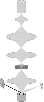
A schematic representation of a scanning electron microscope is depicted in Figure 1. Compared with other electron microscopes, SEM is a less frequently used imaging technique, particularly in liposome research. Nevertheless, several SEM micrographs showing cells with absorbed liposomes have been published, which are very useful in determining mechanisms of cell-liposome interactions (e.g. see Vinay et al 1996).
Complicated sample preparation is necessary for all EM techniques due to the fact that sample investigation may require staining, fixation, high vacuum and/or electrical conductiveness. Although staining procedures may vary, almost all EM techniques are based on embedding the vesicles in a thin film of an electron dense “glass”. When the films are examined by EM, the relatively electron-transparent vesicles will appear as bright areas against a dark background (hence the term negative stain).
Among the above-mentioned techniques NSEM and FFTEM are the most commonly employed techniques. NSEM is a useful method for clarifying questions related to the size distribution of liposomes. It has several advantages, as it is simple to use and necessitates only limited specialized equipment (that can be found easily at any EM laboratory). However, it requires laborious work in order
Electron Gun
Condenser Lenses
Scan Coils
Objective Lens
X-ray
Detector
Secondary
Specimen Stub Electron
Detector
Figure 1. Main components of a scanning electron microscope (SEM) (courtesy of Dr. M. R. Mozafari)
150 |
OZER |
to obtain quantitative data. NSEM was firstly described for visualising viruses, then a wide variety of microorganisms, cells, macromolecules and liposomes. In liposome technology, it provides quantitative data for MLV or ULV type liposomes, niosomes, sphingosomes and the others.
In negative stain methods, a drop of liposome sample at about 0.5–1 mg.ml−1 is dried on the microscopic grid coated with special support (carbon film) and stained with an electron dense solution, such as uranyl UO++2 or Tungsten Molybdate.
Two methods are commonly used in NSEM applications: a) Spray Method, and b) Drop Method. The drop method is the technique most commonly used with liposomes and is the easiest to perform. The spray method is not recomended due to the unreliability of the quality of the preparation. Additionally, the shear forces that the specimen must undergo during atomization may alter the size distribution of liposomes. Nevertheless, NSEM still grossly depends on the preparation of the grid, quality of the grid and hydrophilicity of the grid coat itself. Even when an optimal preparation is done, nobody clearly knows that if the vesicles were fractured or thin sectioned in their middle, or how the vesicles collapsed during drying in the negative stain method. In spite of these disadvantages, the methods are widely used and at the magnifications of up to 200,000 offer a resolution about 10–20 A°.
Introduction of cryoelectron microscopy to the science world provided direct observations of liposomes in their hydrated form. A thin film of the sample is vitrified in a few μm in liquid ethane, and the entire film is investigated in a special cryoholder in the microscope, in a similar way to optical microscopy.
In FFTEM methods, even smaller (compared with NSEM) amounts of sample, at higher concentrations, are quickly frozen and fractured. Platinum shadowing produces a replica which is investigated in the electron beam.
Freeze-fracture and freeze-etching technologies were developed gradually as the ultra-fast freezing technologies. Both sample preparation methods have artifacts; either by drying or by cooling, the system may go into gel or liquid-crystalline lamellar lyotropic phase.
Optical microscopy is the other technique employed for liposome technology. Bright-field and particularly phase-contrast microscopy are the most widely employed techniques. Its resolution is below 0.3 μm. It is a powerful technique for LUV, MLV and especially giant unilamellar vesicles if it is equipped with computer. The artifacts of this method are rather few. The sample thickness is important when getting an idea about the multilamellarity of the liposomes. Larger MLVs are very bright between crossed polarizer and analyzer; but below diameters of 1–2μm, the intensity of the circularly polarized light is too low to be observed birefringence.
Direct optical microscopy gives information about size, homogenity of the sample and lamellarity of MLVs. If there is any large liposome contamination with SUVs, optical microscopy is helpful for assessment. Furthermore, several mechanic characteristics of bilayers can be investigated by optical microscopy.
Resolution has been increased by the introduction of a group of microscopic techniques known as Scanning Probe Microscopy (SPM). Two of the most applied