

Nanomaterials and Nanosystems for Biomedical Applications - M. Reza Mozafari
.pdf130 |
PARADISSIS ET AL. |
A
GROWTH RATE (%)
150 |
|
|
|
|
|
|
|
100 |
|
|
|
|
|
MCF7 |
|
|
|
|
|
|
|
||
50 |
|
|
|
|
|
H460 |
|
0 |
|
|
|
|
|
|
|
0 |
16 |
32 |
48 |
64 |
80 |
96 |
112 |
-50 |
|
|
|
|
|
|
|
-100
-150
TIME (h)
B
GROWTH RATE (%)
150 |
|
|
|
|
|
|
|
100 |
|
|
|
|
|
MCF7 |
|
|
|
|
|
|
|
||
50 |
|
|
|
|
|
H4600 |
|
|
|
|
|
|
|
|
|
0 |
|
|
|
|
|
|
|
0 |
16 |
32 |
48 |
64 |
80 |
96 |
112 |
-50 |
|
|
|
|
|
|
|
-100
-150
TIME (h)
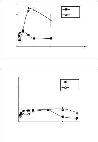
Figure 2. A: Effect of free sclareol on cell growth rate of MCF-7 (black diamonds) and H-460 (triangles) cell lines. Cells were incubated with 100μM of free sclareol. B: Effect of liposomal sclareol on cell growth rate of MCF-7 (black diamonds) and H-460 (triangles) cell lines. Cells were incubated with 100μM of liposomal sclareol
4.DISCUSSION
Extensive literature on the interactions of liposomes with cells has been accumulating over the past several years. However, due to the complex nature of liposomecell interactions, interpretation of experimental results in terms of liposome-cell interactions has proven to be difficult. None of the mechanisms such as endocytosis,
STUDIES OF FREE AND LIPOSOMAL SCLAREOL |
131 |
A
cells |
20 |
|
|
|
|
|
|
|
|
MCF7 |
|
total |
|
|
|
|
|
15 |
|
|
|
H460 |
|
|
|
|
|
||
6 |
|
|
|
|
|
in µg/10 |
10 |
|
|
|
|
Sclareol |
5 |
|
|
|
|
|
|
|
|
|
|
|
0 |
|
|
|
|
|
0 |
24 |
48 |
72 |
96 |
|
|
|
Tim e (h) |
|
|
B
cells |
20 |
|
|
|
|
|
|
|
|
MCF7 |
|
|
|
|
|
|
|
of total |
15 |
|
|
|
H460 |
|
|
|
|
|
|
6 |
10 |
|
|
|
|
in µg/10 |
|
|
|
|
|
5 |
|
|
|
|
|
Sclareol |
|
|
|
|
|
0 |
|
|
|
|
|
|
0 |
24 |
48 |
72 |
96 |
Tim e (h)
Figure 3. A: Uptake of free sclareol by MCF-7 (black cubes) and H-460 (triangles) cell lines. Cells were incubated with 100μM of free sclareol. B: Uptake of liposomal sclareol by MCF-7 (black cubes) and H-460 (triangles) cell lines. Cells were incubated with 100μM of liposomal sclareol
fusion or absorption of liposomes to cells, which are involved in liposome-cell interactions, are mutually exclusive (Allen et al. 1981).
Allen and co-workers (1981) have previously reported that liposome incorporated methotrexate, when tested in cell lines (EMT6 and S49), reduces and mediates the cytotoxicity of the free drug, via the uptake of free drug leaked from liposomes. In another study on the effect of liposomal daunorubicin against leukaemic cells, it has been reported that liposomal daunorubicin was devoid of acute effects such
132 |
PARADISSIS ET AL. |
as ROS production and ATP depletion that resulted in increased necrotic cell death (Liu et al. 2002). However, studies on the uptake of cytotoxic compounds by cells are of considerable importance.
Recently published results from our research group showed that sclareol might possess interesting pharmacological properties as it revealed significant cytostatic and cytotoxic effects against leukemic and solid tumor cell lines (Dimas et al. 2001, 1999; Hatziantoniou et al. 2006). It has been further demonstrated that sclareol induces cell cycle arrest at G0/1 phase of the cycle and kills cells via the mechanism of apoptosis (Dimas et al. 2001, 1999). When tested against colon cancer (HCT116) xenografts developed in NOD/SCID mice, sclareol also exhibited a significant tumor regression in its liposomal form, while the free compound was highly toxic for animals, leading them to death (Hatziantoniou et al. 2006). In continuation of our research on sclareol, this work was focused on determining the effect of free sclareol on cell growth rate of human breast (MCF-7) and lung cancer (H-460) cell lines as well as the role of liposomes to alter the pharmakokinetic parameters of sclareol due to its different rate of uptake by cells. The results showed that liposomal sclareol was less cytotoxic at the concentration of 100μM than that of free sclareol at the same final concentration. At that concentration, free sclareol reduced the growth rate of cells while its incorporation into liposomes largely delayed the appearance of cytotoxic effects for both cell lines These experiments revealed that the reduced appearance of cytotoxicity of the liposomal sclareol could be well correlated with a lower accumulation rate of sclareol into cells (Figure 3B).
5.CONCLUSION
The present study was focused on the uptake of a bioactive compound namely sclareol by MCF-7 and H-460 human cancer cell lines. According to the findings, it has been shown that the liposomal sclareol retains significant growth inhibiting activity and alters the pharmacokinetic parameters. These results should be taken into account in feature in vivo studies.
REFERENCES
Allen TM, McAllister L, Mausolf S, Gyorffy E. A study of the interactions of liposomes containing entrapped anti-cancer drugs with the EMT6, S49 and AE1 (transport-deficient) cell lines. Biochim. Biophys. Acta (1981) 643: 346–362.
Allen TM, Stuart DD. Liposomal pharmacokinetics. Classical, sterically-stabilized, cationic liposomes and immunoliposomes. In: Janoff AS, editor. Liposomes: Rational Design. New York: Marcel Dekker, Inc.; 1999. p. 63–87.
Bligh EG, Dyer WI. A rapid method of total lipid extraction and purification. Canad. J. Biochem. Physiol. (1959) 37: 911–917.
Books H, Lebleu B, Vives E. Tat peptide-mediated cellular delivery: Adv. Drug Deliv. Rev. (2005) 57: 559–577.
Celis J. Tissue culture and Associate Techniques. In: Cell Biology, A Laboratory Handbook. Academy Press, Inc. (1994) p10.
STUDIES OF FREE AND LIPOSOMAL SCLAREOL |
133 |
Demetzos C. A phytochemical study on Cistus incanus subsp. creticus (I). Isolation, structure elucidation and synthesis of a new flavonoid glycoside from Kalanchoe prolifera R. Hamel (II). Ph. D Thesis, Athens, Greece (1990).
Demetzos C, Stahl B, Anastasaki Th., Gazouli, Tzouvelekis L, Rallis M. Chemical analysis and antimicrobial activity of the resin ladano of its essential oil and of the isolated compounds. Planta Med. (1999) 65: 76–78.
Demetzos C, Dimas K. Labdane type diterpenes: Chemistry and Biological Activity. In: Studies in Natural Product Chemistry. Ed. Atta-ur-Rahman, Elsevier, Vol. 25, p. 235–292, (2001).
Dimas K, Demetzos C, Marsellos M, Sotiriadou R, Malamas M, Kokkinopoulos D. Cytotoxic activity of labdane type diterpenes against human leukemic cell lines in vitro. Planta Med. (1998) 64: 208–211.
Dimas K, Kokkinopoulos D, Demetzos C, Vaos B, Marselos M, Malamas M, Tzavaras T. The effect of sclareol on growth and cell cycle progression of human leukemic cell lines. Leuk Res. (1999) 23: 217–234.
Dimas K, Demetzos C, Vaos V, Ioannidis P, Trangas T. Labdane type diterpenes down-regulate the expression of c-Myc protein but not of Blc-2, in human leukemia T-cells undergoing apoptosis. Leukemia Research (2001) 25: 449–454.
Drummond DC, Meyer O, Hong K, Kirpotin DB, Papahadjopoulos D. Optimizing liposomes for delivery of chemotherapeutic agents to solid tumors. Pharmacol. Rev. (1999) 51: 691–743.
Gupta B, Levchenko TS, Torchilin VP. Intracellular delivery of large molecules and small particles by cell-penetrating proteins and peptides. Adv. Drug Deiv. Rev. (2005) 57: 637–651.
Hatziantoniou S, Demetzos C. Qualitative and quantitative one-step analysis of lipids and encapsulated bioactive molecules in liposome preparations by HPTLC/FID (IATROSCAN). J. Liposome Research (2006) 16 (4): 321–330.
Hatziantoniou S, Dimas K, Georgopoulos A, Sotiriadou N, Demetzos C. Cytotoxic and antitumor activity of liposome-incorporated sclareol against cancer cell lines and human colon cancer xenografts.
Pharmacological Research (2006) 53: 80–87.
Liu FT, Kelsey SM, Newland AC, Jia L. Liposomal encapsulation diminishes daunorubicin-induced generation of reactive oxygen species, depletion of ATP and necrotic cell death in human leukaemic cells. Br. J. Haematol. (2002) 117(2): 333–342.
Pakunlu R, Wang Y, Tsao W, Pozharow V, Cook T, Minko T. Enhancement of the efficacy of chemotherapy for lung cancer by simultaneous suppression of multifrug resistance and antiapoptotic cellular defense: novel multicomponent delivery system. Cancer Res. (2004) 64: 6214–6224.
Paradissis A, Hatziantoniou S, Georgopoulos A, Demetzos C. Lipid analysis of Greek broad bean oil: Preparation of liposomes and physicochemical characterization. Eur. J. Lipid Sci. Technol. (2005) 107: 799–804.
Pratt WB, Ruddon RW, Ensminger WD, Maybaum J. The Anticancer Drugs. Oxford University Press (1994).
Green SR, Moehle CM. Basic techniques for mammalian cell tissue culture. In: Curent Protocols in Cell Biology. Wiley J. and Sons, New York, Vol. 1 (1999).

CHAPTER 9
RELEASE ADVANTAGES OF A LIPOSOMAL DENDRIMER-DOXORUBICIN COMPLEX, OVER CONVENTIONAL LIPOSOMAL FORMULATION OF DOXORUBICIN
ARISTARCHOS PAPAGIANNAROS AND COSTAS DEMETZOS†
Department of Pharmaceutical Technology, School of Pharmacy, Panepistimiopolis, University of Athens, Zografou 15771, Athens, Greece
Abstract: Data on the release advantages of a liposomal formulation incorporating a doxorubicin– PAMAM G4 complex in comparison to a liposomal doxorubicin are presented. The liposomes incorporating either doxorubicin-PAMAM complex, or doxorubicin as free drug, were composed of Egg-phosphatidylcholine (EPC): Stearylamine (SA) at a 10:0.1 molar ratio and their size distribution and -potential were characterized. Liposomes incorporating the doxorubicin-PAMAM complex exhibited release properties which were advantageous compared to the conventional type of liposomal doxorubicin in terms of doxorubicin toxicity and its availability to the tumor site. This liposomal formulation may show improved therapeutic properties in vivo
Keywords: Liposome; dendrimer; PAMAM G4; doxorubicin; drug release
1.INTRODUCTION
Liposomes are non-toxic and biocompatible drug delivery systems that have been proven to be very useful in the fight against cancer. Liposomes can increase the therapeutic effectiveness of the encapsulated drugs and decrease their toxicity (Straubinger et al. 2004). One of the best-known liposomal drug delivery systems
This article is dedicated to the memory of Prof. Demetrios Papahadjopoulos (University of California at San Francisco, UCSF) who was my mentor on liposomal technology and a pioneer of nanotechnology.
†Corresponding author: C. Demetzos Department of Pharmaceutical Technology, School of Pharmacy, National and Kapodistrian Panepistimiopolis, University of Athens, Zografou 15571, Athens, Greece. Tel: +30210 7274596; Fax: +30210 7274027. E-mail: demetzos@pharm.uoa.gr
135
M.R. Mozafari (ed.), Nanomaterials and Nanosystems for Biomedical Applications, 135–144. © 2007 Springer.
136 |
PAPAGIANNAROS AND DEMETZOS |
is the liposomal doxorubicin. The high cardiotoxicity of free doxorubicin limits its clinical use, despite its high anticancer activity against a variety of tumours. Liposomal doxorubicin is active against many types of cancer and reduces the toxicity of doxorubicin and it is now in clinical use in USA and Europe (Gabizon 2002). Several clinical trials are currently in progress in order to evaluate the use of doxorubicin liposomes either alone or in combination with other anticancer drugs (Toma et al. 2002; Syrigos et al. 2002).
Despite several advantages, the therapeutic use of liposomes has limitations, which are related to the release of the encapsulated drug that can be only partially delayed by the modification of the membrane composition. Many attempts are made towards a more effective control of the release of the encapsulated drug, using polymers. One novel approach is the entrapment of liposomes in polymeric microspheres and the progressive release of the intact liposomes from the biodegradable matrix (Stenekes et al. 2002). Other approaches are based on the encapsulation of liposomes in microcapsules in order to modulate the release of the encapsulated drug (Dhoot and Wheatley 2003) or to produce liposome-like microspheres (Pan et al. 2004).
Dendrimers are highly branched macromolecules that, contrary to traditional “linear” polymers, possess fractal architecture, nanoscaled size and unique physicochemical properties. They are small in size, and exhibit a low polydispersity that can contribute to a reproducible pharmacokinetic behavior. However, the main characteristics of dendrimers are their multiple reactive groups, a well-defined structure, and their ability to encapsulate drugs in their void spaces (Cloninger 2002; Aulenta et al. 2003). An ideal dendrimer as drug delivery system must be non-toxic, non-immunogenic and biodegradable (Aulenta et al. 2003). The first dendrimer family which has been synthesized, characterized and commercialized is the Poly (amidoamine) (PAMAM) dendrimer. These dendrimers are considered safe regarding toxicity and are non-immunogenic and they have been used in the delivery of drugs, antisense nucleotides and gene therapy, both in vitro and in vivo (Eichman et al. 2001). Dendrimers and dendrons have already been proposed for drug complexation and transport; especially lipidic dendrons that can produce higher order lamellar structures called “dendrisomes” (Khuloud et al. 2003) or can aggregate to form nanosystems (Singh and Florence 2005).
In this paper a liposomal formulation composed of egg phosphatidylcholine and stearylamine (EPC:SA 10:0.1 molar ratio) and a doxorubicin-PAMAM complex attached to liposomes is compared to a conventional liposomal formulation with the same composition encapsulating doxorubicin by the pH gradient method (Papagiannaros et al. 2005; Papagiannaros et al. 2006). The main advantage of the liposomal formulation is the controlled and sustained release of the encapsulated drug; the release of which is controlled by the complexation in the dendrimer’s internal cavity. The liposomal membrane employed in the formulation is useful for the biocompatibility of the liposomal system and it offers advantages of the liposomal drug delivery. This liposomal system is compared to that of the conventional liposomes of the same lipid composition with respect to the % release of the
ADVANTAGES OF A LIPOSOMAL DENDRIMER-DOXORUBICIN COMPLEX |
137 |
encapsulated drug at 37°C, in 50 RPMI culture medium for 48 h period, in order to assess its possible advantages and evaluate its potential applications in cancer therapy.
2.MATERIALS AND METHODS
2.1.Materials
Egg Yolk Phosphatidylcholine (EPC) was purchased from Avanti Polar Lipids (AL, USA). Doxorubicin Hydrochloride was purchased from Pharmacia (NJ, USA). Ammonium sulphate, TES (N-tris (hydroxymethyl) methyl-2-aminoethanesulfonic acid), PAMAM, Poly (amidoamine) 4th generation, Tris (tris (hydroxymethyl) aminomethane), stearylamine (SA), Sephadex G75, chloroform, absolute ethanol and methanol were of spectroscopic grade and were purchased from Sigma (St. Louis, USA).
2.2.Conventional Liposome Preparation, Characterization and Doxorubicin Encapsulation
Liposomes composed of EPC:SA at 10:0.1 molar ratio, were prepared using the reverse phase evaporation method (Szoka et al. 1978) while their size and -potential measurements were performed at 25°C and at an angle of 90° in a photon correlation spectrometer (Zetasizer 3000, Malvern U.K.) and analysed by the CONTIN method (MALVERN software). The liposomes were prepared as follows: EPC, and SA were first dissolved in chloroform / methanol and then transferred into a 100 ml round bottom flask. Then a 150 mM ammonium sulphate (pH=5.3) was added to the flask. The mixture was subsequently sonicated for 15 min in a bath sonicator and the organic solvents were removed using a flash evaporator (Bucchi R-480) at 60°C. The liposomal suspension was finally allowed to anneal at 50°C for 1 hour.
Large Unilamellar Vesicles (LUVs) were prepared by sonicating the liposomal suspension in an ice bath, for two cycles of 5 min each (0.7 cycle and 100% amplitude) interrupted by a 5 min resting period, using a probe sonicator (UP 200S, dr. hielsher GmbH, Berlin, Germany). The 150 mM ammonium sulphate buffer (pH=5.3) of the liposomal suspension was exchanged with a 100 mM TES, 100 mM NaCl buffer (pH=7.5) using a Sephadex G75 column. Doxorubicin was subsequently encapsulated into the liposomes using the pH gradient method (Mayer and Bally 1986). Briefly, 854 μl or 0.015 mmole of doxorubicin was added and the preparation was incubated at 60°C for 30 min. Unentrapped doxorubicin was removed by passing the liposomal suspension through a Sephadex G75 column using 100 mM TES, 100 mM NaCl buffer (pH=7.5).
2.3.Determination of Lipids and Doxorubicin
EPC and SA were determined by high performance thin-layer chromatography coupled with a flame ionization detector (HPTLC-FID, Iatroscan MK-5, Iatron Lab.
138 |
PAPAGIANNAROS AND DEMETZOS |
Inc. Tokyo, Japan) (Goniotaki et al. 2004). Hydrogen flow rate was 160 ml/min, airflow rate 1900 ml/min, scan speed 30 s/scan. As stationary phase Chromarods – SII (Iatron Lab. Inc.) in set of 10 rods was used. Doxorubicin concentration of the liposomal samples was measured on a Perkin Elmer UV-vis spectrometer at=481 nm by adding absolute ethanol to the samples. Prior to determination, the samples were purified using column chromatography (Sephadex G75).
2.4.Release of Doxorubicin from Conventional liposomes in vitro
Equal volumes of liposomal suspension encapsulating doxorubicin in TES (100 mM) and NaCl (100 mM) buffer (pH: 7.5) and in RPMI 1640 culture medium, were mixed and the liposomes were incubated at 37°C. Aliquots of 300 μl were then withdrawn at various time intervals and passed through Sephadex G-75 column, in order to remove the released doxorubicin. Doxorubicin retained in the liposomes was measured by UV-vis spectrometry at =481 nm.
2.5.Incorporation of Doxorubicin in PAMAM Dendrimer and Assessment of Doxorubicin Release
An aqueous solution of doxorubicin (122 μl) was mixed with a PAMAM G4 solution (3:1 and 6:1 molar ratio of doxorubicin-PAMAM) in methanol (2 ml) and the solutions were stirred for 12 hours. The solutions were evaporated to dryness at 30°C in vacuum and the PAMAM dendrimer incorporating doxorubicin was extracted overnight using chloroform. Chloroform was evaporated to dryness, the dry residue was dissolved in TES (10 mM, pH: 7.5) and the absorbance of doxorubicin was measured at =481 nm using UV-vis spectrometry. In the later case acidification of the solution and buffering to pH=4.5 was performed before measuring the absorbance. The release of doxorubicin was studied in TES at 37°C using dialysis bags (molecular weight cut off 13,000).
2.6.Incorporation of Doxorubicin-PAMAM Complex in Liposomes
Liposomes were prepared by using the thin film hydration method (Gabizon 2002). The doxorubicin-PAMAM complex (3:1 molar ratio; 2.1 μmoles of doxorubicin) was attached to liposomes, composed of EPC:SA 10:0.1 (molar ratio). Briefly, the lipid film was prepared by dissolving EPC (73.6 μmole), SA (0.736 μmole) and doxorubicin-PAMAM complex (3:1 molar ratio; 2.1 μmoles of doxorubicin) in chloroform. The solvent was slowly evaporated in a flash evaporator to form a lipid film, which was dried under vacuum for at least 12 h. Multilamellar vesicles (MLVs) were prepared by hydrating the lipid film with TES buffer (10 mM, pH=7.5) and stirring for 1 h. Small unilamellar vesicles (SUVs) were prepared from the resultant liposomal suspension, which was subjected to sonication for two 5 min periods interrupted by a 5 min resting period, in an ice bath using a probe sonicator (amplitude 100, cycle 0,7 – UP 200S, dr. hielsher GmbH, Berlin, Germany). The resultant
ADVANTAGES OF A LIPOSOMAL DENDRIMER-DOXORUBICIN COMPLEX |
139 |
vesicles were allowed for 30 min to anneal any structural defects. Non-incorporated doxorubicin-PAMAM complex was removed by passing the liposomal suspensions through a Sephadex G75 column. The size and -potential of liposomes incorporating the doxorubicin-PAMAM complex (3:1molar ratio; 2.1 μmoles of doxorubicin) were measured using photon correlation spectroscopy (Malvern Zetasizer 3000HS). Doxorubicin concentration was measured on a Perkin Elmer UV-vis spectrometer at =481nm after the addition of absolute ethanol to the samples.
2.7.Release of Doxorubicin from the Liposomes Incorporating Doxorubicin-PAMAM Complex
The release of doxorubicin from the MLCRS incorporating the doxorubicinPAMAM complex (3:1 molar ratio; 2.1 μmoles of doxorubicin) was studied in 50% RPMI culture medium and in TES (100 mM), NaCl (100 mM) buffer (pH 7.5), at 37°C, by placing the liposomal formulations in dialysis bag (molecular weight cut off 13,000). The doxorubicin released at various times, up to 48 h was measured using UV-vis at =481 nm.
2.8.Statistical Analysis
Statistical analysis of the effect of liposome type on the size and -potential was performed using one-way ANOVA followed by a post hoc Tukey’s HSD test (SPSS for Windows release 11). All the results were from four (n=3) independent experiments.
3.RESULTS
3.1.Encapsulation, Physical Properties and Release of Doxorubicin from Conventional Liposomes
Doxorubicin was encapsulated in liposomes composed of EPC:SA (10:0.1 molar ratio) at a doxorubicin to lipid molar ratio of 0.77±0.01 (initial 0.1). The encapsulation efficiency of doxorubicin into liposomes was 99.1%±1.1. Size measurements for liposomes incorporating doxorubicin, indicated an average size of 91.2±0.74 nm and a -potential of –26±3.3 mV (Table 1).
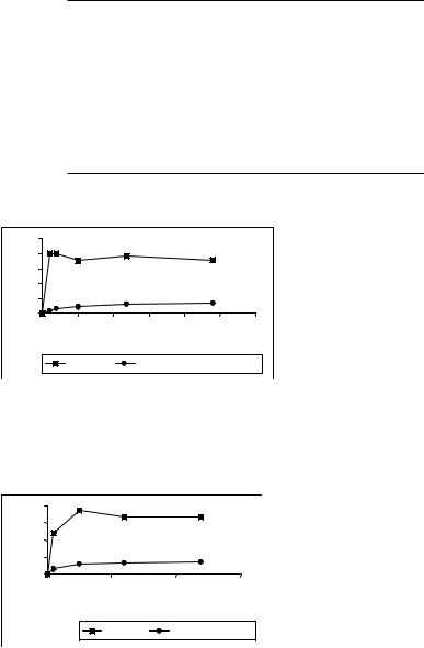
The release of doxorubicin from the conventional liposome EPC:SA 10:0.1 molar ratio in 50% RPMI cell culture medium at 37°C and in TES buffer after 24 hours is quite fast. The liposomes retained 24.5% of the drug in 50% RPMI cell culture medium and 35.5% in buffer at 37°C after 24 hours (Figures 1 and 2).
3.2.Incorporation and Release of Doxorubicin from the Doxorubicin-PAMAM Complex
The doxorubicin-PAMAM complex was formed using two different pH (i.e. 10 mM TES buffer at pH: 7.5 or 10 mM acetate buffer at pH: 4.5) and two different molar
140 |
PAPAGIANNAROS AND DEMETZOS |
Table 1. Physicochemical characteristics of EPC:SA (10:0.1 molar ratio) liposomes encapsulating doxorubicin and of liposomes (EPC:SA 10:0.1 molar ratio), incorporating doxorubicin-PAMAM complex (3:1 molar ratio)
Liposome formulation |
Size (nm) |
-potential (mV) |
|
|
|
Conventional liposomes: |
91.2±0.74 |
−26 0±3.3 |
EPC:SA 10:0.1 (molar ratio) |
|
|
encapsulating doxorubicin |
|
|
Liposomes incorporating |
116.3±7.8 |
−8 7±1.7 |
doxorubicin-PAMAM complex: |
|
|
EPC:SA 10:0.1 (molar ratio) |
|
|
encapsulating doxorubicin as doxorubicin-PAMAM complex (3:1 molar ratio)
release |
100 |
|
|
|
|
|
|
|
80 |
|
|
|
|
|
|
||
60 |
|
|
|
|
|
|
||
doxorubicin |
|
|
|
|
|
|
||
40 |
|
|
|
|
|
|
||
20 |
|
|
|
|
|
|
||
0 |
|
|
|
|
|
|
||
% |
|
|
|
|
|
|
||
0 |
10 |
20 |
30 |
40 |
50 |
60 |
||
|
||||||||
|
|
|
|
time |
|
|
|
|
|
|
liposome |
|
Liposome PAMAM/doxo complex |
||||
Figure 1. Doxorubicin release from liposomes incorporating doxorubicin-PAMAM complex (•) and from conventional liposomes ( ) both composed of EPC:SA 10:0.1 (molar ratio) in 50% RPMI 1640 culture medium at 37°C. Each point represents the mean of three independent experiments (SD never exceeded 5% of the mean value)
nrelease |
80 |
|
|
|
|
60 |
|
|
|
||
40 |
|
|
|
||
doxorubici |
|
|
|
||
20 |
|
|
|
||
0 |
|
|
|
||
% |
|
|
|
||
0 |
20 |
40 |
60 |
||
|
|||||
|
|
time |
|
|
|
|
|
liposomes |
liposome-PAMAM/doxo |
||
|
|
complex |
|
||
|
|
|
|
||
Figure 2. Doxorubicin release from liposomes incorporating doxorubicin-PAMAM complex (•) and from conventional liposomes ( ) both composed of EPC:SA 10:0.1 (molar ratio) in TES buffer at 37°C. Each point represents the mean of three independent experiments (SD never exceeded 5% of the mean value)