

Radiation Physics for Medical Physiscists - E.B. Podgorsak
.pdf8.10 Beta Plus Decay |
329 |
8.10 Beta Plus Decay
8.10.1 General Aspects of the Beta Plus (β+) Decay
The β+ decay is characterized by the production of positrons that appear in a spectral distribution with maximum positron energy specific to the particular β+ decay. As in the β− decay, the daughter recoil kinetic energy in β+ decay is essentially negligible. Radionuclides undergoing β+ decay are often called positron emitters and are used for functional imaging with a special imaging technique called positron emission tomography (PET).
•PET provides information on metabolic function of organs or tissues by detecting how cells process certain compounds such as, for example, glucose. Cancer cells metabolize glucose at a much higher rate than normal tissues. By detecting increased radio-labelled glucose metabolism with a high degree of sensitivity, PET identifies cancerous cells, even at an early stage when other imaging modalities may miss them.
•In a PET study one administers a positron-emitting radionuclide by injection or inhalation. The radionuclide circulates through the bloodstream to reach a particular organ. The positrons emitted by the radionuclide have a very short range in tissue and undergo annihilation with an available electron. This process generally results in emission of two gamma photons, each with energy of 0.511 MeV, moving away from the point of production in nearly opposite directions.
•The radionuclides used in PET studies are produced by bombardment of an appropriate stable radionuclide with protons from a cyclotron (see Sect. 8.4.9) thereby producing positron-emitting radionuclides that are subsequently attached to clinically useful biological markers. The most commonly used positron emitting radionuclides are: carbon-11, nitrogen13, oxygen-15, fluorine-18 and rubidium-82.
•Fluorine-18 radionuclide attached to the biological marker deoxyglucose forms the radiopharmaceutical fluorodeoxyglucose (FDG) that is the most commonly used tracer in studies involving glucose metabolism in cancer diagnosis.
8.10.2 Decay Energy in β+ Decay
The β+ decay can occur to a proton-rich unstable parent nucleus where the mass M (Z, A) of the parent nucleus exceeds the mass M (Z − 1, A) of the daughter nucleus by more than one positron mass me. The decay energy Qβ+ for the β+ decay process is given as
Qβ+ = {M (Z, A) − [M (Z − 1, A) + me]} c2 |
(8.172) |
in terms of nuclear masses M .
330 8 Radioactivity
Adding and subtracting Zmec2 to the right-hand side of (8.172) and neglecting the electron binding energies to the nucleus we obtain
Qβ+ = {M (Z, A) + Zme − [M (Z − 1, A) + me + Zme} c2 |
|
= {M(Z, A) − [M(Z − 1, A) + 2me]} c2 |
(8.173) |
where M(Z, A) and M(Z − 1, A) represent the atomic masses of the parent and daughter, respectively.
We note that the relationships between atomic and nuclear masses of parent and daughter, ignoring the binding energies of orbital electrons, are
M(Z, A) = M (Z, A) + Zme |
(8.174) |
and |
|
M(Z − 1, A) = M (Z − 1, A) + (Z − 1)me . |
(8.175) |
For the β+ decay to occur the atomic mass of the parent M(Z, A) must exceed the atomic mass of the daughter M(Z − 1, A) by more than two electron rest masses, or in rest energies
M(Z, A)c2 > M(Z − 1, A)c2 + 2mec2 , |
(8.176) |
where mec2 is the electron rest energy of 0.5110 MeV.
8.10.3 Beta Plus (β+) Decay of Nitrogen-13 into Carbon-13
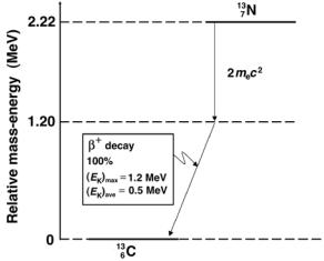
An example for a simple β+ decay is the decay of nitrogen-13 into carbon13 with a half-life of 10 minutes. Nitrogen-13 is a proton-rich radionuclide produced in a cyclotron. The decay scheme is shown in Fig. 8.23 and the basic equation for the decay is as follows:
713N → 613C + e+ + νe + Qβ+ (1.2 MeV) . |
(8.177) |
The decay energy Qβ+ for the β+ decay of nitrogen-13 into carbon-13 is calculated as follows, with the atomic masses for the two nuclides listed in Table 8.5:
Qβ+ = M(137N) − [M(136C) + 2me] c2
=(13.005739u − 13.003355u)c2 − 2mec2
=0.002383u × 931.5 MeV/u = 2.220 MeV − 1.022 MeV
= 1.2 MeV. |
(8.178) |
The energy di erence between the ground state of nitrogen-13 and carbon-13 is 2.22 MeV; however, only 2.22 MeV − 2mec2 = 1.2 MeV is available for the maximum energy of the positron.
Ammonia is the substance that can be labeled with the nitrogen-13 radionuclide for use in functional imaging with positron emission tomography (PET) scanning. The nitrogen-13 labeled ammonia is injected intravenously and is mainly used for cardiac imaging for diagnosis of coronary artery disease and myocardial infarction. It is also occasionally used for liver and brain imaging.
8.10 Beta Plus Decay |
331 |
Fig. 8.23. The decay scheme for β+ decay of nitrogen-13 into carbon-13. The relative mass-energy levels of the ground states of the two nuclides are calculated from atomic masses listed in Table 8.6 on page 353
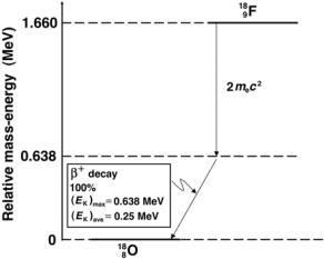
8.10.4 Beta Plus (β+) Decay of Fluorine-18 into Oxygen-18
The β+ decay of fluorine-18 into oxygen-18 with a half-life of 110 min is an important practical example of the β+ decay. Fluoro-deoxy-glucose (FDG) labeled with radionuclide fluorine-18 is a sugar compound that can be injected intravenously into a patient for use in positron emission tomography (PET) functional imaging. Based on demonstrated areas of increased glucose metabolism the FDG PET scan:
1.Can detect malignant disease.
2.Can distinguish benign from malignant disease.
3.Can be used for staging of malignant disease.
4.Can be used for monitoring response to therapy of malignant disease.
The decay energy Qβ+ for the β+ decay of fluorine-18 into oxygen-13 is calculated as follows:
Qβ+ = M(189F) − [M(188O) + 2me] c2
=(18.000937u − 17.999160u)c2 − 2mec2
=0.001777u × 931.5 MeV/u
=1.660 MeV − 1.022 MeV
= 0.638 MeV. |
(8.179) |
The energy di erence between the ground states of fluorine-18 and oxygen-18 is 1.66 MeV; however, only 1.66 MeV − 2mec2 = 0.638 MeV is available for
332 8 Radioactivity
Fig. 8.24. The decay scheme for β+ decay of fluorine-18 into oxygen-18. The relative mass-energy levels for the ground states of the two nuclides are calculated from atomic masses listed in Table 8.6 on page 353
the maximum energy of the positron, as shown schematically in Fig. 8.24 and in (8.180) below
918F → 818O + e+ + νe + Qβ+ (0.638 MeV) . |
(8.180) |
8.11 Electron Capture (EC)
8.11.1 Decay Energy in Electron Capture
Electron capture (EC) radioactive decay may occur when an atomic electron ventures inside the nuclear volume, is captured by a proton, and triggers a proton to neutron transformation. Of all atomic electrons, the K-shell electrons have the largest probability for venturing into the nuclear volume and thus contribute most often to the EC decay process. Typical ratios EC(K shell)/EC(L shell) are of the order of 10:1.
Electron capture can occur in proton-rich, unstable parent nuclei, when the mass M (Z, A) of the parent nucleus combined with the mass of one electron me exceeds the mass of the daughter nucleus M (Z − 1, A). The decay energy QEC for electron capture is given as
QEC = {[M (Z, A) + me] − M (Z − 1, A)} |
|
= {M (Z, A) − [M (Z − 1, A) − me} c2 |
(8.181) |
in terms of nuclear masses M . Adding and subtracting Zme to the right-hand side of (8.181) and neglecting the electron binding energies to the nucleus we
8.11 Electron Capture (EC) |
333 |
obtain the decay energy QEC in terms of atomic masses M |
|
QEC = {M(Z, A) − M(Z − 1, A)} c2 . |
(8.182) |
For electron capture to occur, the atomic mass of the parent M(Z, A) must exceed the atomic mass of the daughter M(Z − 1, A); i.e., M(Z, A) > M(Z − 1, A). The atomic rest energy di erence between the parent and the daughter gives the energy released to the neutrino and the daughter atom in an EC radioactive decay event.
Electron capture is a competing process to β+ decay; however, the conditions on electron capture as far as relative atomic masses of parent and daughter are concerned are less restrictive than those imposed on β+ decay that results in positron emission and subsequent positron annihilation with emission of annihilation quanta. The condition on EC decay is that the parent atomic mass M(P) simply exceeds the daughter atomic mass M(D), while the condition on β+ decay is that the parent atomic mass exceeds that of the daughter by a minimum of two electron masses.
•When the condition QEC > 0 is satisfied but Qβ+ of (8.172) is negative, the β+ decay will not happen because it is energetically forbidden and EC decay will happen alone.
•When Qβ+ > 0 then QEC is always positive and both decays (β+ and EC) can happen. The branching ratios λEC/λβ+ vary considerably from one nuclide to another; for example, from a low of 0.03 for fluorine-18 to several hundred for other proton-rich radionuclides.
•In contrast to the β− and β+ decay processes in which three decay products share the decay energy and produce a continuous spectral distribution, in the EC decay the two decay products do not have a continuous spectral distribution; rather they are given discrete (monoenergetic) energies. The monoenergetic neutrinos produce a line spectrum with energy Eν , while the daughter has the recoil kinetic energy (EK)D discussed below.
8.11.2 Recoil Kinetic Energy of the Daughter Nucleus in Electron Capture Decay
The recoil kinetic energy (EK)D of the daughter nucleus in electron capture decay is determined in two steps:
1.First, we determine the momenta of the daughter pD = M (D)υD and the neutrino pν = Eν /c. The two momenta are identical in magnitude but opposite in direction, so we can write
pD = M (D)υD = pν = |
Eν |
, |
(8.183) |
|
c |
||||
|
|
|
where

334 |
8 Radioactivity |
|
|
Eν |
is the neutrino energy |
|
M (D) |
is the mass of the daughter nucleus |
|
υD |
is the velocity of the daughter nucleus |
2.The recoil kinetic energy of the daughter (classically) is given as follows after inserting υD from (8.183):
(EK)D = |
M (D)υD |
= |
Eν2 |
. |
(8.184) |
|
2 |
2M (D)c2 |
|||||
|
|
|
|
The energy available for sharing between the daughter nucleus and neutrino is equal to the electron capture decay energy QEC decreased by the binding energy EB of the captured electron, i.e.,
E2
QEC − EB = Eν + (EK)D = Eν + ν 2 (8.185) 2M (D)c
or
E2
ν 2 + Eν − (QEC − EB) = 0 . (8.186) 2M (D)c
Equation (8.186) results in the following expression for the energy of the monoenergetic neutrino emitted in electron capture:
|
ν |
− |
|
|
M (D)c2 |
|
≈ |
|
EC − |
|
B |
|
|
E |
|
= |
1 + |
1 + |
2(QEC − EB) |
|
M (D)c2 |
|
Q |
|
E |
|
. (8.187) |
|
|
|
|
|
|||||||||
In the first approximation the recoil kinetic energy (EK)D of the daughter is neglected and so is the binding energy EB of the captured electron. The energy of the monoenergetic neutrino in electron capture is then approximated by the electron capture decay energy, i.e., Eν ≈ QEC.
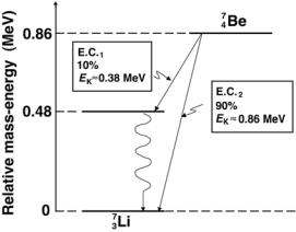
8.11.3 Electron Capture Decay of Beryllium-7 into Lithium-7
An example for EC decay is given in Fig. 8.25 that shows a decay scheme for beryllium-7 decaying through EC into lithium-7. Beryllium-7 has too many protons for nuclear stability, so it achieves better stability by transforming a proton into a neutron. However, it can do so only through EC and not through β+ decay, because the atomic rest energy of beryllium-7 exceeds that of lithium-7 by only 0.86 MeV and not by a minimum of 1.02 MeV required for β+ decay to be energetically feasible.
The decay energy for the electron capture decay of berillium-7 into
lithium-7 is calculated as follows: |
|
|
||
QEC = M(47Be) − M(37Li) |
c2 = (7.0169292u − 7.016004u)c2 |
|
||
0.0009252u |
× |
931.5 MeV/u = 0.862 MeV. |
(8.188) |
|
= |
|
|
||
8.11 Electron Capture (EC) |
335 |
Fig. 8.25. The decay scheme for electron capture decay of berillium-7 into lithium- 7. The relative mass-energy levels for the ground states of the two nuclides are calculated from the respective atomic masses listed in Table 8.6 on page 353
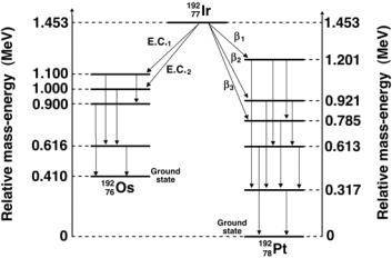
8.11.4 Decay of Iridium-192
Iridium-192 serves as an important radioactive source for use in brachytherapy with remote afterloading techniques. It decays with a half-life of 74 days into stable platinum-192 by β− decay and into stable osmium-192 by electron capture decay. The source is produced through a neutron activation process on iridium-191 in a nuclear reactor (see Sect. 8.4.8). The natural abundance of stable iridium-191 is 37.3% in a mixture with 62.7% of stable iridium-193. The cross section σ for thermal neutron capture is 954 b.
As shown in Fig. 8.26, iridium-192 has a very complicated γ ray spectrum with 14 γ energies ranging from 0.2 MeV to 0.9 MeV, providing e ective photon energy of 0.38 MeV. Because of the relatively short half-life, the iridium-192 source requires a source change in remote afterloading machines every 3 to 4 months.
With M representing atomic masses, the β− decay energy Qβ− for iridium192 decaying into platinum-192 is given as follows:
Qβ− = |
M(19277Ir) − M(19278Pt) c2 |
|
||
= |
|
− |
191.96104u)c2 |
|
|
191.96260u |
|
|
|
= |
0.00156u × 931.5 MeV/u = 1.453 MeV. |
(8.189) |
||
The electron capture decay energy QEC for iridium-192 decaying into osmium192 is
QEC = |
M(19277Ir) − M(19276Os) c2 |
|
||
= |
|
− |
191.96148u)c2 |
|
|
(191.96260u |
|
|
|
= |
0.00112u × 931.5 MeV/u = 1.043 MeV |
(8.190) |
||
336 8 Radioactivity
Fig. 8.26. The decay scheme for decay of iridium-192 into platinum-192 through β− decay and into osminum-192 through electron capture decay. The relative massenergy levels for the ground states of the three nuclides are calculated from the respective atomic masses given in Table 8.6 on page 353
8.12 Gamma Decay
8.12.1 General Aspects of Gamma (γ) Decay
The α decay as well as the three beta decay modes may produce a daughter nucleus in an excited state without expending the full amount of the decay energy available.
The daughter nucleus will reach its ground state (i.e., it will de-excite) through one of the following two processes:
1.Emit the excitation energy in the form of a γ photon (pure γ decay).
2.Transfer the excitation energy to one of its orbital electrons in a process called internal conversion.
In most radioactive α or β decays the daughter nucleus de-excitation occurs instantaneously (i.e., within 10−12 s), so that we refer to the emitted γ rays as if they were produced by the parent nucleus. For example, for the cobalt-60 β− decay into nickel-60, the γ rays following the β− decay actually originate from nuclear de-excitations of nickel-60, yet for convenience, we refer to these γ rays as the cobalt-60 γ rays. Similarly, we refer to γ photons following the β− decay of cesium-137 into barium-137m as cesium-137 γ rays even though the γ photons actually originate from a transition in the barium-137 nucleus.
In certain α or β decays, the excited daughter nucleus does not immediately decay to its ground state; rather, it de-excites with a time delay:
•The excited state of the daughter is then referred to as a metastable state and the process of de-excitation is called an isomeric transition.
8.12 Gamma Decay |
337 |
The metastable states are characterized by their own half-lives t1/2 and mean (average) lives τ .
•The nucleus in a metastable state is identified with a letter m next to the
atomic mass number designation (e.g, barium-137m or 13756 mBa with a halflife of 2.552 min; technetium-99m or 9943mTc with a half-life of 6.01 hours).
•The term isomer is used for designation of nuclei that have the same atomic number Z and same atomic mass number A but di er in energy states.
In addition to α and β decay there are many other modes for producing nuclei in excited states that subsequently undergo γ decay. For example, excited states with energies up to 8 MeV may be produced with neutron capture (n, γ) reactions as well as with other nuclear reactions, such as (p, γ) and (α, γ), etc. Examples of γ rays following α and β decays are given in Fig. 8.19 for α decay and Figs. 8.21 and 8.22 for β− decay.
8.12.2 Emission of Gamma Rays in Gamma Decay
In a general sense, γ decay stands for nuclear de-excitation either by emission of a γ ray photon or by internal conversion. In a more narrow sense, γ decay only implies emission of γ photons. The energy of γ rays emitted by a particular radionuclide is determined by the energy level structure of the radinuclides and can range from a relatively low value of 100 keV up to about 3 MeV.
The γ decay process may be represented as follows:
ZAX → ZAX + γ + Qγ , |
(8.191) |
where AZ X strands for an excited state of the nucleus AZ X and Qγ is the gamma decay energy.
8.12.3 Gamma Decay Energy
The decay energy Qγ in γ emission is the sum of the γ photon energy Eγ and the recoil kinetic energy of the daughter (EK)D, i.e.,
Qγ = Eγ + (EK)D . |
(8.192) |
Since the magnitudes of the momenta of the daughter recoil nucleus pD = M (D)υD and the γ photon pγ = Eγ /c are equal, i.e., pD = pγ , we can determine the partition of energy between Eγ = pγ c = M (D)υDc and (EK)D = M (D)υD2 /2 as
|
M (D)υ2 |
Eγ2 |
|
|
||
(EK)D = |
D |
= |
|
, |
(8.193) |
|
2 |
2M (D)c2 |
|||||
|
|
|
|
|||
where M (D) and υD are the rest mass and recoil velocity, respectively, of the daughter nucleus.
338 8 Radioactivity
The gamma decay energy Qγ may now be written as
|
Eγ |
|
|
Qγ = Eγ + (EK)D = Eγ 1 + |
2M (D)c2 |
. |
(8.194) |
Equation (8.194) shows that the recoil kinetic energy of the daughter (EK)D represents less than 0.1% of the gamma photon energy Eγ . The recoil energy of the daughter nucleus is thus negligible for most practical purposes. The label for daughter in gamma decay is used in parallel with the same label used in other nuclear decays that are clearly defined with a parent decaying into a daughter. In gamma decay the parent and daughter represent the same nucleus, except that the parent nucleus is in an excited state and the daughter nucleus is in a lower excited state or the ground state.
8.12.4 Resonance Absorption and the M¨ossbauer E ect
The question of resonance absorption is of importance and deserves a brief discussion. The resonance absorption is a phenomenon in which a photon produced by a nuclear or atomic transition is re-absorbed by the same type of nucleus or atom, respectively. Since the photon shares the de-excitation energy with the atom or nucleus (recoil energy), it is quite possible that its energy will not su ce to allow triggering the reverse interaction and undergoing resonance absorption. However, if the recoil energy of the daughter atom or nucleus is not excessive, the resonance absorption is possible because of the natural width of the photon energy distribution and the finite lifetime of atomic and nuclear states, where the width and lifetime are governed by the uncertainty principle (see Sect. 1.23).
The photons’ emission and absorption spectra di er because of the atomic or nuclear recoil energy that makes the emission energy slightly smaller than ∆E, the energy di erence between the two states. However, if there is a region of overlap between the emission and absorption spectrum, resonance absorption is possible.
For atomic transitions that are of the order of eV to keV the resonance absorption is not hindered. On the other hand, for nuclear transitions that are of the order of 10 MeV, there is no overlap between the emission and the absorption photon spectrum and resonance absorption is not possible. However, there is a way around this problem. In 1957 Rudolph M¨ossbauer discovered that nuclear transitions occur with negligible nuclear recoil, if the decaying nucleus is embedded into a crystalline lattice. Here, the crystal as a whole rather than only the daughter nucleus absorbs the recoil momentum. This e ect, called M¨ossbauer e ect, minimizes the recoil energy and makes nuclear resonance absorption possible.