

Metabolic Engineering - T. Scheper and Jens Nielsen
.pdf110 |
M. McIntyre et al. |
Fig. 3. Macroscopic and microscopic morphology of filamentous fungi. Macroscopic morphology describes the gross morphology, while microscopic morphology describes the properties (dimensions and compartmentalization) of the gross morphological forms
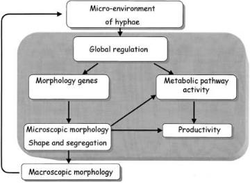
is the availability of nutrients and oxygen that determines the global regulation of genes. This in turn has influence on the genes directly controlling morphology or productivity. The resulting micromorphology can have a direct effect on metabolic pathway activity through the co-regulation of genes and can influence productivity due to the segregation of hyphae.Not all hyphal compartments are likely to have the same level of activity [7, 54]. Microscopic morphology also has other, indirect effects on productivity, with differentiation and hyphal dimensions influencing the secretion pathway. The processes of clumping and pelleting, and thus macromorphology, have significant influence on the measured mean activities or specific productivities of the cultures investigated [55]. Macroscopic morphology also determines the micro-environment of hyphae through effects on mixing, mass transfer, and culture rheology. Pellets may have dense and inactive cores due to poor diffusion of nutrients [51, 56], which may lead to cell lysis and thereby loss of the interior pellet structure [51]. Furthermore, the products of autolysis, which may be growth inhibitors, could diffuse through the pellets into the medium and inhibit the growth of the culture. Thus, development of macromorphologies indirectly affects the productivity of a culture.
If we are to consider metabolic engineering of the morphology of Aspergilli, or indeed filamentous fungi in general, efforts should be concentrated on understanding the processes that are represented within the shaded area on Fig. 4.
Metabolic Engineering of the Morphology of Aspergillus |
111 |
Fig. 4. Schematic representation of the interactions between process conditions, morphology, and productivity
It is only once the regulation and control of morphology is better understood that we can begin to engineer strains with better performance in submerged cultivation, with regard to productivity and physical properties of the culture.
Previous research has focused on the influence of morphology on either enzyme or secondary metabolite production, i.e., the major products from industrial bioprocesses utilizing filamentous organisms. With many advanced analysis tools in place (discussed in Sect. 2), detailed information on hyphal growth and kinetics can be obtained in a rapid and reproducible manner. Thus, effects of environmental changes or mutations on morphology can be quantified, allowing the relevance of these changes for process optimization to be assessed.
3.2.1
Penicillin Production
The effect of agitation on morphology and penicillin production by Penicillium chrysogenum has been the subject of a number of studies [55, 57–59]. Lower penicillin production was observed when agitation rates were high, a phenomenon which was attributed to the fact that mycelia were shorter and less branched. High agitation has been shown to promote rapid mycelial fragmentation [58, 59] and a higher branching frequency [58] for freely dispersed hyphal elements. Fragmentation of hyphal elements occurs when the local shearing forces become larger than the tensile strength of the cell wall [50]. The influence of the clumping of hyphae on rheology, and subsequently on penicillin production, has been widely discussed in the literature [55, 57–59]. The aim of many of the studies has been to optimize penicillin production by improved mixing, the resultant morphologies being quantified in an attempt to explain the results.
112 |
M. McIntyre et al. |
While these studies provide valuable information on the influence of environmental factors on the penicillin production process, they are of limited value from the viewpoint of metabolic engineering of morphology. This is because they aimed at describing the complex interactions between mechanical forces, growth,and rheology,rather than the influence of micromorphology on the production process.
In P. chrysogenum, the process of hyphal differentiation complicates studies correlating morphology and productivity. Understanding of this process is essential if we are to consider optimization of secondary metabolite production via morphological engineering. Great progress in this area has been made possible by development of automated image analysis routines written specifically for the purpose of quantifying differentiation [11, 60]. Application of these routines has shown that penicillin production is correlated with the fraction of subapical cells in the mycelia [50,61],and an increase in the relative area of these regions (rather than an increase in tips) is likely to result in elevated productivity.
3.2.2
Enzyme Production
Protein secretion has been shown to occur at or very close to the tips of fungal hyphae [52, 62–64]. There have been a number of studies, therefore, attempting to correlate tip number with enzyme production [65, 66] and to investigate protein secretion by morphological mutants [52, 67, 68]. On investigating heterologous enzyme secretion by Aspergillus niger during continuous cultivations, Wongwicharn et al. [65] found that production was correlated with tip number as the concentration of oxygen was increased in the cultures. However, as metabolism, physiology (and thus protein secretion), and morphology are likely to be affected by the change in O2 levels, such correlations should be treated with caution. The resultant changes in production may not be due to the changes in morphology alone; both physiology and morphology have been affected by the same external influence. Of more interest is the fact that these workers showed a further correlation between the active area (determined by biological staining) of hyphae and protein secretion, which is a more meaningful indication of the effect of increased oxygen in the influent gas. Fungal hyphae are not uniform, with respect to physiology, over their length [69]. Therefore, it is the observed changes in the active length, rather than overall length, of the hyphae that are likely to be responsible for alterations to growth and enzyme production [7].
Similarly, agitation rates [57, 58, 70] and biomass concentrations [55] are known to influence the physiological properties of a culture in addition to resulting in altered morphologies. Controlling such variables has been the strategy employed to alter morphology and investigate the subsequent effect on heterologous protein production in cultures of Aspergillus awamori [66]. Mean total hyphal length was found to decrease concomitant with increases in stirrer speed or increases in inoculum spore concentration. However, a reduced inoculum resulted in a more branched mycelium and an optimum stirrer speed was observed to result in a higher number of tips. In terms of productivity, the morphological differences had only a limited effect on product formation.
Metabolic Engineering of the Morphology of Aspergillus |
113 |
A clear picture of the effect of tip number on protein secretion is not apparent from the studies described above. Perhaps more insightful are the studies which have been carried out with morphological mutants, where comparisons of the effects of different morphologies may be more valid, being made without the influence of changes in environmental conditions. Spohr et al. [68] compared the a-amylase production in three strains of A. oryzae – a wild type, a transformed strain with an increased copy number of the a-amylase gene, and a morphological mutant of the transformed strain (which had a dense mycelium with more tips, relative to the other strains). The morphological mutant was found to be more efficient in producing a-amylase.
In a similar study [67], highly branched mutants of two strains of A. oryzae were investigated in submerged cultivation and morphology and protein secretion monitored. However, specific enzyme production was only improved in few of the highly branched strains, and the effect was dependent on the mode of cultivation. The authors concluded there was no clear correlation between branch frequency and the ability to secrete protein. The somewhat conflicting evidence presented above, concerning enzyme production in different morphological mutants,may be a result of the different types of morphological analysis applied in each of the studies. In general, only the freely dispersed (micromorphologies) were analyzed, and while these may be the predominant morphological form, they may not represent the total biomass. The relative amounts of the morphological forms are likely to be dependent on strain and cultivation conditions.
The observations from the studies above are further complicated by the fact that morphology also has an influence on broth rheology [71, 72] and thus can additionally affect production due to altered mixing and mass transfer in the culture fluid [55, 56, 73]. A linear relationship has been shown to exist between the degree of branching and the culture viscosity, with cultures of highly branched mutants being less viscous than wild type strains [67]. Mycelial morphology may not have a direct effect on protein secretion [70]; however, the relationship between agitation, morphology, and productivity must be considered when metabolic engineering of morphology is to be carried out. Changes in environmental conditions or mutant strains may appear to result in desirable morphological characteristics for improved productivity (e.g., increased number of tips). However, the performance of these strains in bioreactors remains to be the critical measure of their worth in process optimization.
From the evidence gathered, it is apparent that morphology has a significant role to play, influencing protein secretion either directly (tip number) or indirectly (by affecting mixing and mass transfer). Despite the conflicting results from submerged cultivations, direct evidence exists for protein secretion at the tips of fungal hyphae. Using immunogold labeling, Wösten et al. [64] localized secretion of glucoamylase in A. niger to the tips of actively growing hyphae. Further, staining with FITC conjugated antibody against a-amylase resulted in intense fluorescence of new tips and extending branches of A. oryzae [68]. Recently,visualization of proteins using GFP-fusions has allowed products of interest to be localized within hyphae [74, 75], providing additional evidence that protein secretion is an apical phenomenon. The importance of physiological information in addition to morphological data cannot be overstated when corre-
114 |
M. McIntyre et al. |
lations between morphology and productivity are being formulated. In particular, fluorescence microscopy with biologically active stains has added greatly to our knowledge regarding the role of morphology in protein and secondary metabolite production. This is clearly an interesting route to pursue in morphological engineering.
4
Molecular Aspects of Morphological Control
4.1
Filamentous Fungi
Many genes have been identified in filamentous fungi where deletion or disruption results in morphological aberration.In many cases the gene product has not been identified, and in other cases has been shown to be a protein with a regulatory function. In fewer cases,the gene has been cloned,the function of the protein identified, and the morphological phenotype after disruption/deletion of the gene has been fully characterized. In the fewest of cases the organism has been studied in submerged cultivation and perhaps the effect of the morphological defect on productivity has been examined.
In the following section we have attempted to give an overview of the genes involved in controlling morphology in Aspergilli, illustrating with examples from the fewest studies where a clearer picture of the role genes in morphological development is available. It is using these examples that allows discussion of metabolic engineering of morphology and where we can begin to relate genetic manipulation of morphology genes to bioreactor performance and productivity.
4.1.1
Genes Involved in Morphology
Table 1 lists the genes of A. nidulans that have been identified as having a role in morphology, where the role of the protein encoded is known. The functions of the proteins listed are mainly related to the establishment and maintenance of hyphal polarity with inactivation of the corresponding genes resulting in swollen hyphae or aberrant branching patterns. Clearly, the interest is in exploiting this information for the improvement of submerged bioprocesses. It may be desirable to obtain a homogenous culture of filamentous fungal cells where polarity has been lost, thus leading to a culture giving rise to a lower medium viscosity and thereby an improved mixing of the culture, compared to a truly filamentous culture (see also Fig. 4). On the other hand, a culture which is hyperbranched may be desirable for the production of heterologous proteins where increased tip number may result in increased secretion and improved yields. Certainly, what still remains to be determined is the performance of many such morphological mutants in submerged culture with respect to growth and production characteristics
Protein kinases have proven to belong to ever-expanding gene/protein families and some of these have been shown to be very important in directing the tip

Metabolic Engineering of the Morphology of Aspergillus |
115 |
Table 1. Genes involved in morphological development of Aspergilli and subsequent effect on morphology following gene disruption
Gene |
Protein function |
Morphology obtained on |
Reference |
|
|
gene inactivation |
|
hypA/podA |
Establishment and mainten- |
|
ance of hyphal polarity. |
|
Activation of growth arrest |
|
in subapical cells |
hypC |
Cell size control and control |
|
of spacing of septa. |
podB |
Establishment and mainten- |
|
ance of hyphal polarity. |
|
Required for cytoskeletal |
|
organization in tip cells |
Wide hyphae with thick |
37, 39 |
lateral cell walls. High |
|
frequency of dichotomous |
|
apical branching |
|
Short subapical cells. |
37 |
High branching frequency |
|
Swollen hyphae |
39 |
sepA |
Formin. Control and organ- |
|
ization of actin filaments at |
|
sites of localized cell wall |
|
deposition |
Aseptated, wide hyphae. |
76 |
High frequency of dichotomous apical branching.
swoA |
Maintenance of hyphal |
Swollen hyphae |
77 |
|
polarity |
|
|
swoF |
Establishment and main- |
Swollen hyphae |
77 |
|
tenance of hyphal polarity |
|
|
|
|
|
|
extension of hyphal cells. Protein kinases mediate the phosphorylation that regulates protein function directly, or via signal transduction, in many areas of the cell metabolism. The analysis of protein kinases in filamentous fungi is still in its early stages; however, it has already become clear that protein kinases are essential in linking signal transduction cascades, protein modification, and fungal morphogenesis. In Saccharomyces cerevisiae computer-based sequence analysis of the genome has revealed 113 genes which can be identified as protein kinases [78]. In filamentous fungi, and in eucaryotes in general, the protein kinases that phosphorylate either serine or threonine (Ser/Thr kinases) represent virtually all of the kinases described and this group includes cAMP-dependent kinases (PKA), protein kinase type C (PKC), mitogen-activated kinases (MAP), and p21activated kinases (PAK) [79].
Of these, PKAs seem to have an important role in fungal development. However, caution should be exercised about specific function since no direct substrates for PKA have been identified yet [79]. In the plant smut fungus Ustilago maydis, cAMP signaling controls the dimorphic switch between the budding yeast form and (virulent) filamentous growth and it is also known to be involved in virulence of the rice blast fungus Magnaporthe grisea [80].
From an industrial perspective, an interesting study was carried out in N. crassa with the temperature-sensitive mcb mutant, which has a mutation in a regulatory subunit of the cAMP dependent protein kinase A. The strain displayed a complete loss in growth polarity at the restrictive temperature [81] and also in minimal medium supplemented with carboxymethyl cellulose (CMC)
116 |
M. McIntyre et al. |
and sucrose [82]. This resulted in a considerable increase in the growing surface area of the fungus. It was hypothesized that protein secretion was limited by the amount of growing surface area; the protein secretion of the mcb mutant in liquid medium had a threefold higher yield of extracellular protein on biomass than the wild-type (50 mg/l to 15 mg/l). In addition, in the supplemented medium the yield of units CMCase on biomass was 20-fold higher. CMCase is mainly produced late in the cultivation and, therefore, it was stated that the level of protein production was not likely to be linked with the hyphal growth rate. However, hyphal growth rate was not measured, and it is likely that the CMCase production might be induced only when sucrose is depleted and as a result of the growth kinetics determined by the medium. (The wild-type grows fast to a high biomass concentration and experiences sucrose depletion more suddenly than the mcb mutant.) Therefore,it might have stopped its growth before it could produce the necessary proteins for CMCase production. CMCase production is complex to examine and, in addition, the protein secretion capacity of N. crassa is very low compared to the levels of Trichoderma or Aspergilli (g/l). As such, it may be very interesting to examine the effect of an mcb mutation on industriallevel protein producing strains of these species.
4.1.2
Engineering Hyphal Architecture
As discussed in Sect. 3, chitin synthesis is important in determining fungal cell shape and this process, in combination with embedding of polymers in the cell wall, is central in determining tip growth, branching, and differentiation of cell walls. For these reasons, the chitin synthases of A. nidulans and Aspergillus. fumigatus have been studied in some detail as targets for antifungal drugs. Additionally, Aspergillus strains disrupted in one or more chitin synthases have been shown to have altered morphologies and, therefore, it may be possible to regulate morphology by genetic manipulation of chitin synthases.
Chitin synthases catalyze the polymerization of N-acetylglucosamine (NAG) residues linked by b(1–4) glycosidic bonds. The product is chitin, which is an unbranched polysaccharide that in fungi is aggregated into microfibrils with hydrogen bonds cross-linking adjacent chains [83]. In yeast, the chain length has been reported to be about 100 residues [84]. The microfibrils are located at the innermost part of the fungal cell walls where they exist as a rigid three-dimen- sional web capable of retaining its shape even when the matrix materials in which it is embedded are removed [85].
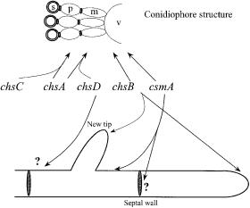
In A. nidulans, four chitin synthases have been cloned (chsA, chsB, chsC, chsD) as well as a gene, csmA, encoding a chitin synthase with a myosin motorlike domain fused at the N-terminus [86]. These chitin synthase genes are classified as class II, III, I, IV, and V, respectively, according to the amino acid similarity system of Bowen et al. [87]. Classes III and V of chitin synthases have been found exclusively in filamentous fungi, signifying a need for chitin synthases with specialized functions, perhaps because of the diversity of the processes requiring chitin deposition. The sites of chitin synthesis in A. nidulans are shown in Fig. 5, which also indicates where the gene products are most active.
Metabolic Engineering of the Morphology of Aspergillus |
117 |
Systematic studies with A. nidulans have shown, that the gene products of chsA, chsC, and chsD are involved in conidiophore formation (conidiation) and consequently spore production [88] (Fig. 5). Double mutants with chsA/chsC and chsA/chsD disruptions severely reduce spore production,signifying that the genes have functional overlap, but surprisingly, no effect was found in a chsC/chsD disruption. This points to the fact that chsA plays a main role in conidiation while chsC and chsD might be supplementary enzymes for two different parts of the conidiation. The two other known chitin synthases, chsB and csmA, are also important in spore production, signifying that all chitin synthases are involved in the complex conidiation process. However, Borgia et al. [89] found it probable (based on heterocaryon studies) that chsB does not take part in synthesis of the conidia itself. In the case of csmA disrupted strains, Horiuchi et al. [90] observed short stalks on the conidiophore vesicle, indicating a role for this chitin synthase in organizing the conidiophore vesicle.
Little is known about the in vivo regulation of chitin synthases in filamentous fungi. Spatial regulation requires either a mechanism for proper targeting of the active chitin synthase and/or a strictly localized activation of random dispersed chitin synthases at the site where chitin synthesis is required. In yeast, localization and activation of chitin synthases are affected not only by ions,metabolites, and zymogenicity but, as has been demonstrated with CHS3, by a large number of proteins such as activator proteins, translocational proteins, and septins [91] and perhaps also phosphorylation [92]. It seems that chitin synthase activity is regulated in a similar complex manner in filamentous fungi, for example, in both yeast and A. fumigatus the major part of chitin is synthesized by a non-zy- mogenic form [93].
Fig. 5. Sites of chitin synthesis in A. nidulans. Conidiophore vesicle (v), metulae or sterigmata (m), phialides (p), and spore (s). The arrows suggest site of chitin synthesis based on observed mutant phenotypes. The products of the genes chsA, chsC, chsA, and chsD seem to have functional overlap
118 |
M. McIntyre et al. |
The formation of new branches requires considerably localized chitinase and glucanase activity, which must be both directed and activated precisely. Regulation of chitin synthase activity has been postulated to occur in the following way. Chitinases, located in lysosomal vesicles [94], may be released through the plasma membrane to the cell wall, lysing the chitin present there. This in turn could be broken down by N-acetyl glucosaminidase to yield N- acetyl glucosamine,which may activate the local chitin synthases [95] in the new tip. However, this mechanism has yet to be verified in vivo.
The effects of chitin synthase gene inactivation are summarized in Table 2. In A. nidulans disruptants of chsA, chsC,and chsD there are no phenotypic changes reported during hyphal growth although a chsD disruptant has been reported to have reduced cell wall chitin [96]. However, chsA/chsC double mutants were sensitive to salts, SDS, the chitin-binding dyes Calcofluor White and Congo red, and chitin synthase inhibitors [88] indicating ill-defined roles for all three chitin synthases in hyphal growth. In csmA disruptions it was found [90] that septa were irregularly positioned, a trait that was remedied when the full gene (including the myosin motor) was expressed driven by the alcA promoter but not when only the chitin synthase part of csmA was expressed. This indicates that the myosin motor domain is important for spatial regulation of this chitin synthase and for septum formation. The csmA disruption also displayed swelling of older parts of the hyphal cell walls, abnormal conidiophores, hypersensitivity to Calcofluor White, and low chitin content [96], indicating a general interference in chitin synthesis in the strain. Therefore, CSMA seems to have a role in maintaining hyphal cell wall integrity and establishing polarized cell wall (or septal) synthesis.
The chsB mutant of A. nidulans had a very reduced specific growth rate and produced stunted and bulging, highly branched hyphae suggesting that the chsB product is very important for the synthesis of chitin at the apical tips in A. nidulans [89, 100]. The chsB gene product only synthesizes a minor chitin sub-frac- tion (Table 2) but it has been shown to be important for correct organization of the hyphal growth. Studies using heterocaryons show that the chsB gene product is not readily diffusible in the hyphae and that individual chitin synthase molecules act in areas of the mycelium in close proximity to the nucleus encoding the molecule [89]. In contrast to the severe phenotype observed in the chsB mutant of A. nidulans, a disruption of the highly similar (88.9% similarity) chsG mutant of A. fumigatus was not as severely inhibiting to growth. The hyphae were hyper-branched but not stunted or bulging [93], indicating that other chitin synthases are capable of maintaining well-organized polar growth. Interestingly, it does not seem to be the other class III chitin synthase (chsC) of A. fumigatus since disruption of chsC/chsG had the same effect as the chsG mutant.
So far there have been no reports of chitin synthase manipulated strains grown in submerged bioprocesses, despite evidence suggesting direct morphological changes may be generated by manipulating chitin synthases. This may make them interesting to examine in connection with fermentation rheology and product secretion. It might be possible that the increased number of tips seen in the chsB mutant could enhance enzyme secretion or that other chitin

Metabolic Engineering of the Morphology of Aspergillus |
119 |
Table 2. Phenotypic effect of single and double chitin synthase gene inactivation in A. nidulans. The nomenclature of Horiuchi et al. [90] has been used as opposed to that of Specht et al. [96]. For clarity chsD [90] = chsE [96] and csmA [90] = chsD [96]
Chitin |
Effect of gene inactivation |
Reference |
|
|
|
|
|
|
On hyphal growth |
On chitin content and |
|
|
|
conidia formation |
|
|
|
|
|
chsA |
No observed effect |
10% decrease in chitin content |
97 |
|
|
30–40% loss in conidia formation |
98 |
chsC |
No observed effect |
No observed effect |
99 |
chsD |
No observed effect |
No effect on chitin content |
99 |
|
|
45% loss of conidia formation |
|
|
|
No loss in conidia formation |
97 |
|
|
30–40% decrease in chitin content. |
96 |
chsB |
Stunted and bulging |
No effect on chitin content |
89 |
|
highly branched |
|
|
|
hyphae |
Reduced (55%) conidia formation |
100 |
|
|
||
csmA |
Intrahyphal hyphae |
Swollen conidiophore vesicles |
90 |
|
and disturbance of |
|
|
|
septation |
|
|
|
Ballooned cell walls |
40% decrease in chitin content |
96 |
|
at subapical regions |
80% loss in conidia formation |
|
chsA and |
No observed effect |
Conidia formation almost totally |
88 |
chsC |
|
lost (99.9%) |
|
chsA and |
No observed effect |
~30% decrease in chitin content |
99 |
chsD |
|
90%–97% loss in conidia formation |
97 |
chsC and |
No observed effect |
Same effect as in chsD inactivated |
99 |
chsD |
|
strain |
|
csmA and |
Same effect as in csmA |
Same effect as in csmA inactivated |
90 |
chsD |
inactivated strain |
strain |
|
|
|
|
|
synthases could be manipulated, making the hyphal structure in such a way that the viscosity of the fermentation culture may be lowered.
4.2
Dimorphic Organisms
The study of dimorphic organisms is extremely relevant when considering the factors controlling and regulating morphology, particularly as investigations of these organisms may give further insight into the control of cell shape and how growth is directed either isotropically or polarly. The dimorphic fungi are defined as those organisms in which vegetative growth can occur in either a hyphal or budding mode depending on the environmental conditions [20]. The list of environmental effectors is rather exhaustive, the effect is often strain specific, and studies dealing with this aspect of dimorphism are numerous in the literature [101–105]. The following review section will consider the regulation of the